. 2023 Aug;31(8):887-894.
doi: 10.1038/s41431-023-01327-7. Epub 2023 Mar 20.

Ribonuclease inhibitor 1 (RNH1) deficiency cause congenital cataracts and global developmental delay with infection-induced psychomotor regression and anemia

Affiliations

Ribonuclease inhibitor 1 (RNH1) deficiency cause congenital cataracts and global developmental delay with infection-induced psychomotor regression and anemia

Carola Hedberg-Oldfors et al. Eur J Hum Genet. 2023 Aug.

Abstract

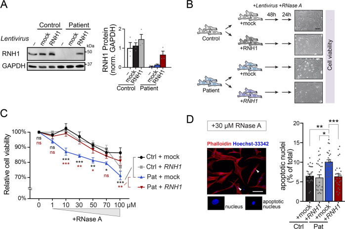
Ribonuclease inhibitor 1, also known as angiogenin inhibitor 1, encoded by RNH1, is a ubiquitously expressed leucine-rich repeat protein, which is highly conserved in mammalian species. Inactivation of rnh1 in mice causes an embryonically lethal anemia, but the exact biological function of RNH1 in humans remains unknown and no human genetic disease has so far been associated with RNH1. Here, we describe a family with two out of seven siblings affected by a disease characterized by congenital cataract, global developmental delay, myopathy and psychomotor deterioration, seizures and periodic anemia associated with upper respiratory tract infections. A homozygous splice-site variant (c.615-2A > C) in RNH1 segregated with the disease. Sequencing of RNA derived from patient fibroblasts and cDNA analysis of skeletal muscle mRNA showed aberrant splicing with skipping of exon 7. Western blot analysis revealed a total lack of the RNH1 protein. Functional analysis revealed that patient fibroblasts were more sensitive to RNase A exposure, and this phenotype was reversed by transduction with a lentivirus expressing RNH1 to complement patient cells. Our results demonstrate that loss-of-function of RNH1 in humans is associated with a multiorgan developmental disease with recessive inheritance. It may be speculated that the infection-induced deterioration resulted from an increased susceptibility toward extracellular RNases and/or other inflammatory responses normally kept in place by the RNase inhibitor RNH1.


Conflict of interest statement

The authors declare no competing interests.

Figures

Fig. 1
Fig. 1. Hematological and neuroradiological investigations.
A, B Both siblings developed macrocytic anemia (B-MCV > 86) with worsening during episodes of infections associated with levels of reticulocytes in the lower part of the normal level range suggestive of impaired erythropoiesis. In patient II:8, the baseline hemoglobin level tended to decrease over time. B-Hb = Blood-Hemoglobin, B-MCV = Blood-Mean Corpuscular Volume. Normal range of B-Hb, B-MCV and reticulocytes are given within parenthesis. CF CT of the brain in patient II:7 at 8 months of age showing reduced attenuation of the entire supratentorial gray matter, compressed lateral ventricles and flattened extracerebral CSF spaces compatible with edema, loss of gray and white matter differentiation suggestive of ischemia and herniation (E, F) through the tentorium and foramen magnum. GI In patient II:8 MRI of the brain at 8 months of age showed symmetrically increased T2 signal in thalamus, globus pallidus and in patchy cortical/subcortical fronto-parietal, paramedian and occipital areas.
Fig. 2
Fig. 2. Pedigree and molecular genetics.
A Pedigree of the family. Filled symbols indicate affected individuals. Segregation of the c.615-2A > C variant in the family is indicated by + (wild type) and – (variant). B Sequencing chromatograms showing the splice-site variant c.615-2A > C in RNH1 indicated by an arrow. C Schematic illustration of the exon-intron structure of RNH1 (NM_203387.3), including the novel splice-site variant identified in this study (in red, c.615-2A > C). Domains encoded by each exon are indicated.
Fig. 3
Fig. 3. Analysis of aberrant RNH1 splicing in muscle and fibroblasts of patient II:8.
A Schematic illustration showing the splice-site effect of the c.615-2A > C variant as investigated with reverse transcriptase polymerase chain reaction (RT-PC) followed by PCR and Sanger sequencing using forward primer located in exon 6 and reverse primer located in exon 8 on RNA extracted from muscle tissue (for forward primer located in exon 5 and reverse primer located in exon 9 see Supplementary Fig. 3A). In patient II:8 a band with lower molecular size was identified compared to a control sample using and Sanger sequencing chromatograms showing transcript lacking exon 7. B Sashimi-plots illustrating splicing of RNH1 exons 5-9 (black boxes), generated from RNA-sequencing of patient-derived fibroblasts (II:8) (mock-transduced fibroblasts; Supplementary Fig. 5B). The numbers display junctions information for regions within the current IGV view. The “bridges” crossing exons indicate junction reads and the numbers of junction reads are shown on the “bridges”. Genome coordinates by GRCh38/hg38.
Fig. 4
Fig. 4. Functional investigations of patient-derived fibroblasts.
A Western blot demonstrating lack of RNH1 protein in patient-derived fibroblasts and re-expression of RNH1-V5 by following lentiviral transduction. Data represented as mean ± standard error of the mean (s.e.m.; n = 3). B Outline of the RNase A tolerance assay. Inserts, DIC images of cells at time of analysis. Scale bar, 100 μm. C Relative cell viability following exposure to an increasing concentration of recombinant RNase A. Data represented as mean ± s.e.m. (n = 6). Statistical comparisons between (by brackets) indicated groups were done by two-way ANOVA with Holm-Sidak tests. *, p < 0.05; **, p < 0.01; ***, p < 0.001; ns, not significant. D Quantitative analysis of the proportion of apoptotic cell nuclei, detected by nuclear Hoechst-staining, after 24 h exposure to 30 μM RNase A. Representative phalloidin- and Hoechst-stained cells (left) with apoptotic nuclei indicated by arrowheads, and summary data (right), represented as mean ± s.e.m. (n = 25–28 fields of view from a total of four independent experiments). Scale bar, 100 μm. Statistical comparisons by one-way ANOVA with Bonferroni correction. *, p < 0.05; **, p < 0.01; ***, p < 0.001.

Comment in

References

    1. Dickson KA, Haigis MC, Raines RT. Ribonuclease inhibitor: structure and function. Prog Nucleic Acid Res Mol Biol. 2005;80:349–74. doi: 10.1016/S0079-6603(05)80009-1. - DOI - PMC - PubMed
    1. Lee FS, Fox EA, Zhou HM, Strydom DJ, Vallee BL. Primary structure of human placental ribonuclease inhibitor. Biochemistry. 1988;27:8545–53. doi: 10.1021/bi00423a007. - DOI - PubMed
    1. Kobe B, Deisenhofer J. Crystal structure of porcine ribonuclease inhibitor, a protein with leucine-rich repeats. Nature. 1993;366:751–6. doi: 10.1038/366751a0. - DOI - PubMed
    1. Dickson KA, Kang DK, Kwon YS, Kim JC, Leland PA, Kim BM, et al. Ribonuclease inhibitor regulates neovascularization by human angiogenin. Biochemistry. 2009;48:3804–6. doi: 10.1021/bi9005094. - DOI - PMC - PubMed
    1. Sarangdhar MA, Allam R. Angiogenin (ANG)-Ribonuclease Inhibitor (RNH1) System in Protein Synthesis and Disease. Int J Mol Sci. 2021;22:1287. doi: 10.3390/ijms22031287. - DOI - PMC - PubMed

Publication types