. 2023 Mar 20;42(1):68.
doi: 10.1186/s13046-023-02644-x.

CLDN6 inhibits breast cancer metastasis through WIP-dependent actin cytoskeleton-mediated autophagy

Affiliations

CLDN6 inhibits breast cancer metastasis through WIP-dependent actin cytoskeleton-mediated autophagy

Yuan Dong et al. J Exp Clin Cancer Res. .

Abstract

Background: As a breast cancer suppressor gene, CLDN6 overexpression was found to inhibit breast cancer metastasis in our previous studies, but the specific mechanism remains unclear. This study aimed to clarify the role and mechanism of CLDN6 in inhibiting breast cancer metastasis.

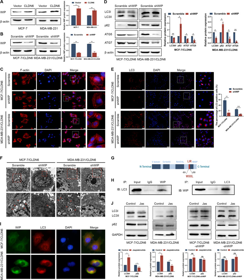
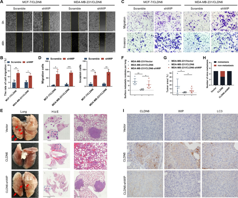
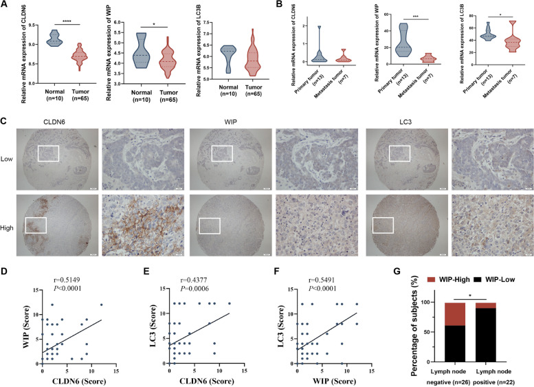
Methods: Western blot, immunofluorescence and transmission electron microscopy were performed to detect autophagy. Wound healing, transwell assays and lung metastasis mouse models were used to examine breast cancer metastasis. Phalloidin staining and immunofluorescent staining were used to observe actin cytoskeleton. mRNA seq, RT-PCR, western blot, chromatin immunoprecipitation, dual luciferase reporter assay, co-immunoprecipitation and immunofluorescence were performed to define the molecular mechanism. The expression levels and clinical implication of CLDN6, WIP and LC3 in breast cancer tissues were evaluated using immunohistochemistry.

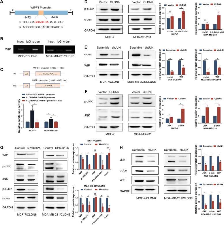
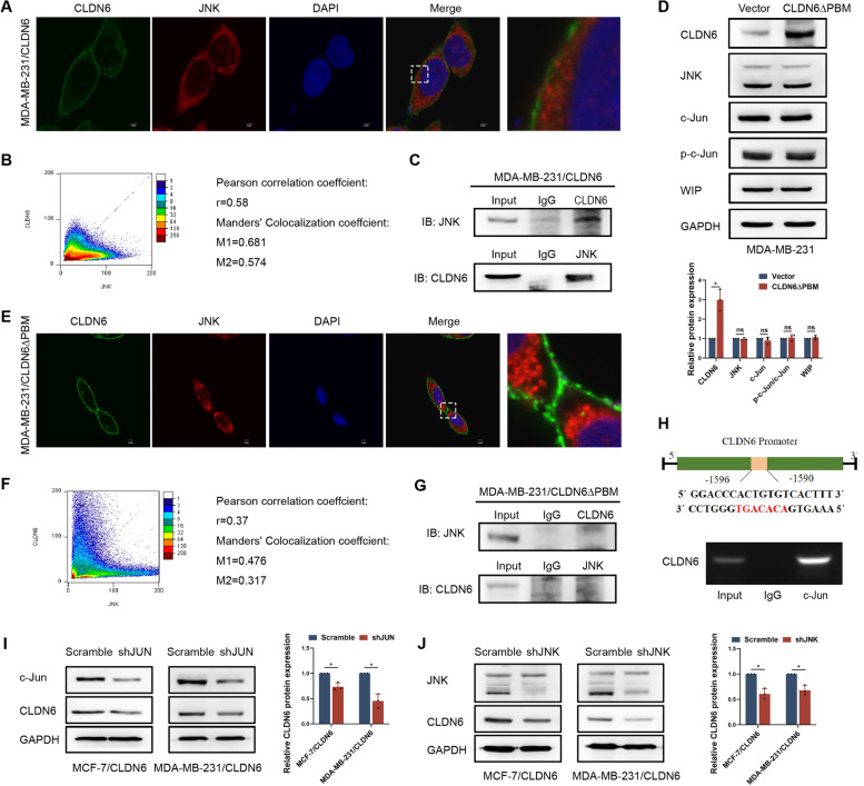
Results: We demonstrated that CLDN6 inhibited breast cancer metastasis through autophagy in vitro and vivo. We unraveled a novel mechanism that CLDN6 regulated autophagy via WIP-dependent actin cytoskeleton assembly. Through its PDZ-binding motif, overexpressed CLDN6 interacted with JNK and upregulated JNK/c-Jun pathway. C-Jun promoted WIP expression at the transcriptional level. Notably, we observed c-Jun transcriptionally upregulated CLDN6 expression, and there was a positive feedback loop between CLDN6 and JNK/c-Jun. Finally, we found that CLDN6, WIP and LC3 expression correlated with each other, and WIP expression was significantly associated with lymph node metastasis of breast cancer patients.

Conclusions: The data provide a new insight into the inhibitory effects of CLDN6-mediated autophagy on breast cancer metastasis, and revealed the new mechanism of CLDN6 regulating autophagy through WIP-dependent actin cytoskeleton. Our findings enrich the theoretical basis for CLDN6 as a potential biomarker for breast cancer diagnosis and therapy.

Keywords: Actin cytoskeleton; Autophagy; Breast cancer; CLDN6; Metastasis; WIP.


Conflict of interest statement

The authors declare that they have no competing interests.

Figures

Fig. 1
Fig. 1
CLDN6 regulates breast cancer metastasis via autophagy in vitro and vivo. A Western blot showed levels of autophagy-related proteins in MCF-7 and MDA-MB-231 cells with or without CLDN6 overexpression. B LC3 puncta was visualized by IF analysis, and the percentage of LC3 positive cells was quantified, zoomed-in image (right). Scale bar: 50 μm. C Western blot showed levels of autophagy after treated with 2 mM 3-MA or 25 μM CQ for 24 h. (D-E) Wound healing assay and transwell migration and invasion assays were performed in CLDN6-overexpression cells treated with 3-MA or CQ. Scale bar: 200 μm (D) and 50 μm (E). Results of (A − E) were from three independent experiments. F Bioluminescence imaging (BLI) was used to monitor metastases of BALB/c-nu mice. Representative lung metastatic nodes (red arrowheads). Representative H&E staining of lung sections from the three groups (n = 5 mice/group), Scale bar: 2 mm (left), 200 μm (right). G The number of surface metastatic nodes per lung in each group. H The areas were quantified with Image-Pro Plus and the tumor ratio (%) was determined as (tumor area)/(total lung area) × 100%. I The number of mice with lung metastasis was counted in each group. *P < 0.05, **P < 0.01, ***P < 0.001, ****P < 0.0001 and ns: no significance
Fig. 2
Fig. 2
CLDN6 mediates autophagy by regulating actin cytoskeleton. A F-actin labeled with Rhodamine phalloidin in MCF-7 and MDA-MB-231 cells with or without CLDN6 overexpression, zoomed-in image (right). Scale bar: 20 μm. B IF analysis was performed to detect the colocalization of LC3 (green) with p-Arp3 (red) in MCF-7/CLDN6 and MDA-MB-231/CLDN6 cells. Scale bar: 20 μm. C ImageJ was used for colocation analysis of p-Arp3 and LC3. Pearson correlation analysis showed that r > 0.5. Manders, colocation coefficient showed that M1 > 0.5 and M2 > 0.5. D The expression of LC3 and p62 was detected by western blot in MCF-7/CLDN6 and MDA-MB-231/CLDN6 cells treated with or without 150 μM CK666 for 4 h. E IF analysis showed that the percentage of LC3 positive cells was decreased after CK666 treatment. All results were from three independent experiments. Scale bar: 20 μm. *P < 0.05, **P < 0.01 and ***P < 0.001
Fig. 3
Fig. 3
CLDN6 increases WIP expression to regulate actin cytoskeleton-mediated autophagy. A Western blot showed levels of WIP protein in MCF-7 and MDA-MB-231 cells with or without CLDN6 overexpression. B Western blot showed levels of WIP protein in MCF-7/CLDN6 and MDA-MB-231/CLDN6 cells with or without WIP knockdown. C F-actin labeled with Rhodamine phalloidin in MCF-7/CLDN6 and MDA-MB-231/CLDN6 cells with or without WIP knockdown, zoomed-in image (right). Scale bar: 20 μm. D Western blot showed levels of autophagy-related proteins in MCF-7/CLDN6 and MDA-MB-231/CLDN6 cells with or without WIP knockdown. Results were from three independent experiments. E IF analysis showed LC3 positive cells in MCF-7/CLDN6 and MDA-MB-231/CLDN6 cells with or without WIP knockdown, zoomed-in image (right). Scale bar: 50 μm. F TEM analysis was performed on MCF-7/CLDN6 and MDA-MB-231/CLDN6 cells with or without WIP knockdown. Control group displayed several autophagic vacuoles (red arrowheads) which were not observed in WIP knockdown cells. Scale bar: 2 μm (top) and 1 μm (bottom). G The potential LIR motifs of the WIP. H The interaction of WIP and LC3 was detected by Co–IP assay in MDA-MB-231/CLDN6 cells. I IF analysis was performed to detect the colocalization of WIP (green) with LC3 (red) in MCF-7/CLDN6 and MDA-MB-231/CLDN6 cells. Scale bar: 20 μm. J Western blot showed the expression of LC3 and p62 in MCF-7/CLDN6, MDA-MB-231/CLDN6, MCF-7/CLDN6-shWIP and MDA-MB-231/CLDN6-shWIP cells treated with or without 100 nM Jasplakinolide for 2 h. Results were from three independent experiments. *P < 0.05, **P < 0.01, ***P < 0.001 and ns: no significance
Fig. 4
Fig. 4
CLDN6 inhibits breast cancer metastasis via the upregulation of WIP expression in vitro and vivo. A-D Wound healing assay and transwell migration and invasion assays were performed in MCF-7/CLDN6 and MDA-MB-231/CLDN6 cells with or without WIP knockdown. Results were from three independent experiments. Scale bar: 200 μm (A) and 50 μm (C). E Representative examples of lungs with metastatic foci (red arrowheads) are shown. Representative H&E staining of lung sections from the three groups (n = 5 mice/group), zoomed-in image (right). Scale bar: 5 mm (left), 500 μm (right). F The number of surface metastatic nodes per lung in each group. G The areas were quantified with Image-Pro Plus and the tumor ratio (%) was determined as (tumor area)/(total lung area) × 100%. H The number of mice with lung metastasis was counted in each group. I Representative IHC images of CLDN6, WIP, and LC3 in the lung metastatic tumor tissues. Scale bar: 20 μm. *P < 0.05, **P < 0.01 and ***P < 0.001
Fig. 5
Fig. 5
CLDN6 upregulates WIP at transcriptional level via JNK/c-Jun pathway. A The potential transcriptional binding site of c-Jun on the WIP promoter. B ChIP experiment determined the combination of c-Jun with the predicted site in the WIP promoter. C Cells were transfected with Wt or Mut WIP promoter reporter plasmid. At 48 h after transfection, promoter activity was analyzed using dual-luciferase assay. D Western blot showed levels of total and phosphorylated c-Jun in MCF-7 and MDA-MB-231 cells with or without CLDN6 overexpression. E Western blot showed levels of WIP protein in MCF-7/CLDN6 and MDA-MB-231/CLDN6 cells with or without c-Jun knockdown. F Western blot showed levels of total and phosphorylated JNK in MCF-7 and MDA-MB-231 cells with or without CLDN6 overexpression. G Western blot showed levels of WIP and JNK/c-Jun in MCF-7/CLDN6 and MDA-MB-231/CLDN6 cells treated with or without 15 μM SP600125 for 4 h. H Western blot showed levels of WIP and JNK/c-Jun in MCF-7/CLDN6 and MDA-MB-231/CLDN6 cells with or without JNK knockdown. Results were from three independent experiments. *P < 0.05, **P < 0.01, and ns: no significance
Fig. 6
Fig. 6
CLDN6 interacts with JNK via the PDZ-binding motif. A IF analysis was performed to detect the co-localization of CLDN6 (green) with JNK (red) in MDA-MB-231/CLDN6 cells, zoomed-in image (right). Scale bar: 3 μm. B ImageJ was used for colocation analysis. Pearson correlation analysis showed that r > 0.5. Manders, colocation coefficient showed that M1 > 0.5 and M2 > 0.5. C The interaction of CLDN6 and JNK was detected by Co–IP assay in MDA-MB-231/CLDN6 cells. D Western blot showed levels of JNK/c-Jun pathway and WIP in MDA-MB-231 cells with or without CLDN6ΔPBM overexpression. Results were from three independent experiments. E The interaction of CLDN6 and JNK was detected by Co–IP assay in MDA-MB-231/CLDN6ΔPBM cells. F IF analysis was performed to detect the co-localization of CLDN6 (green) with JNK (red) in MDA-MB-231/CLDN6ΔPBM cells. Scale bar: 3 μm. G ImageJ was used for colocation analysis. Pearson correlation analysis showed that r < 0.5. Manders, colocation coefficient showed that M1 < 0.5 and M2 < 0.5. H The potential transcriptional binding site of c-Jun on the CLDN6 promoter, and was verified by ChIP assays. I-J JNK/c-Jun feedback regulates CLDN6 expression. I Western blot showed levels of CLDN6 protein in MCF-7/CLDN6 and MDA-MB-231/CLDN6 cells with or without c-Jun knockdown. J Western blot showed levels of CLDN6 protein in MCF-7/CLDN6 and MDA-MB-231/CLDN6 cells with or without JNK knockdown. Results were from three independent experiments. *P < 0.05 and ns: no significance
Fig. 7
Fig. 7
The clinical correlation between CLDN6, WIP and LC3 expression in breast cancer patients. A The mRNA expression of CLDN6, WIP and LC3 in adjacent normal and breast cancer tissues in the GEO database GSE103512. B The mRNA expression of CLDN6, WIP and LC3 in primary breast tumor tissues and metastatic tissues in the GEO database GSE191230. C Representative IHC images of low and high CLDN6, WIP and LC3 expression in breast cancer tissues. Scale bar, 200 µm (left), 20 µm (right). D-F Correlation analysis between CLDN6 and WIP (D), CLDN6 and LC3 (E), and WIP and LC3 (F) in breast cancer tissue microarray. Pearson correlation test, n = 58. G Percentage of high and low expression of WIP in breast cancer patients with lymph node-negative (n = 26) and node-positive (n = 22). *P < 0.05, ***P < 0.001, and ****P < 0.0001
Fig. 8
Fig. 8
A proposed model for the regulatory mechanism of CLDN6-mediated autophagy inhibiting breast cancer metastasis

References

    1. Lin D, Guo Y, Li Y, Ruan Y, Zhang M, Jin X, et al. Bioinformatic analysis reveals potential properties of human Claudin-6 regulation and functions. Oncol Rep. 2017;38(2):875–885. doi: 10.3892/or.2017.5756. - DOI - PMC - PubMed
    1. Sas D, Hu M, Moe OW, Baum M. Effect of claudins 6 and 9 on paracellular permeability in MDCK II cells. Am J Physiol Regul Integr Comp Physiol. 2008;295(5):R1713–R1719. doi: 10.1152/ajpregu.90596.2008. - DOI - PMC - PubMed
    1. Jimenez FR, Lewis JB, Belgique ST, Milner DC, Lewis AL, Dunaway TM, et al. Cigarette smoke and decreased oxygen tension inhibit pulmonary claudin-6 expression. Exp Lung Res. 2016;42(8–10):440–452. doi: 10.1080/01902148.2016.1261309. - DOI - PubMed
    1. Kitajiri SI, Furuse M, Morita K, Saishin-Kiuchi Y, Kido H, Ito J, et al. Expression patterns of claudins, tight junction adhesion molecules, in the inner ear. Hear Res. 2004;187(1–2):25–34. doi: 10.1016/S0378-5955(03)00338-1. - DOI - PubMed
    1. Turksen K, Troy TC. Permeability barrier dysfunction in transgenic mice overexpressing claudin 6. Development (Cambridge, England) 2002;129(7):1775–1784. doi: 10.1242/dev.129.7.1775. - DOI - PubMed