. 2023 Apr;42(2):183-202.
doi: 10.14366/usg.22130. Epub 2022 Oct 5.

Top applications of dermatologic ultrasonography that can modify management

Affiliations

Top applications of dermatologic ultrasonography that can modify management

Ximena Wortsman. Ultrasonography. 2023 Apr.

Abstract

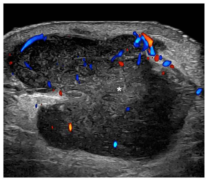
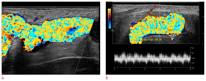
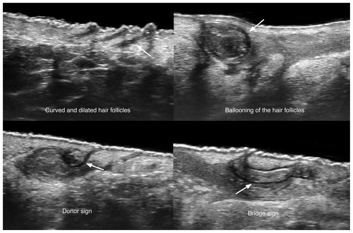
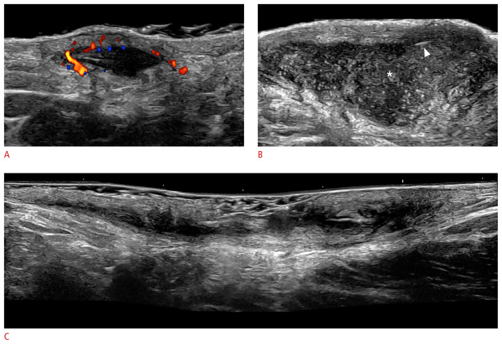
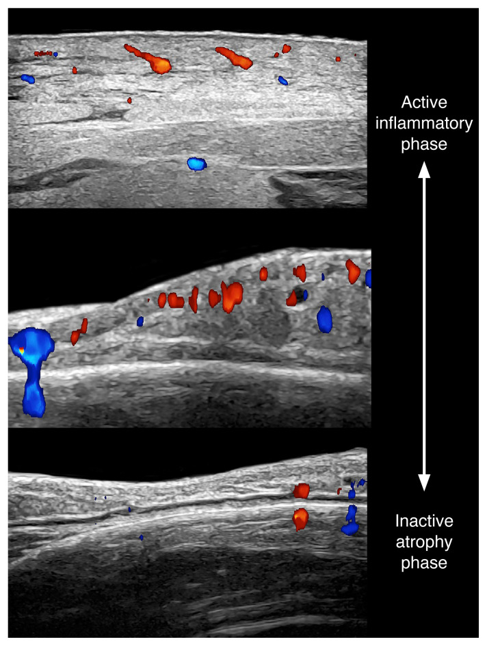
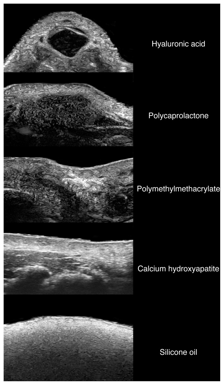
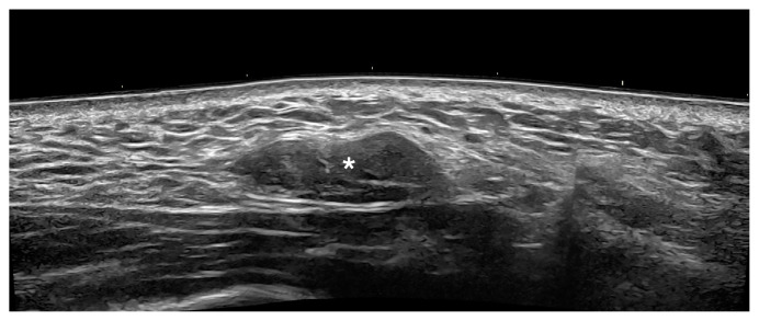
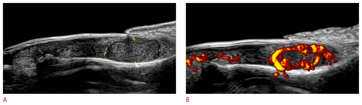
Dermatologic ultrasonography is a new field that has been growing exponentially in the last 10 years. It has multiple applications that can modify patient management, such as the assessment of benign and malignant cutaneous tumors, vascular anomalies, inflammatory dermatologic entities, aesthetic complications, and nail lesions. Compared with other imaging techniques such as computed tomography or magnetic resonance imaging, ultrasonography has the highest axial spatial resolution and has benefited from the development of high- and ultra-high-frequency probes that could even reach 70 MHz. The daily use of ultrasonography in dermatology has been reported to improve the accuracy of diagnoses, the tracking of activity, and the assessment of severity in common dermatologic conditions, which certainly can support better treatment of patients.

Keywords: Cancer; Dermatology; Hidradenitis suppurativa; Skin; Ultrasonography.


Conflict of interest statement

No potential conflict of interest relevant to this article was reported.

Figures

Fig. 1
Fig. 1
Normal ultrasound anatomy of the skin. These images (A, forearm skin at 70 MHZ; B, plantar skin at 24 MHz) present the ultrasonographic patterns of the epidermis, dermis and hypodermis at different frequencies. Notice the echostructure of the hair follicle in A.
Fig. 2
Fig. 2
Normal ultrasound anatomy of the nail (longitudinal view; index finger) at 24 MHz. The vertical arrow shows the dorsal plate and the oblique arrow presents the ventral plate.
Fig. 3
Fig. 3
Normal hair tract (arrow) within a dermal hair follicle before emerging into the surface at 70 MHz.
Fig. 4
Fig. 4
Epidermal cyst (24 MHz). A. Intact (left cheek): grayscale ultrasonography with a color filter presents a well-defined, round-shaped, hypoechoic dermal and hypodermal structure (*). Posterior acoustic enhancement is noted at the bottom of the cyst (arrows). B. Ruptured (right dorsal forearm): color Doppler ultrasonography presents a partially lobulated hypoechoic dermal and hypodermal structure with some ill-defined areas at the bottom and a more intense hypoechoic band on the surface suggestive of a fragment of the capsule. The cyst generates a posterior acoustic enhancement artifact at the bottom (arrows). The hypervascularity in the periphery of the cyst should be noticed. There is also increased echogenicity of the surrounding hypodermis due to inflammation.
Fig. 5
Fig. 5
Pilomatrixoma of the “target type” (24 MHz; right arm). Color Doppler (A) and three-dimensional grayscale reconstruction with a color filter (B) show a well-defined, oval-shaped dermal and hypodermal structure. There are multiple hyperechoic focal areas suggestive of calcifications, some of them with posterior acoustic shadowing artifact. On color Doppler, there is hypervascularity in the periphery of the tumor.
Fig. 6
Fig. 6
Dermatofibroma. Grayscale (A) and color Doppler ultrasound (B) images (24 MHz; dorsum of the right foot) present hypoechoic ill-defined dermal focal zone (between markers), slightly fusiform, with a more deeply hypoechoic central part. On color Doppler, there are few vessels within the structure.
Fig. 7
Fig. 7
Basal cell carcinoma (low risk of recurrence; macronodular type; 70 MHz; nasal tip). Ultrasonography (grayscale) presents a hypoechoic dermal lesion (*) with irregular borders and a few hyperechoic spots (arrowheads).
Fig. 8
Fig. 8
Squamous cell carcinoma (24 MHz; left cheek). Grayscale (A) and power Doppler (B) show a hypoechoic lesion (*) with irregular borders located in the dermis and upper hypodermis. There is an upward displacement of the epidermis and increased echogenicity of the surrounding hypodermis. On power Doppler, there is prominent hypervascularity within the lesion and its periphery.
Fig. 9
Fig. 9
Melanoma in-transit metastasis (24 MHz; right arm). Color Doppler ultrasound demonstrates a hypoechoic hypodermal nodule, slightly heterogeneous with lobulated borders and irregular vascularity. There is increased echogenicity of the surrounding hypodermis. The asterisk marks the metastasis.
Fig. 10
Fig. 10
Infantile hemangioma in the partial regression phase (24 MHz; scalp). A, B. Heterogeneous dermal and hypodermal mass that presents hypoechoic and hyperechoic areas. On power Doppler (B), there is prominent vascularity in the mass.
Fig. 11
Fig. 11
Arteriovenous vascular malformation (24 MHz; left gluteal region). Color Doppler (A) and pulsed Doppler (B) show a tortuous vascular hypodermal structure with turbulent flow (aliasing artifact). The pulsed Doppler (spectral curve analysis) demonstrated a to-and-fro blood flow.
Fig. 12
Fig. 12
Ultrasonographic signs of early hidradenitis suppurativa at 70 MHz (axillary and groin regions). The arrows on the left show the donor sign and the arrow on the right demonstrates the bridge sign.
Fig. 13
Fig. 13
Key ultrasound lesions of hidradenitis suppurativa (24 MHz; axillary and groin regions). Pseudocyst (color Doppler) (A), fluid collection (*) (B), and fistulous tract, also called tunnel (grayscale) (C) are shown. The hypervascularity in the periphery of the pseudocyst in A and the presence of fragments of hair tracts in the fluid collection (B, arrowhead) should be noted. The pseudocyst is in the dermis, and the fluid collection and tunnel are located in the dermis and upper hypodermis.
Fig. 14
Fig. 14
Morphea (i.e., cutaneous form of scleroderma; 24 MHz; top, right breast; middle and bottom, frontal part of the face). Color Doppler ultrasound images demonstrate the different phases of activity of morphea from active (top) to inactive and atrophy (bottom).
Fig. 15
Fig. 15
Ultrasonographic patterns of common cosmetic fillers (24 MHz; facial region).
Fig. 16
Fig. 16
Granuloma after the injection of hyaluronic acid in the right cheek (24 MHz). Ultrasonography (grayscale; transverse panoramic view) presents an oval-shaped and slightly lobulated nodule (*) in the hypodermis. This site was previously injected with hyaluronic acid.
Fig. 17
Fig. 17
Glomus tumor of the nail (24 MHz; left thumb). Grayscale (A) and Power Doppler (B) images show an oval-shaped, well-defined, hypoechoic nodule (between markers in A) in the proximal part of the nail bed. There is a slight upward displacement of the nail plate and scalloping of the underlying bony margin of the distal phalanx. On power Doppler, there is prominent vascularity within the nodule.
Fig. 18
Fig. 18
Onychomatricoma (24 MHz, right index finger). Grayscale (A) and three-dimensional reconstruction (B) of the nail (with a color filter) show an ill-defined, hypoechoic structure (*) in the proximal nail bed that presents multiple hyperechoic lines protruding into the nail plate (arrowheads).
Fig. 19
Fig. 19
Periungual myxoid (synovial) cyst (24 MHz; left middle finger). Ultrasound grayscale images (longitudinal [A] and transverse view of the proximal nail fold [B], transverse view of the middle of the nail [C]) demonstrate an oval-shaped, well-defined anechoic cystic structure (*) that is compressing the proximal part of the nail plate (arrows) and therefore, the matrix region. There is a concavity in the nail plate (C) on the same axis of the cyst.
Fig. 20
Fig. 20
Ultrasound patterns of nail psoriasis (color Doppler; 24 MHz; fingernails). The hypervascularity of the nail beds should be noticed, as well as the morphological alterations. A–C. On ultrasonography, loss of definition of the ventral plate, hyperechoic deposits in the ventral nail plate (A), thickening and decreased echogenicity of the nail bed (B), and a wavy and thick nail plate (C, arrows) are suggestive of nail psoriasis.
Fig. 21
Fig. 21
Retronychia at 24 MHz. A. Power Doppler (left first toe) presents thickening and hypoechogenicity of the nail bed, fragmentation of the proximal nail plate (onychomadesis), thickening and hypoechogenicity of the proximal nail fold. There is a halo sign (*, hypoechoic band) in the periphery of the origin of the nail plate with hypervascularity of the proximal nail fold. B. Grayscale comparative views side-by-side show a decreased distance between the origin of the nail plate and the base of the distal phalanx on the left side (retronychia) versus the right side (4.8 mm vs. 9.2 mm).

References

    1. Wortsman X. Top advances in dermatologic ultrasound. J Ultrasound Med. 2022;42:521–545. - PubMed
    1. Alfageme F, Wortsman X, Catalano O, Roustan G, Crisan M, Crisan D, et al. European Federation of Societies for Ultrasound in Medicine and Biology (EFSUMB) position statement on dermatologic ultrasound. Ultraschall Med. 2021;42:39–47. - PubMed
    1. Almuhanna N, Wortsman X, Wohlmuth-Wieser I, Kinoshita-Ise M, Alhusayen R. Overview of ultrasound imaging applications in dermatology [formula: see text] J Cutan Med Surg. 2021;25:521–529. - PMC - PubMed
    1. Catalano O, Varelli C, Sbordone C, Corvino A, De Rosa D, Vallone G, et al. A bump: what to do next? Ultrasound imaging of superficial soft-tissue palpable lesions. J Ultrasound. 2020;23:287–300. - PMC - PubMed
    1. Mandava A, Ravuri PR, Konathan R. High-resolution ultrasound imaging of cutaneous lesions. Indian J Radiol Imaging. 2013;23:269–277. - PMC - PubMed

LinkOut - more resources