

doi: 10.1016/j.crmeth.2023.100412.
eCollection 2023 Feb 27.
Illuminating cellular and extracellular vesicle-mediated communication via a split-Nanoluc reporter in vitro and in vivo
Affiliations
- PMID: 36936071
- PMCID: PMC10014296
- DOI: 10.1016/j.crmeth.2023.100412
Item in Clipboard
Illuminating cellular and extracellular vesicle-mediated communication via a split-Nanoluc reporter in vitro and in vivo
Cell Rep Methods.
.
Abstract
Tools to effectively demonstrate and quantify functional delivery in cellular communication have been lacking. This study reports the use of a fluorescently labeled split Nanoluc reporter system to demonstrate and quantify functional transfer between cells in vitro and in a subcutaneous tumor mouse model. Our construct allows monitoring of direct, indirect, and specifically extracellular vesicle-mediated functional communication.
Keywords: cancer; cellular communication; extracellular vesicles; functional transfer.
© 2023 The Authors.
Conflict of interest statement
The authors declare no competing interests.
Figures
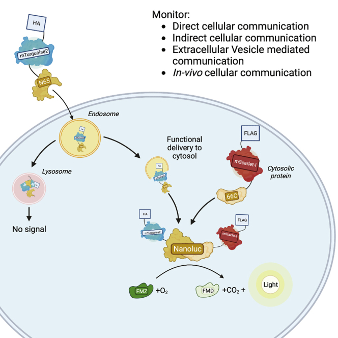
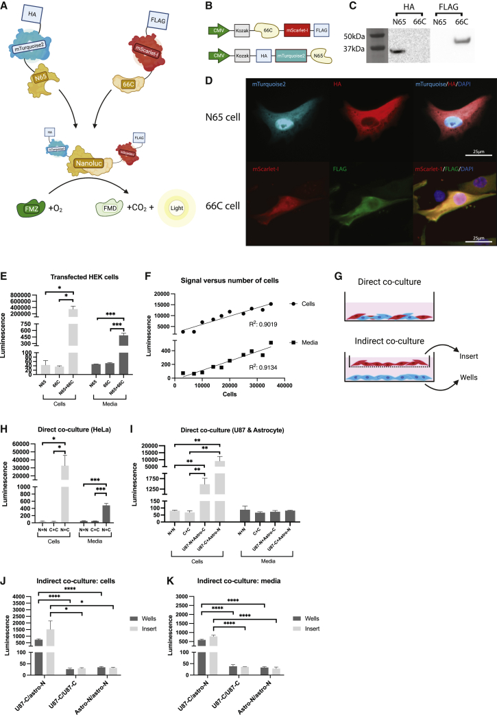
Validation of the split Nanoluc construct and functional delivery (A) Schematic illustration of the mechanism. The split halves of the Nanoluc fuse when in proximity to one another. Nanoluc then oxidizes furimazine (FMZ) to furimamide (FMD), carbon dioxide (CO2), and light. HA, human influenza hemagglutinin. (B) Design of the construct. CMV, cytomegalo virus. (C) Western blot of HEK cell lysates stained with anti-FLAG and anti-HA. kDa, kilodalton. (D) Fluorescent imaging of N65 and 66C transduced HeLa cells. DAPI, 4′,6-diamidino-2-phenylindole. Scale bar: 25 . (E) HEK cells transfected with either one or both constructs. Signal measured in media and in cell lysate after adding FMZ for 1 min (n = 3 per condition, SEM). Student’s independent t test. ∗p < 0.05, ∗∗∗p < 0.001. (F) Correlation between luminescence and cell number in transfected HEK cells, as measured in cell lysate and media. One replicate per condition. Linear regression analysis, p < 0.0001 for both. (G) Schematic of direct and indirect co-culture of cells. (H) Direct co-culture of transduced HeLa cells. N65 (N) or 66C (C) was cultured. Luminescence could be detected in the cell lysate and media after adding FMZ. Student’s independent t test. ∗p < 0.05, ∗∗∗p < 0.001 (n = 3 per condition, SEM). (I) Direct co-culture of U87 cells and astrocytes transduced with either N65 (N) or 66C (C). Strong signal was detected in the cell lysate but not media. Student’s independent t test. ∗∗p < 0.01 (n = 3 per condition, SEM). (J) Indirect co-culture of U87-66C and astrocyte-N65 cells. Luminescent signal in cells. Student’s independent t test. ∗p < 0.05, ∗∗∗∗p < 0.0001 (n = 3 per condition, SEM). (K) Indirect co-culture of U87-66C and astrocyte-N65 cells. Luminescent signal in media. Student’s independent t test. ∗∗∗∗p < 0.0001 (n = 3 per condition, SEM).
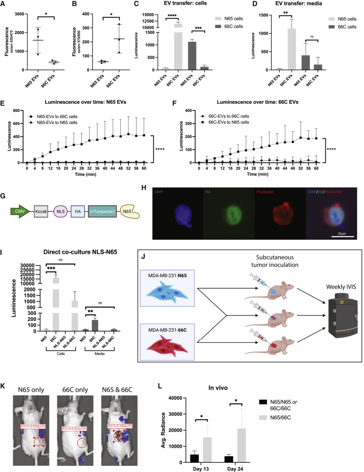
Functional delivery in EVs, modification of the construct, and in vivo possibilities (A) Fluorescence of EVs isolated from transfected HEK cells via size exclusion (SE) excited at 435 nm. Student’s independent t test. ∗p < 0.05 (n = 3 per condition, SEM). (B) Fluorescence of EVs excited at 570 nm. Student’s independent t test. ∗p < 0.05 (n = 3 per condition, SEM). (C) EVs isolated from transfected HEK cells and added to transduced HEK-N65 and HEK-66C cells. Luminescence in cells after addition of FMZ. Student’s independent t test. ∗∗∗p < 0.001, ∗∗∗∗p < 0.0001 (n = 3 per condition, SEM). (D) EVs isolated from transfected HEK cells and added to transduced HEK-N65 and HEK-66C cells. Luminescence in media after addition of FMZ. Student’s independent t test. ns, not significant. ∗∗p < 0.01 (n = 3 per condition, SEM). (E) Luminescence over time upon adding HEK-N65-derived EVs. EVs and FMZ were added at t = 0. Difference between HEK-66c and HEK-N65 cells at t = 60 min, independent Student’s t test: ∗∗∗∗p < 0.0001 (n = 3 per condition, SEM). (F) Luminescence over time upon adding HEK-66C-derived EVs. EVs and FMZ were added at t = 0. Difference between HEK-66c and HEK-N65 cells at t = 60 min, Student’s t test: ∗∗∗∗p < 0.0001 (n = 3 per condition, SEM). (G) Nuclear-localizing construct for N65 cells. A nuclear-localizing signal (NLS) was added to the HA-mTurquoise2-N65 construct, fusing it to the split Nanoluc. (H) HEK cells transduced with the NLS-N65 construct. The signal is localized to the nucleus, with no fluorescence seen in the cell membrane, as visualized with phalloidin staining. 25 . (I) Direct co-culture of NLS and normal constructs. When the protein is contained in the nucleus, a decrease in signal is seen. Student’s independent t test. ns, not significant. ∗∗p < 0.01, ∗∗∗p < 0.001 (n = 3 per condition, SEM). (J) Setup for the in vivo experiment. IVIS, in vivo imaging system. (K) Examples of nude mice injected with MDA-MB-231-N65, MDA-MB-231-66c, or both tumor lines. Measurement of luminescence 10 min after injection of fluorofurimazine. (L) Average radiance 10 min after injection of fluorofurimazine on days 13 and 24 of tumor injection. Student’s independent t test. ∗p < 0.05 (n = 4 per condition, SEM).
References
Publication types
MeSH terms
Substances
Grants and funding
LinkOut - more resources
Full Text Sources
