Review
. 2023 Mar 1:14:1117749.
doi: 10.3389/fimmu.2023.1117749. eCollection 2023.

IgA-producing B cells in lung homeostasis and disease

Affiliations
Review

IgA-producing B cells in lung homeostasis and disease

Youri Bertrand et al. Front Immunol. .

Abstract

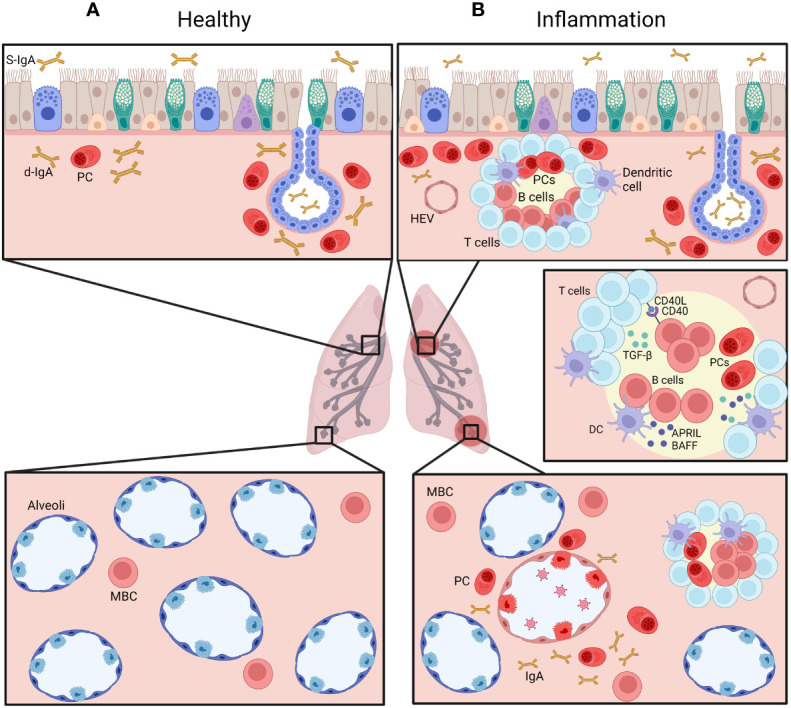
Immunoglobulin A (IgA) is the most abundant Ig in mucosae where it plays key roles in host defense against pathogens and in mucosal immunoregulation. Whereas intense research has established the different roles of secretory IgA in the gut, its function has been much less studied in the lung. This review will first summarize the state-of-the-art knowledge on the distribution and phenotype of IgA+ B cells in the human lung in both homeostasis and disease. Second, it will analyze the studies looking at cellular and molecular mechanisms of homing and priming of IgA+ B cells in the lung, notably following immunization. Lastly, published data on observations related to IgA and IgA+ B cells in lung and airway disease such as asthma, cystic fibrosis, idiopathic pulmonary fibrosis, or chronic rhinosinusitis, will be discussed. Collectively it provides the state-of-the-art of our current understanding of the biology of IgA-producing cells in the airways and identifies gaps that future research should address in order to improve mucosal protection against lung infections and chronic inflammatory diseases.

Keywords: IgA+ B cells; airway disease; lung B cells; lung mucosal immunity; upper airway immunity.


Conflict of interest statement

The authors declare that the research was conducted in the absence of any commercial or financial relationships that could be construed as a potential conflict of interest.

Figures

Figure 1
Figure 1
Proportion of B-cell subsets in blood. In peripheral blood, transitional (CD24+ CD38+) represent 2.4%; naive (CD27-; IgD+) 65%; memory unswitched (CD27+; IgD+) 15%; memory switched (CD27+; IgD-) 13; plasmablasts/plasma cells (CD24-; CD38+) 1%; and others 2.5%. Out of memory switched B cells (inset), 23% are IgG+, 21% are IgA+: 21%, and 52% are IgM+ (IgM+ only or IgM+/IgD+).
Figure 2
Figure 2
Distribution of IgA+ B cells in the airways, at homeostasis (A) and during chronic inflammatory condition (B). Plasma cells (PC) are normally mainly found beneath the airway epithelium and in submucosal glands (upper panels), representing a pro-IgA niche, and secrete d-IgA, which is transported through pIgR-mediated routing to the mucosal lumen. IgA+ memory B cells are also found in the alveolar septa (lower panels). During chronic inflammation, accumulation of IgA+ PCs is observed, along increased numbers of IgA+ B cells within lymphoid follicles, following cognate interactions with T cells as well as innate signaling from dendritic cells (DC). Created with BioRender.com.
Figure 3
Figure 3
T cell-dependent pathway to IgA synthesis. Th cells may secrete active TGFβ1 which binds to TGFβ-R, triggering the phosphorylation and dimerization of Smad2 and Smad3 that are interacting with Smad4 and RunX3 to promote the transcription of Iα and Cα. The second signal consists of CD40 that interacts with CD40L expressed by T cells, to recruit a complex of TRAFs protein that activates IκB kinase. The phosphorylation of IκB by IKK unleashes NFκB and allows its translocation to the nucleus where it promotes the transcription of AID and CSR to IgA. Created with BioRender.com.
Figure 4
Figure 4
T cell-independent pathway to IgA synthesis. Epithelial and dendritic cells may secrete BAFF that binds to BAFF-R, inducing an interaction between BAFF-R and TRAF3 which induces ubiquitinylation of NIK. Accumulation of NIK leads to activation of NFkB that promotes B-cell survival. BAFF-R also interacts with BCR and Lyn, activating the PI3 kinase and Akt/mTOR pathways to promote survival and maturation of naive B cells. Secondly, BCMA receptor can also bind BAFF as well as APRIL, inducing the recruitment of TRAFs and the activation of NFkB as well as of Elk1 and MAP Kinase pathways, promoting the survival of plasma cells. Third, ligation of TACI by BAFF or APRIL activates the canonical NFkB pathway via its interaction with MyD88, leading to the differentiation of B cells into PCs and class switch to IgA. It may also induce a negative regulation of B cells through an unknown mechanism, or the differentiation of B cells into Bregs when the binding by APRIL occurs in the absence of BAFF. Created with BioRender.com.

References

    1. Mistry D, Stockley RA. IgA1 protease. Int J Biochem Cell Biol (2006) 38(8):1244–8. doi: 10.1016/j.biocel.2005.10.005 - DOI - PMC - PubMed
    1. Woof JM, Russell MW. Structure and function relationships in IgA. Mucosal Immunol (2011) 4(6):590–7. doi: 10.1038/mi.2011.39 - DOI - PubMed
    1. Burnett D. Immunoglobulins in the lung. Thorax (1986) 41(5):337. doi: 10.1136/thx.41.5.337 - DOI - PMC - PubMed
    1. Burnett D, Crocker J, Stockley RA. Cells containing IgA subclasses in bronchi of subjects with and without chronic obstructive lung disease. J Clin Pathol (1987) 40(10):1217–20. doi: 10.1136/jcp.40.10.1217 - DOI - PMC - PubMed
    1. Pilette C, Ouadrhiri Y, Godding V, Vaerman JP, Sibille Y. Lung mucosal immunity: immunoglobulin-a revisited. Eur Respir J (2001) 18(3):571–88. doi: 10.1183/09031936.01.00228801 - DOI - PubMed

Publication types

Substances