. 2023 Mar 2:14:1099799.
doi: 10.3389/fimmu.2023.1099799. eCollection 2023.

Skeletal myotube-derived extracellular vesicles enhance itaconate production and attenuate inflammatory responses of macrophages

Affiliations

Skeletal myotube-derived extracellular vesicles enhance itaconate production and attenuate inflammatory responses of macrophages

Atomu Yamaguchi et al. Front Immunol. .

Abstract

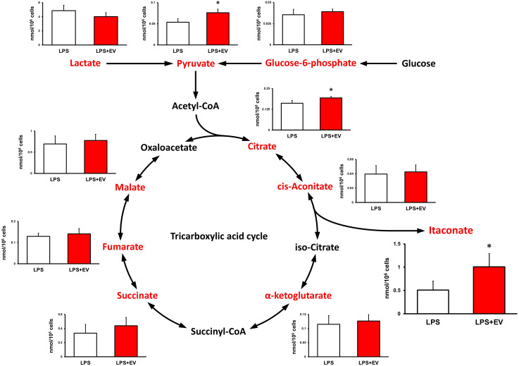
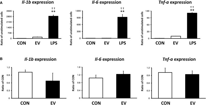
Introduction: Macrophages play an important role in the innate immunity. While macrophage inflammation is necessary for biological defense, it must be appropriately controlled. Extracellular vesicles (EVs) are small vesicles released from all types of cells and play a central role in intercellular communication. Skeletal muscle has been suggested to release anti-inflammatory factors, but the effect of myotube-derived EVs on macrophages is unknown. As an anti-inflammatory mechanism of macrophages, the immune responsive gene 1 (IRG1)-itaconate pathway is essential. In this study, we show that skeletal muscle-derived EVs suppress macrophage inflammatory responses, upregulating the IRG1-itaconate pathway.

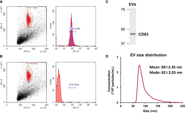
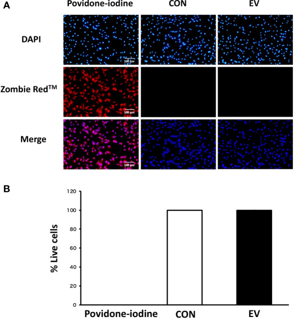
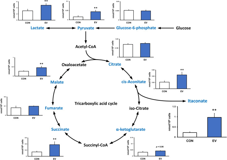
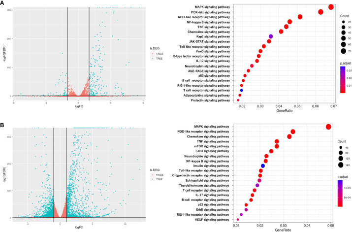
Methods: C2C12 myoblasts were differentiated into myotubes and EVs were extracted by ultracentrifugation. Skeletal myotube-derived EVs were administered to mouse bone marrow-derived macrophages, then lipopolysaccharide (LPS) stimulation was performed and inflammatory cytokine expression was measured by RT-qPCR. Metabolite abundance in macrophages after addition of EVs was measured by CE/MS, and IRG1 expression was measured by RT-PCR. Furthermore, RNA-seq analysis was performed on macrophages after EV treatment.

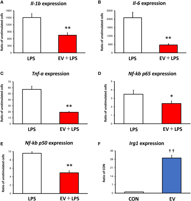
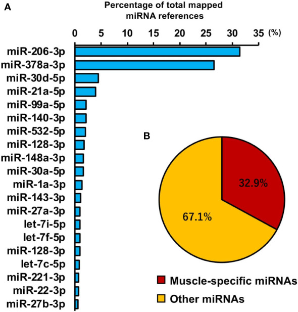
Results: EVs attenuated the expression of LPS-induced pro-inflammatory factors in macrophages. Itaconate abundance and IRG1 expression were significantly increased in the EV-treated group. RNA-seq analysis revealed activation of the PI3K-Akt and JAK-STAT pathways in macrophages after EV treatment. The most abundant miRNA in myotube EVs was miR-206-3p, followed by miR-378a-3p, miR-30d-5p, and miR-21a-5p.

Discussion: Skeletal myotube EVs are supposed to increase the production of itaconate via upregulation of IRG1 expression and exhibited an anti-inflammatory effect in macrophages. This anti-inflammatory effect was suggested to involve the PI3K-Akt and JAK-STAT pathways. The miRNA profiles within EVs implied that miR-206-3p, miR-378a-3p, miR-30d-5p, and miR-21a-5p may be responsible for the anti-inflammatory effects of the EVs. In summary, in this study we showed that myotube-derived EVs prevent macrophage inflammatory responses by activating the IRG1-itaconate pathway.

Keywords: IRG1; extracellular vesicle; itaconate; macrophage; skeletal muscle.


Conflict of interest statement

The authors declare that the research was conducted in the absence of any commercial or financial relationships that could be construed as a potential conflict of interest.

Figures

Figure 1
Figure 1
Extracellular vesicle (EV) characterization. (A) The presence of CD63 on isolated EVs was verified by flow cytometry. (B) CD63 was absent in particles treated with the isotype control. (C) Western blot analysis of CD63. (D) Size distribution of isolated EVs was measured by tunable resistive pulse sensing. n = 3.
Figure 2
Figure 2
mRNA expression levels in macrophages by qPCR. (A-E) Bone marrow-derived macrophages were stimulated with 100 ng/mL lipopolysaccharide (LPS) 24 h after extracellular vesicle (EV) treatment and the expression levels of pro-inflammatory genes were measured. (F) Irg1 expression level in macrophages was measured after EV treatment. *p < 0.05, **p < 0.01 compared with LPS, **p < 0.01 compared with LPS, †† p < 0.01 compared with CON (Student’s t-test). n = 4. Mean ± SD shown.
Figure 3
Figure 3
Cell viability 24 h after extracellular vesicle (EV) treatment. (A) Bone marrow-derived macrophages were stained with Zombie Red™ immunofluorescence reagent. After fixation, the cells were counter-stained with DAPI. (B) The percentage of live cells to total cells was calculated. Triplicate cell cultures were analyzed and 5 random fields of each well were examined. Mean ± SD shown.
Figure 4
Figure 4
Metabolite profile in bone marrow-derived macrophages treated by myotube-derived extracellular vesicles. **p < 0.01 compared with CON (Student’s t-test). n = 4. Mean ± SD shown.
Figure 5
Figure 5
Metabolite profile in bone marrow-derived macrophages treated by myotube-derived extracellular vesicles. *p < 0.05 compared with LPS (Student’s t-test). n = 4. Mean ± SD shown.
Figure 6
Figure 6
RNA sequencing analysis of BMDMs after extracellular vesicle (EV) treatment. (A) Left: Volcano plot of differentially expressed RNAs in control group vs. EV group. Blue dots represent RNAs with statistically significant difference and red dots show RNAs with no statistically significant difference between EV group vs. control group. Right: Kyoto Encyclopedia of Genes and Genomes (KEGG) analysis on differentially expressed RNAs; the 20 most enriched pathways linked to signaling transduction are presented. (B) Left: Volcano plot of differentially expressed RNAs in control group vs. LPS group. Blue dots represent RNAs with statistically significant difference and red dots show RNAs with no statistically significant difference between control group vs. LPS group. Right: Kyoto Encyclopedia of Genes and Genomes (KEGG) analysis on differentially expressed RNAs; the 20 most enriched pathways linked to signaling transduction are presented. n = 3.
Figure 7
Figure 7
miRNA profile in skeletal myotube-derived extracellular vesicles (EVs). miRNA-sequencing analysis was performed on miRNAs extracted from the EVs. (A) The 20 most abundant miRNAs in myotube EVs. (B) Ratio of muscle-specific miRNAs to total miRNAs. n = 2.
Figure 8
Figure 8
Macrophage responses to myotube-derived extracellular vesicle (EV) treatment in inflammation-related genes. (A) mRNA expression levels of pro-inflammatory factors were measured by qPCR after 1.5 h-treatment by skeletal muscle EVs or LPS. **p < 0.01 compared with CON, †† p < 0.01 compared with LPS (Tukey’s multiple comparison test). (B) mRNA expression levels of pro-inflammatory factors were measured by qPCR 24 h after EV treatment. n = 4. Mean ± SD shown.

References

    1. Weiss G, Schaible UE. Macrophage defense mechanisms against intracellular bacteria. Immunol Rev (2015) 264(1):182–203. doi: 10.1111/imr.12266 - DOI - PMC - PubMed
    1. Orecchioni M, Ghosheh Y, Pramod AB, Ley K. Corrigendum: Macrophage polarization: Different gene signatures in M1(LPS+) vs. classically and M2(LPS-) vs. alternatively activated macrophages. Front Immunol (2020) 11:234. doi: 10.3389/fimmu.2020.00234 - DOI - PMC - PubMed
    1. Funes SC, Rios M, Escobar-Vera J, Kalergis AM. Implications of macrophage polarization in autoimmunity. Immunology (2018) 154(2):186–95. doi: 10.1111/imm.12910 - DOI - PMC - PubMed
    1. Harrell CR, Jovicic N, Djonov V, Arsenijevic N, Volarevic V. Mesenchymal stem cell-derived exosomes and other extracellular vesicles as new remedies in the therapy of inflammatory diseases. Cells (2019) 8(12):1605. doi: 10.3390/cells8121605 - DOI - PMC - PubMed
    1. Andaloussi S EL, Mäger I, Breakefield XO, Wood MJ. Extracellular vesicles: Biology and emerging therapeutic opportunities. Nat Rev Drug Discov (2013) 12(5):347–57. doi: 10.1038/nrd3978 - DOI - PubMed

Publication types

MeSH terms