. 2023 Mar 3:10:1097609.
doi: 10.3389/fmolb.2023.1097609. eCollection 2023.

sRNA expedites polycistronic mRNA decay in Escherichia coli

Affiliations

sRNA expedites polycistronic mRNA decay in Escherichia coli

Heung Jin Jeon et al. Front Mol Biosci. .

Abstract

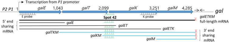
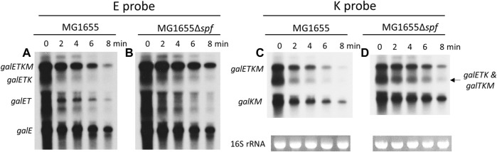
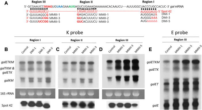
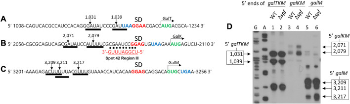
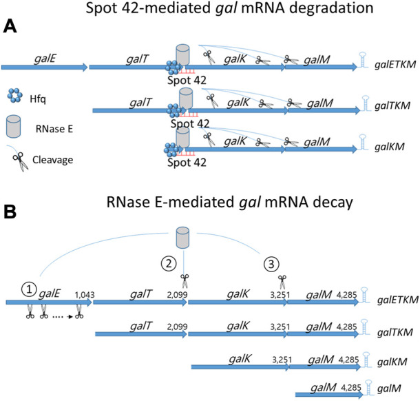
In bacteria, most small RNA (sRNA) elicits RNase E-mediated target mRNA degradation by binding near the translation initiation site at the 5' end of the target mRNA. Spot 42 is an sRNA that binds in the middle of the gal operon near the translation initiation site of galK, the third gene of four, but it is not clear whether this binding causes degradation of gal mRNA. In this study, we measured the decay rate of gal mRNA using Northern blot and found that Spot 42 binding caused degradation of only a specific group of gal mRNA that shares their 3' end with full-length mRNA. The results showed that in the MG1655Δspf strain in which the Spot 42 gene was removed, the half-life of each gal mRNA in the group increased by about 200% compared to the wild type. Since these mRNA species are intermediate mRNA molecules created by the decay process of the full-length gal mRNA, these results suggest that sRNA accelerates the mRNA decaying processes that normally operate, thus revealing an unprecedented role of sRNA in mRNA biology.

Keywords: RNase E; Spot42; galactose operon; mRNA decay; polarity; sRNA.


Conflict of interest statement

The authors declare that the research was conducted in the absence of any commercial or financial relationships that could be construed as a potential conflict of interest.

Figures

FIGURE 1
FIGURE 1
Map of gal operon and the 3′ and 5′end-sharing mRNA. P1 and P2 are the two promoters of the gal operon of Escherichia coli which are separated by five nucleotides. Numbers indicate gal nucleotide residue coordinates, where one is the start of transcription from the P1 promoter. The number at the end of each gene belongs to the last nucleotide of the stop codon of that gene. Six nucleotides downstream of the stop codon of the galM gene is a 17-nucleotide inverted repeat sequence (head-to-head arrows) that forms the terminator hairpin (red) that terminates transcription and protects the full-length galETKM mRNA from 3′→5′ exonuclease digestion. Two DNA probes, E and K (500 bp) (underlines) used in the Northern blots are presented. The galETKM operon produces two kinds of mRNA groups: 3′ end-sharing and 5′ end-sharing. The 3′ end-sharing mRNA shares the same 3′ end with the full-length galETKM mRNA. The 5’end-sharing mRNA shares the same 5′ end with the galETKM mRNA. The E and K probes were used to detect the 5′end-sharing and 3′ end-sharing mRNA, respectively. Spot 42 (green) binds at the translation initiation region of the galK gene. Thus, Spot 42 binds to multiple mRNAs.
FIGURE 2
FIGURE 2
Spot 42 accelerates the mRNA decay rate of the 3′ end-sharing mRNA only. (A, B) Northern blots of gal mRNA from wild type (WT) MG1655 and MG1655Δspf in the presence of rifampicin (100 μg/mL) with E probe. (C, D) Northern blots of gal mRNA from wild type (WT) MG1655 and MG1655Δspf in the presence of rifampicin (100 μg/mL) with K probe.
FIGURE 3
FIGURE 3
Region III of Spot 42 is involved in mRNA degradation of the 3′ end-sharing mRNA only. (A) Nucleotide sequence of gal, Spot 42, and Spot 42 mutants. The Shine and Dalgarno sequence for translation initiation of galK is presented in red. The translation stop codon of galT and the translation initiation codon of galK are presented in blue and green, respectively. Also, about 50 nucleotide sequences of the 5′ portion of Spot 42 are presented in red below the gal sequence. The three regions of Spot 42 that form perfect base pairings with the gal nucleotide sequences are identified as regions I, II, and III. Black dots represent base pairing. Spot 42 mutations in each region are listed under the corresponding region by name. Nucleotide changes are marked in red. The region I mutations are nucleotide deletions denoted as Δ in red. (B) K-probed Northern blot of the region I mutant; (C) K-probed Northern blot of the region II mutant; (D) K-probed Northern blot of the region III mutants; (E) E-probed Northern blot of the region III mutants.
FIGURE 4
FIGURE 4
Spot 42-mediated gal mRNA degradation needs Hfq. (A) K-probed Northern blot of MG1655 (lane 1), MG1655Δhfq (lane 2), and MG1655Δhfq harboring the Hfq expression plasmid pHfq (lane 3). (B) The relative amount of hfq mRNA was measured by using qRT-PCR in MG1655 and MG1655Δhfq harboring pHfq plasmid. Error bars represent the mean fold-change ±standard deviation from three independent experiments. *p-value ≤ 0.05 (statistically significant).
FIGURE 5
FIGURE 5
Spot 42 negatively regulates RNase E cleavage that generates galKM. Nucleotide sequences upstream of the translation initiation codons of galT (A), galK (B), and galM (C), show the correspondence of the cleavage positions (inverted arrows) of the RNase E consensus cleavage sites (thick underlines). Also highlighted are the putative Shine–Dalgarno sequences (in bold red) and the initiation (green) and termination (blue) codons. The RNase E cleavages in (A), (B), and (C) result in the generation of the 5′ end of galTKM, galKM, and galM, respectively. Base pairing of region III to the corresponding regions of gal are presented with dots. (D) 5′RACE results show the 5′ ends of galTKM at 1,031 and 1,039 in wild type (WT) (lane 1) and Δspf (lane 2), the 5′ ends of galKM at 2,071 and 2,079 in wild type (WT) (lane 3), and Δspf (lane 4), and the 5′ ends of galM at 3,209, 3,211, and 3,217 in wild type (WT) (lane 5) and Δspf (lane 6).
FIGURE 6
FIGURE 6
mRNA decay of the multicistronic galETKM. (A) Spot 42-mediated gal mRNA degradation: Hfq-mediated region III base pairing to galETKM, galTKM, and galKM recruit RNase E and subsequently induce RNase E cleavage downstream. (B) RNase E-mediated gal mRNA decay: 1) Generation of galTKM mRNA: The first gene of the gal operon, galE, is removed by progressive and successive RNase E cleavages initiated at the 5′ end of galETKM mRNA. This progressive RNase E cleavage is followed by the 3→5′ exo-ribonucleolytic digestion of the released RNA fragment stops at the translation initiation site of the next galT gene, generating galTKM mRNA. The sites of RNase E cleavage that generate the 5′ ends of galTKM are presented in Figure 5A. The 5′ ends are experimentally shown (lane 1; Figure 5D). 2) Generation of galKM mRNA: Sometimes RNase E cleaves in the middle of galETKM mRNA, several nucleotides upstream from the translation initiation site of the galK gene, followed by the 3→5′ exo-ribonucleolytic digestion of the 5′ portion RNA fragment, which generates galKM mRNA, galKM. The sites of the RNase E cleavages that generate the 5′ ends of galKM are presented in Figure 5A. The 5′ ends are experimentally shown (lane 3; Figure 5D). 3) Generation of galM mRNA: RNase E cleavages on the upstream sites from the translation initiation of the last gene of the operon, galM, followed by the 3→5′ exo-ribonucleolytic digestion of the released RNA fragment generates galM mRNA, galM. The sites of the RNase E cleavages that generate the 5′ ends of galM are presented in Figure 5A. The 5′ ends are experimentally shown (lane 5; Figure 5D). RNase E also cleaves a few nucleotides upstream from the transcription terminator hairpin at the 3′ end of galM mRNA. Exoribonucleases rapidly remove the 5′ portion of the galM mRNA upstream of the hairpin structure, completing the decay process of the multicistronic full-length mRNA down to the nucleotides. Thus, the decay of the full-length galETKM mRNA proceeds by generating the 3′ end-sharing mRNA (galTKM, galKM, and galM), which we defined as ‘decay intermediates’ (Jeon et al., 2020).

References

    1. Adhya S. (2003). Suboperonic regulatory signals. Sci. STKE 2003 (185), pe22. 10.1126/stke.2003.185.pe22 - DOI - PubMed
    1. Aiba H. (2007). Mechanism of RNA silencing by Hfq-binding small RNAs. Curr. Opin. Microbiol. 10 (2), 134–139. 10.1016/j.mib.2007.03.010 - DOI - PubMed
    1. Beisel C. L., Storz G. (2011). The base-pairing RNA spot 42 participates in a multioutput feedforward loop to help enact catabolite repression in Escherichia coli . Mol. Cell 41 (3), 286–297. 10.1016/j.molcel.2010.12.027 - DOI - PMC - PubMed
    1. Belasco J. G., Beatty J. T., Adams C. W., von Gabain A., Cohen S. N. (1985). Differential expression of photosynthesis genes in R. capsulata results from segmental differences in stability within the polycistronic rxcA transcript. Cell 40 (1), 171–181. 10.1016/0092-8674(85)90320-4 - DOI - PubMed
    1. Bossi L., Schwartz A., Guillemardet B., Boudvillain M., Figueroa-Bossi N. (2012). A role for Rho-dependent polarity in gene regulation by a noncoding small RNA. Genes Dev. 26 (16), 1864–1873. 10.1101/gad.195412.112 - DOI - PMC - PubMed

LinkOut - more resources