Review
. 2023 Mar 2:17:1073511.
doi: 10.3389/fncel.2023.1073511. eCollection 2023.

What type of cell death occurs in chronic cerebral hypoperfusion? A review focusing on pyroptosis and its potential therapeutic implications

Affiliations
Review

What type of cell death occurs in chronic cerebral hypoperfusion? A review focusing on pyroptosis and its potential therapeutic implications

Yuxuan He et al. Front Cell Neurosci. .

Abstract

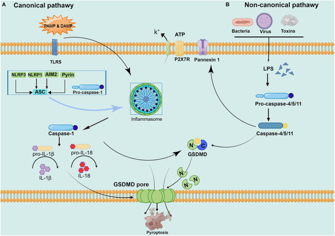
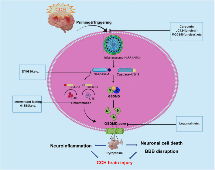
Chronic cerebral hypoperfusion (CCH) is a major global disease with chronic cerebral blood flow reduction. It is also the main cause of cognitive impairment and neurodegenerative diseases. Pyroptosis, a novel form of cell death, is characterized by the rupture of the cell membrane and the release of pro-inflammatory mediators. In recent years, an increasing number of studies have identified the involvement of pyroptosis and its mediated inflammatory response in the pathological process of CCH. Therefore, preventing the activation of pyroptosis following CCH is beneficial to inhibit the inflammatory cascade and reduce brain injury. In this review, we discuss the research progress on the relationship between pyroptosis and CCH, in order to provide a reference for research in related fields.

Keywords: chronic cerebral hypoperfusion; inflammasome; inflammatory cytokine; pyroptosis; review; therapeutic implications.


Conflict of interest statement

The authors declare that the research was conducted in the absence of any commercial or financial relationships that could be construed as a potential conflict of interest.

Figures

Figure 1
Figure 1
Pathways of pyroptosis activation. (A) The canonical pyroptosis pathway: Under PAMP/ DAMP signal stimulation, NOD, ASC, and pro-caspase-1 are successfully assembled into multicomplex inflammasomes. ASC recruits the enzyme ogen caspase-1, which causes automatic cleavage and activation. Activated caspase-1 cleaves inactive pro-IL-1 and proIL-18, turning them into biologically active IL-1 and IL-18, promoting the maturation and secretion of inflammatory cytokines and inducing cell death under inflammatory and stressed pathological conditions. Cell membrane rupture is induced by cleaving GSDMD to form the GSDMD-N and forming pores in the plasma membrane. (B) The non-canonical pyroptosis pathway: Stimulated by signals such as LPS, the pro-caspase-11 is activated. On the one hand, activated caspase-11 cleaves GSDMD, forms the GSDMD-N and forms a pyroptosis pore in the plasma membrane, inducing cell membrane rupture and releasing contents. On the other hand, activated caspase-11 induces activated cleaving of caspase-1 to turn pro-IL-1β and pro-IL-18 into IL-1β and IL-18, expanding the inflammatory response and intensifying organismal injury.
Figure 2
Figure 2
Graphical description of pathological mechanism of pyroptosis following CCH brain injury and potential drugs targeting pyroptosis-regulated cell death in CCH. During the onset of CCH, NLRP3, and AIM2 inflammasome are activated and then stimulate caspase-1/4/5/11. Caspases-1 and -4/5/11 are both responsible for the processing of GSDMD, which leads to the release of the GSDMD-NT. The formation of pores by GSDMD-NT in the plasma membrane and organelle membranes is what causes pyroptosis to occur. In the case of the pores in the plasma membranes of endothelium and neuronal cells directly could cause cell lysis, which induces BBB disruption and neuronal cell death. At the same time, inflammatory cytokines are released via pores and contribute to the regulation of neuroinflammation. These underlying pathophysiological processes will aggravate CCH-related brain injury. To date, several inhibitors of NLRP3, AIM2 inflammasome, Caspase-1, and GSDMD have been described for promising in alleviating CCH-related brain injury.

References

    1. Aachoui Y., Sagulenko V., Miao E. A., Stacey K. J. (2013). Inflammasome-mediated pyroptotic and apoptotic cell death and defense against infection. Curr. Opin. Microbiol. 16, 319–326. 10.1016/j.mib.2013.04.004 - DOI - PMC - PubMed
    1. Ashok A., Rai N. K., Raza W., Pandey R., Bandyopadhyay S. (2016). Chronic cerebral hypoperfusion-induced impairment of Aβ clearance requires HB-EGF-dependent sequential activation of HIF1α and MMP9. Neurobiol. Dis. 95, 179–193. 10.1016/j.nbd.2016.07.013 - DOI - PubMed
    1. Bai B., Yang Y., Wang Q., Li M., Tian C., Liu Y., et al. . (2020). NLRP3 inflammasome in endothelial dysfunction. Cell Death Dis. 11:776. 10.1038/s41419-020-02985-x - DOI - PMC - PubMed
    1. Bakker F. C., Klijn C. J., Jennekens-Schinkel A., Kappelle L. J. (2000). Cognitive disorders in patients with occlusive disease of the carotid artery: a systematic review of the literature. J. Neurol. 247, 669–676. 10.1007/s004150070108 - DOI - PubMed
    1. Bellut M., Papp L., Bieber M., Kraft P., Stoll G., Schuhmann M. K. (2021). NLPR3 inflammasome inhibition alleviates hypoxic endothelial cell death in vitro and protects blood-brain barrier integrity in murine stroke. Cell Death Dis. 13:20. 10.1038/s41419-021-04379-z - DOI - PMC - PubMed

LinkOut - more resources