. 2023 Mar 8:2023:7829290.
doi: 10.1155/2023/7829290. eCollection 2023.

Molecular Identification, Pathogenesis, and Life Cycle of Sarcocystis cruzi from Cattle (Bos taurus) in New Valley Governorate, Egypt

Affiliations

Molecular Identification, Pathogenesis, and Life Cycle of Sarcocystis cruzi from Cattle (Bos taurus) in New Valley Governorate, Egypt

Mohammed B M El-Mahdi et al. J Parasitol Res. .

Abstract

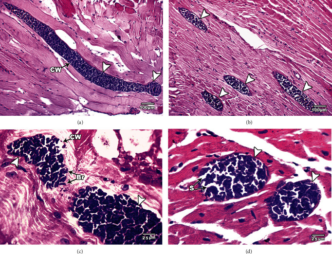
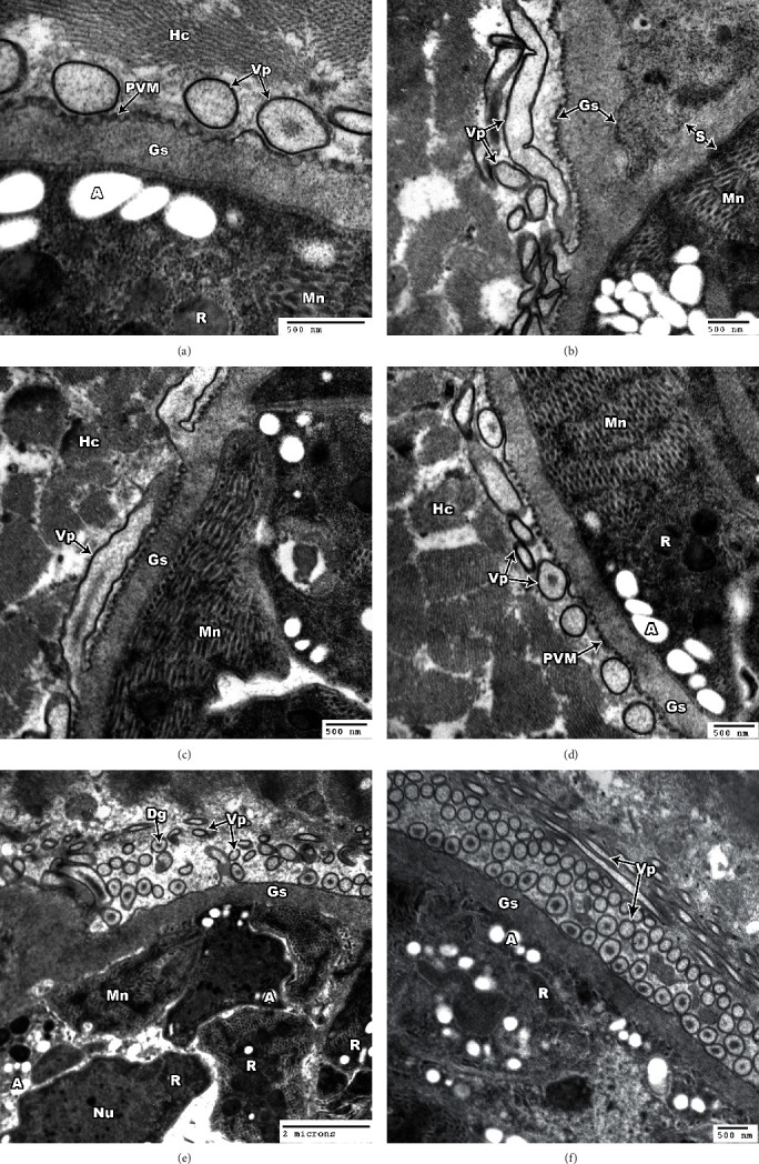
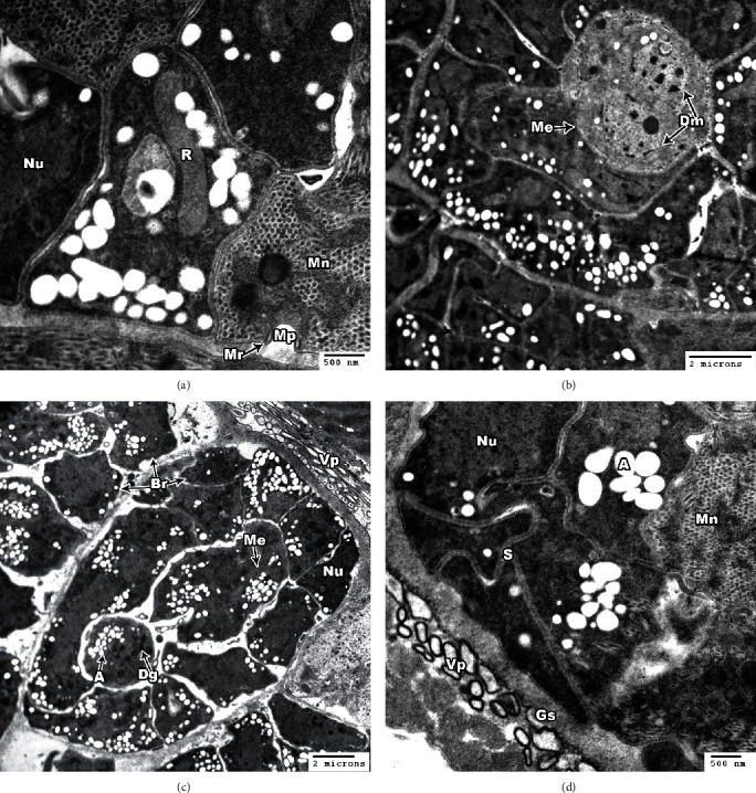
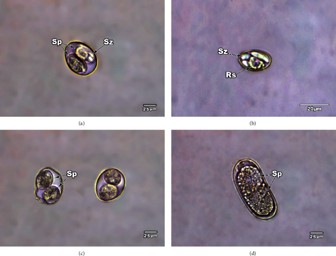
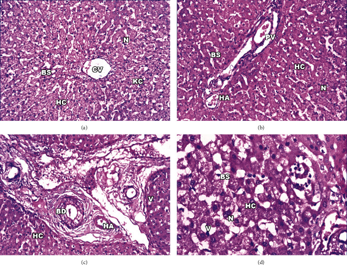
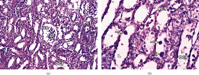
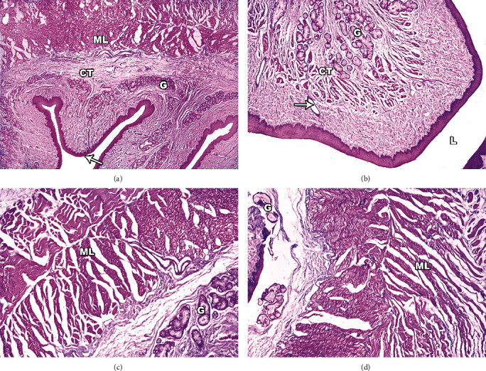
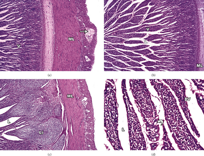
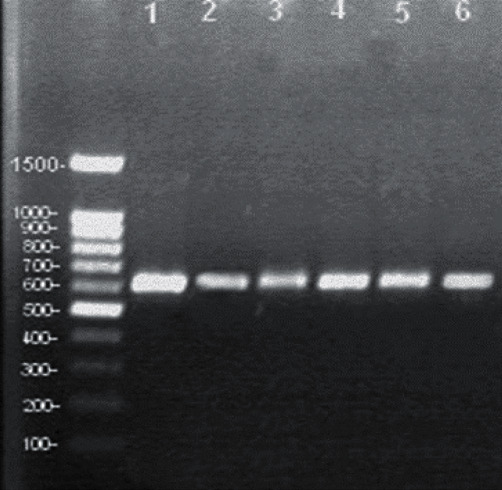
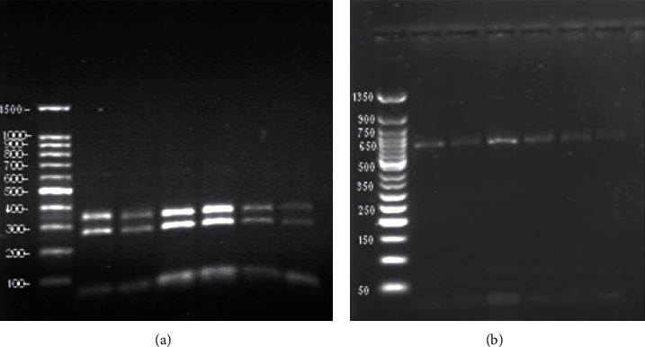
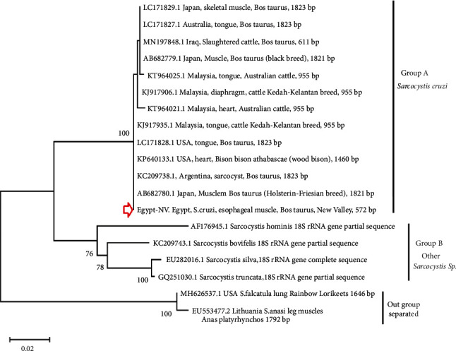
Sarcocystis cruzi was identified by molecular methods from an intermediate host, cattle (Bos taurus), in El-Kharga, New Valley Governorate, Egypt, and its life cycle and pathogenicity were studied in the final host, dogs (Canis familiaris). 600 slaughtered cattle aged 6-8 years (480/120 males/females) were included. In addition, three laboratory-bred, coccidian-free puppies aged 2-3 months were fed infected bovine muscles to locate the definitive host and analyze sporogony. 18S rRNA-specific gene primers were used for DNA amplification from esophageal muscles. These polymerase chain reaction (PCR) amplicons were subjected to restriction fragment length polymorphism (RFLP) and molecular sequence analysis. Infection was detected in 78.8% (473/600; 95% CI, 75.56-82.11%). Histopathological examination of esophageal muscles showed oval- to spherical-shaped cysts, 96.7 μm wide by 326.9 μm long; cysts in cardiac muscles were ovoid and smaller. Infected puppies began shedding sporocysts in feces 7 days post-inoculation and showed distorted organ architecture, severe cellular damage, and inflammatory lesions in liver, kidney, esophagus, and stomach. Three oocysts with different shapes and sizes were identified. Partial 18S rRNA gene sequences of isolated New Valley sarcocysts were identical to S. cruzi isolated from different areas, verifying their genetic relatedness. Our analysis suggests that S. cruzi is the most prevalent in slaughtered cattle in New Valley Governorate, Egypt.


Conflict of interest statement

The author(s) declare(s) that they have no conflicts of interest.

Figures

Figure 1
Figure 1
Light micrographs of S. cruzi in muscle tissues of cattle, hematoxylin and eosin-stained images. Elongated sarcocysts are seen embedded in the esophagus (a–c) and cardiac (d) muscles. Arrows refer to the sarcocyst. Note the cyst wall (CW) underlined by a layer of ground substance extended into the interior of the cyst as septa dividing it into compartments enclosing bradyzoites (Br). Original magnification: 100× (a and b) and 400× (c and d).
Figure 2
Figure 2
TEM of S. cruzi wall. (a) Parasitophorous vacuolar membrane (PVM) and villar protrusions (Vp). (b) Villar protrusions (Vp) and ground substance (Gs). (c) Cyst wall protrusions (Vp) with no microfilaments. (d) Cyst wall protrusions (Vp) adjacent to the sarcoplasm of host cell (Hc). Note magnified protrusion. (e) Cyst wall with hair-like protrusions (Vp); the protrusion contains coarse or fine electron dense granule (Dg). (f) Villar protrusions cut in different directions.
Figure 3
Figure 3
TEM of S. cruzi. (a) Section through the micropore (Mp); the pore is surrounded by a micropore ring (Mr). Note the clear micronemes (Mn), nucleus (Nu), and rhoptry (R). (b) The architecture of a dividing metrocyte (Me) with two daughter merozoites (Dm) in the inside of a Sarcocystis cruzi cyst. (c) A high concentration of bradyzoites is enclosed by a thin cyst wall and septa. Note the bradyzoites containing several dense granules (Dg), in addition to several amylopectin granules (A), nucleus (Nu), and mitochondrion (Mc). (d) Cross section of bradyzoite showing nucleus (Nu), amylopectin granules (A), micronemes (Mn), septa (S), and ground substance (Gs).
Figure 4
Figure 4
Unstained mature globular oocysts of Sarcocystis from feces of the infected dogs. (a) Oocysts with two elongated sporocysts (Sp), which lie transversely in their position inside the oocyst. Note the sporozoites (Sz) inside the sporocyst. (b) Elongated ellipsoidal sporocyst, with each sporocyst contain four sporozoites (Sz) and one large internal residual body (Rs). (c) Oocyst containing two rounded sporocysts, which lie longitudinally at their position inside the oocyst. (d) Oocyst with two incompletely divided sporocysts, which were composed of numerous small granules. Original magnification: 400×.
Figure 5
Figure 5
Light micrograph of infected dogs' liver sections. (a) Hepatocytes (Hc) with variable size and shape nuclei (N). Note the dilated central vein (Cv), blood sinusoid (Bs), and activated Kupffer cells (Kc). (b) Hepatocytes (Hc) with nuclei (N), hepatic portal vein (Pv), and blood sinusoid (Bs). (c) Portal area with dilated and congested portal vein (Pv) and numerous branches of bile ducts (Bd). (d) Disorganized liver hepatocytes (Hc) with cytoplasmic vacuolization (V). Original magnification: 400×.
Figure 6
Figure 6
Light micrographs of infected dog's kidney sections stained with H&E. (a) Section of renal cortex showing highly deteriorated renal corpuscle with degenerated glomerulus (G), dilated urinary space (US), and proximal (PCT) and distal (DCT) convoluted tubules. (b) Kidney tissue with cellular abnormalities and leukocyte infiltration. Note the tubular necrosis, epithelial lining degeneration, necrotic foci, distorted proximal (PCT) and distal (DCT) convoluted tubules with dilated lumen (L), and segregated nuclei with red blood cells (RBCS) (arrows) in its lumen. Original magnification: 400×.
Figure 7
Figure 7
Light micrographs of infected dogs' esophagus sections stained with H&E. (a) Full-thickness section of esophagus showing four layers: mucosa (thin arrow), submucosa, muscular layer (ML), adventitia and submucosal glands (G), and ducts and keratinized stratified squamous epithelium (small arrow). (b) Marked degenerative changes of muscle fibres with dispersing edema dispersing muscle fibres and esophageal glands (G) located in submucosa form ducts (arrow) separated by connective tissue (CT). (c) Muscular layer (ML), esophageal mucous glands (G), and connective tissue separating between glands. (d) Hypertrophied muscle fibres (ML) and damage of myofibrils. Note the increased space between the myofibrils. Original magnification: 400×.
Figure 8
Figure 8
Light micrographs of infected dogs' stomach sections stained with H&E. (a) Full-thickness section of stomach showing sloughing of the simple columnar epithelium (E), absence of the normal villus (V) architecture, with damaged muscular layer (ML) and degenerated muscle fibres with marked edema (arrow). (b) Some villi show distorted architecture with dilatation of their middle part. (c) Marked leucocytic infiltration (Li) and edema widen the submucosa. (d) Dissolved villi tips with damaged villi (DV); some oxyntic cells are shrunken with pyknotic nucleus, while others are vacuolated. There is also widening of gastric glands (black arrow).
Figure 9
Figure 9
Amplification of 18S ribosomal gene of Sarcocystis sp. from selected random cattle samples, which demonstrate a band of 620 bp with similar length in all investigated six samples used that belonged to S. cruzi. The 100 bp DNA ladder Ready-to-Use (RTU) is shown.
Figure 10
Figure 10
The RFLP restriction digest analysis by Mbo1 endonuclease enzyme on the amplified 18S targeted region from S. cruzi. (a) The Mbo1 enzyme produced two fractionated regions of approximately 275 and 350 base pair in length. The 100 bp DNA ladder Ready-to-Use (RTU) is shown. (b) The Hinf enzyme has no cutting effects. The BERUS 50 bp DNA ladder is shown.
Figure 11
Figure 11
Phylogenetic analysis of S. cruzi isolated in the present study along with different Sarcocystis spp. recorded in cattle from other countries. The program MegaX V.10.2.6 was used to construct the tree using maximum likelihood method based on 18S rRNA gene sequences and under distant measurement of T92 + G + I. The Egypt-NV obtained nucleotide sequence has been deposited in the GenBank with accession number: OL305830.

Similar articles

Cited by

References

    1. Máca O. Molecular identification of Sarcocystis lutrae (Apicomplexa: Sarcocystidae) from the raccoon dog, Nyctereutes procyonoides, and the common raccoon, Procyon lotor, in the Czech Republic. Parasites & Vectors . 2020;13(1):p. 231. doi: 10.1186/s13071-020-04108-z. - DOI - PMC - PubMed
    1. Fayer R. Sarcocystis spp. in human infections. Clinical Microbiology Reviews . 2004;17(4):894–902. doi: 10.1128/CMR.17.4.894-902.2004. - DOI - PMC - PubMed
    1. Dubey J., Lindsay D. S. Neosporosis, toxoplasmosis, and sarcocystosis in ruminants. The Veterinary Clinics of North America. Food Animal Practice . 2006;22(3):645–671. doi: 10.1016/j.cvfa.2006.08.001. - DOI - PubMed
    1. Dubey J. T. A review of Sarcocystis of domestic animals and of other coccidia of cats and dogs. Journal of the American Veterinary Medical Association . 1976;169(10):1061–1078. - PubMed
    1. Dubey J. P., Speer C., Fayer R. Sarcocystosis of Animals and Man . Boca Raton: CRC Press, Inc.; 1989. p. p. 215.

LinkOut - more resources