. 2023 Mar 1:10:1061118.
doi: 10.3389/fcvm.2023.1061118. eCollection 2023.

Impact of severe secondary tricuspid regurgitation on rest and exercise hemodynamics of patients with heart failure and a preserved left ventricular ejection fraction

Affiliations

Impact of severe secondary tricuspid regurgitation on rest and exercise hemodynamics of patients with heart failure and a preserved left ventricular ejection fraction

Claudia Baratto et al. Front Cardiovasc Med. .

Abstract

Background: Both secondary tricuspid regurgitation (STR) and heart failure with preserved ejection fraction (HFpEF) are relevant public health problems in the elderly population, presenting with potential overlaps and sharing similar risk factors. However, the impact of severe STR on hemodynamics and cardiorespiratory adaptation to exercise in HFpEF remains to be clarified.

Aim: To explore the impact of STR on exercise hemodynamics and cardiorespiratory adaptation in HFpEF.

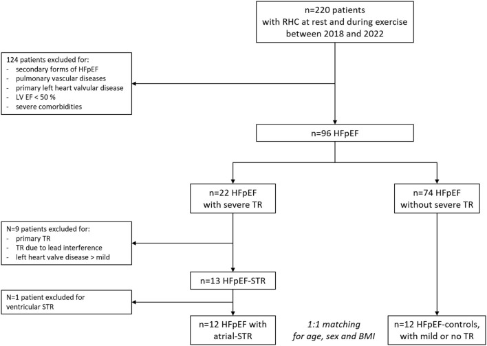
Methods: We analyzed invasive hemodynamics and gas-exchange data obtained at rest and during exercise from HFpEF patients with severe STR (HFpEF-STR), compared with 1:1 age-, sex-, and body mass index (BMI)- matched HFpEF patients with mild or no STR (HFpEF-controls).

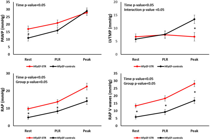
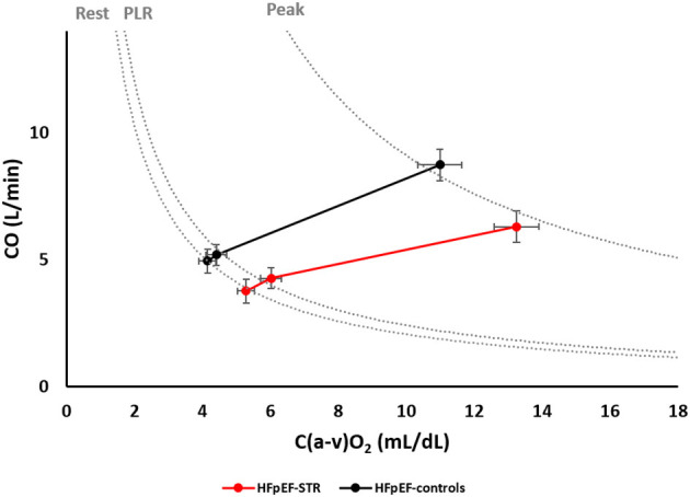
Results: Twelve HFpEF with atrial-STR (mean age 72 years, 92% females, BMI 28 Kg/m2) and 12 HFpEF-controls patients were analyzed. HFpEF-STR had higher (p < 0.01) right atrial pressure than HFpEF-controls both at rest (10 ± 1 vs. 5 ± 1 mmHg) and during exercise (23 ± 2 vs. 14 ± 2 mmHg). Despite higher pulmonary artery wedge pressure (PAWP) at rest in HFpEF-STR than in HFpEF-controls (17 ± 2 vs. 11 ± 2, p = 0.04), PAWP at peak exercise was no more different (28 ± 2 vs. 29 ± 2). Left ventricular transmural pressure and cardiac output (CO) increased less in HFpEF-STR than in HFpEF-controls (interaction p-value < 0.05). This latter was due to lower stroke volume (SV) values both at rest (48 ± 9 vs. 77 ± 9 mL, p < 0.05) and at peak exercise (54 ± 10 vs. 93 ± 10 mL, p < 0.05). Despite these differences, the two groups of patients laid on the same oxygen consumption isophlets because of the increased peripheral oxygen extraction in HFpEF-STR (p < 0.01). We found an inverse relationship between pulmonary vascular resistance and SV, both at rest and at peak exercise (R 2 = 0.12 and 0.19, respectively).

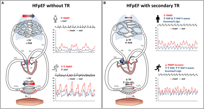
Conclusions: Severe STR complicating HFpEF impairs SV and CO reserve, leading to pulmonary vascular de-recruitment and relative left heart underfilling, undermining the typical HFpEF pathophysiology.

Keywords: exercise; heart failure with preserved ejection faction; hemodynamics; oxygen consumption; right heart catheterization; tricuspid regurgitation.


Conflict of interest statement

The authors declare that the research was conducted in the absence of any commercial or financial relationships that could be construed as a potential conflict of interest. The reviewer GI declared a shared parent affiliation with the authors CB, SC, GC, DS, AZ, MG, FH, MT, NR, FP, GPe, GPa, LB, and DM to the handling editor at the time of the review.

Figures

Figure 1
Figure 1
Patients' selection flow-chart. BMI, body mass index; HFpEF, heart failure with preserved ejection fraction; LV EF, left ventricular ejection fraction; RHC, right heart catheterization; STR, secondary tricuspid regurgitation; TR, tricuspid regurgitation.
Figure 2
Figure 2
Evolution of left and right heart hemodynamics during exercise in our patients' population. HFpEF, heart failure with preserved ejection fraction; LVTMP, left ventricular transmural pressure; PAWP, pulmonary artery wedge pressure; PLR, passive leg raising; RAP, right atrial pressure; STR, secondary tricuspid regurgitation. *P < 0.05.
Figure 3
Figure 3
Relative weight of cardiac output and arteriovenous oxygen difference in determining oxygen consumption at rest, during passive leg raising and at peak exercise. Dotted lines represent oxygen consumption isophlets, i.e., cardiac output and arteriovenous oxygen difference coordinates whose product is a given oxygen consumption, at rest, during passive leg raising, and at peak exercise. C(a-v)O2, arteriovenous oxygen difference; CO, cardiac output; HFpEF, heart failure with preserved ejection fraction; PLR, passive leg raising; STR, secondary tricuspid regurgitation.
Figure 4
Figure 4
Exercise pathophysiology of secondary tricuspid regurgitation (STR) in heart failure with preserved ejection fraction (HFpEF). The typical HFpEF patient without STR (panel on the left) may present with (A) high pulmonary artery wedge pressure, either at rest or during exercise, with a steep pulmonary artery wedge pressure rise, and mildly increased right atrial pressure. (B) substantially normal stroke volume, pulmonary vascular resistance and peripheral oxygen extraction. This HFpEF pathophysiology is altered in the presence of STR (panel on the right), being characterized by: (i) high right atrial pressure that lacks inspiratory decrease or may present with overt Kussmaul's sign, tall V waves in the right atrium, right atrial enlargement and venous congestion; (ii) reduced forward stroke volume with pulmonary vascular derecruitment (increasing pulmonary vascular resistance), left ventricular underfilling, flatter pulmonary artery wedge pressure rise during exercise, and high ratio between right atrial pressure and pulmonary artery wedge pressure; (iii) higher reliance upon peripheral oxygen extraction to exercise despite low stroke volumeAo, aorta; C(a-v)O2, arteriovenous oxygen difference; CaO2, arterial oxygen content; CvO2, venous oxygen content; IVC, inferior vena cava; LA, left atrium; LV, left ventricle; O2, oxygen; PA, pulmonary artery; PAWP, pulmonary artery wedge pressure; PV, pulmonary vein; PVR, pulmonary vascular resistance; RAP, right atrial pressure; SV, stroke volume; TR, tricuspid regurgitation.

References

    1. Vahanian A, Beyersdorf F, Praz F, Milojevic M, Baldus S, Bauersachs J, et al. ESC/EACTS scientific document group. ESC/EACTS guidelines for the management of valvular heart disease. Eur Heart J. (2022) 43:561–632. 10.1093/ejcts/ezac209 - DOI - PubMed
    1. Praz F, Muraru D, Kreidel F, Lurz P, Hahn RT, Delgado V, et al. Transcatheter treatment for tricuspid valve disease. EuroIntervention. (2021) 17:791–808. 10.4244/EIJ-D-21-00695 - DOI - PMC - PubMed
    1. Russo G, Taramasso M, Pedicino D, Gennari M, Gavazzoni M, Pozzoli A, et al. Challenges and future perspectives of transcatheter tricuspid valve interventions: adopt old strategies or adapt to new opportunities? Eur J Heart Fail. (2022) 24:442–54. 10.1002/ejhf.2398 - DOI - PubMed
    1. Ricci F, Bufano G, Galusko V, Sekar B, Benedetto U, Awad WI, et al. Tricuspid regurgitation management: a systematic review of clinical practice guidelines and recommendations. Eur Heart J Qual Care Clin Outcomes. (2022) 8:238–48. 10.1093/ehjqcco/qcab081 - DOI - PubMed
    1. Borlaug BA. Evaluation and management of heart failure with preserved ejection fraction. Nat Rev Cardiol. (2020) 17:559–73. 10.1038/s41569-020-0363-2 - DOI - PubMed

LinkOut - more resources