Case Reports
. 2023 Feb 17:32:e01724.
doi: 10.1016/j.idcr.2023.e01724. eCollection 2023.

Cytomegalovirus pneumonitis in infants: The challenge in diagnosis among pediatricians

Affiliations
Case Reports

Cytomegalovirus pneumonitis in infants: The challenge in diagnosis among pediatricians

Djatnika Setiabudi et al. IDCases. .

Abstract

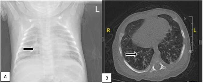
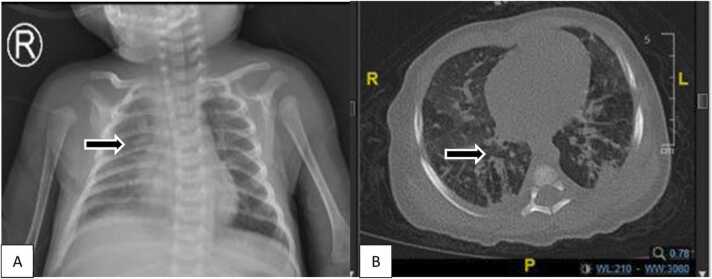
Cytomegalovirus (CMV) pneumonitis infections might present mild or severe illnesses and need sophisticated diagnostic tools, so it remains a diagnostic challenge. We reported five infants diagnosed with CMV pneumonitis who were initially and undiagnosed by the pediatrician in secondary private or public health hospitals with no improvement with standard and escalation of antibiotics treatment for bronchopneumonia as the initial diagnoses. As all cases occurred during the COVID-19 pandemic, they proved negative COVID-19 identified by polymerase chain reaction (PCR) SARS-CoV-2. We diagnosed acquired perinatal pneumonitis CMV in all claims based on clinical criteria, imaging studies, CMV serology, and PCR-CMV urinary tests as diagnostic tools. They showed clinical improvement after two weeks of valganciclovir therapy. Other organs' involvement was considered to be evaluated, including brain-evoked response audiometry (BERA) and eye examination. The physician should consider the possibility of CMV pneumonitis, who did not respond to standard and escalation of antibiotics treatment after initial diagnoses of bronchopneumonia.

Keywords: CMV pneumonitis; Case report; PCR-CMV urinary; Respiratory infection.


Figures

Fig. 1
Fig. 1
A. Chest X-ray showed bilateral bronchopneumonia. B. HRCT showed consolidation in the apical segment of the superior lobe, the superior segment of the inferior lobe, a posterodorsal segment of the inferior lobe of the right lung as well as in the anterior segment of the superior lobe, superior lingula, posterodorsal inferior lobe of the left lung; patchy consolidation in the superior-inferior lobe and posterodorsal segments of the inferior lobe of the left lung; Fibrosis in the laterodorsal and posterodorsal segments of the inferior lobes of the lung bilaterally suggestive of bilateral pneumonia with pulmonary interstitial involvement.
Fig. 2
Fig. 2
A. Chest X-ray showed bilateral bronchopneumonia. B HRCT showed ground glass opacity in the apical, anterior, and posterior segments of the superior lobe, medial segment, lateral lobe medius, superior segment, anterobasal, laterodorsal, posterodorsal, and mediobasal inferior lobe of the right and left lung. Interlobular septal thickening in the apical, anterior, and posterior segments of the superior lobe, laterodorsal segment, posterodorsal inferior lobe of the right lung; anterior segment, apicoposterior superior lobe, laterodorsal segment, anterobasal, posterodorsal inferior lobe of the left lung.
Fig. 3
Fig. 3
A. Chest X-ray showed bilateral bronchopneumonia. B. HRCT showed ground glass opacity in the apical, anterior, and posterior segments of the superior lobe, medial segment, lateral lobe medius, superior segment, anterobasal, laterodorsal, posterodorsal, and mediobasal inferior lobe of the right and left lung. Consolidation and fibrosis within interstitial lung involvement suggest pneumonia with interstitial participation.
Fig. 4
Fig. 4
A. Chest X-ray showed bilateral bronchopneumonia. B. HRCT showed ground glass opacity in the apical lobe superior, anterobasal, posterodorsal inferior lobe of the right lung, superior lobe apicoposterior, lingula superior, lingula inferior, superior, posterodorsal inferior lobe of the left lung; Multifocal consolidation in the posterior segment of the superior lobe, superior, inferior lobe of the right lung, anterobasal segment of inferior lobe of left lung e.c. Suggestive of pneumonia. Fibrosis in the apical segment, superior posterior lobe, posterodorsal inferior lobe of the right lung, apicoposterior, superior anterior lobe, laterodorsal, posterodorsal inferior lobe of the left lung.
Fig. 5
Fig. 5
A. Chest X-ray showed bilateral bronchopneumonia and cardiomegaly. B. HRCT showed consolidation with air bronchogram (+) in the posterior and anterior segments of the superior lobe and the entire inferior lobe of the right lung; throughout the superior and inferior lobes of the left lung accompanied by ground-glass opacities and surrounding fibrosis, Appears emphysema in the anterior segment of the superior lobe of the right lung.

References

    1. Gantt S., Bitnun A., Renaud C., Kakkar F., Vaudry W. Diagnosis and management of infants with congenital cytomegalovirus infection. Paediatr Child Health. 2017;22(2):72–74. doi: 10.1093/pch/pxx002. - DOI - PMC - PubMed
    1. Martinez-Gomez E., Perez-Carpena P., Flook M., Lopez-Escamez J.A. A Systematic review on the association of acquired human cytomegalovirus infection with hearing loss. J Clin Med. 2020;9(12) 1–14.10.3390/jcm9124011. - PMC - PubMed
    1. Brito L.F., Brune W., Stahl F.R. Cytomegalovirus (CMV) pneumonitis: Cell tropism, inflammation, and immunity. Int J Mol Sci. 2019;20(16):1–14. doi: 10.3390/ijms20163865. - DOI - PMC - PubMed
    1. Bardanzellu F., Fanos V., Reali A. Human breast milk-acquired cytomegalovirus infection: Certainties, doubts, and perspectives. Curr. Pedia Rev. 2018;15(1):30–41. doi: 10.2174/1573396315666181126105812. - DOI - PMC - PubMed
    1. Styczynski Jan. Who is the patient at risk of CMV recurrence: A review of the current scientific evidence focusing on hematopoietic cell transplantation. Infect Dis Ther. 2018;7:1–16. doi: 10.1007/s40121-017-0180-z. - DOI - PMC - PubMed

Publication types