Review
. 2023 Jul;12(17):e2202110.
doi: 10.1002/adhm.202202110. Epub 2023 Apr 2.

Keeping It Organized: Multicompartment Constructs to Mimic Tissue Heterogeneity

Affiliations
Review

Keeping It Organized: Multicompartment Constructs to Mimic Tissue Heterogeneity

Alvaro Sanchez-Rubio et al. Adv Healthc Mater. 2023 Jul.

Abstract

Tissue engineering aims at replicating tissues and organs to develop applications in vivo and in vitro. In vivo, by engineering artificial constructs using functional materials and cells to provide both physiological form and function. In vitro, by engineering three-dimensional (3D) models to support drug discovery and enable understanding of fundamental biology. 3D culture constructs mimic cell-cell and cell-matrix interactions and use biomaterials seeking to increase the resemblance of engineered tissues with its in vivo homologues. Native tissues, however, include complex architectures, with compartmentalized regions of different properties containing different types of cells that can be captured by multicompartment constructs. Recent advances in fabrication technologies, such as micropatterning, microfluidics or 3D bioprinting, have enabled compartmentalized structures with defined compositions and properties that are essential in creating 3D cell-laden multiphasic complex architectures. This review focuses on advances in engineered multicompartment constructs that mimic tissue heterogeneity. It includes multiphasic 3D implantable scaffolds and in vitro models, including systems that incorporate different regions emulating in vivo tissues, highlighting the emergence and relevance of 3D bioprinting in the future of biological research and medicine.

Keywords: 3D bioprinting; hydrogels; in vitro models; multicompartment models; tissue engineering.


Conflict of interest statement

The authors declare no conflict of interest.

Figures
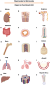
Figure 1
Figure 1
Multicompartmentalization and heterogeneity in biological systems. From top to bottom (different systems) and left to right (reducing scale): kidney to nephron, intestine to villi, bone to osteon, lung to alveoli, liver to hepatic sinus. Figure made with BioRender.
Figure 2
Figure 2
In vitro modeling, single and multiple compartment in biological systems. First row: a) breast cancer biological system component versus b) breast cancer cells‐based spheroid (parenchymal) within a fibroblast‐laden gel. (stromal). Second row: c) muscle biological system component versus d) muscle mimicking construct with tendon region at the sides to provide anchorage and a more rigid environment. e) Bone biological system component versus f) homogeneous mixture of stem cells and vascular cells. g) Brain tumor biological system component versus h) brain tumor model where brain tumor microgels (parenchymal) are embedded in neural hydrogel (stromal). Figure made with BioRender.
Figure 3
Figure 3
Approaches for compartmentalization. First row: NoECM direct contact. A first compartment is created, once the initial conformation is achieved a second compartment might be formed over it. Second row: NoECM indirect contact. A first compartment is created, within the same culture space a second compartment is created allowing them to secrete signaling molecules but without direct contact between them. Third row: gel–gel. A combination of different hydrogels leads to a two or more hydrogel system. Hydrogel microparticles can be used to obtain compartments in different scales. Fourth row: hybrid multimaterial approach, where a combination of thermoplastics, cements or hydrogels might be used together in order to generate the different compartments. Figure made with BioRender.
Figure 4
Figure 4
Technologies used for fabrication of compartmentalized models. a) Masks and Stamps are used to create patterned surfaces with indented or differently treated compartments. b) Different cell types are placed on different compartments to create multiple juxtaposed compartments. c) Microfluidic chips can be produced with neighboring compartments. d) Different cell types with or without support materials can be placed in each of these compartments. e) Bioprinting is able to combine several bioinks within the same construct, creating complex architectures that include f) different materials and cell types.
Figure 5
Figure 5
3D Bioprinting modalities and multicompartment examples. a) Extrusion bioprinting: illustrated representation of a hydrogel constructs of osteochondral tissues,[ 32 ] a minibrain with inner glioblastoma compartment surrounded by macrophages,[ 31 ] and an ex vivo glioblastoma‐on‐a‐chip model.[ 49 ] b) Laser‐assisted bioprinting: depiction of a confocal reconstruction of different layers of apical (green) and endothelial cells (red) and a cross‐section illustrating different layering of constructs. An example of horizontal patterning at different time points containing patterned apical (pore) and endothelial cells (grid).[ 176 ] c) Inkjet bioprinting: diagram showing process for horizontal patterning of a 2‐cell 3D microtissue array,[ 177 ] and examples of this patterning technique using green/red/blue labeled cells.[ 178 ] d) Stereolithography bioprinting: representation of a perfusable network model within hydrogel and a complex lung alveoli model containing air sac and oxygenating vascular network.[ 168 ] Figure made with BioRender.
Figure 6
Figure 6
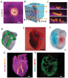
Multicompartment printing of the human brain. a) Cross‐section top view of an endothelial cell vessel (CD31; green) surrounded by fibroblasts (red) within cardiac tissue compartment with cardiomyocytes (actinin; pink). b) Diagram showing bifurcating blood vessel within engineered cardiac tissue (coordinates in mm), and c) the lumens sections of each indicated region of the printed construct. d) Human heart CAD model showing cardiac muscle (gray) and vessel (green) regions. e) A printed heart within a support bath. f) Obtained printed heart after removing of support bath, with left and right ventricles injected with red and blue dyes to demonstrate separated hollow chambers. g) 3D confocal image of the printed human heart (cardiomyocytes in pink, endothelial cells in orange). h) Immunostained cross‐sections of the printed heart showing cardiac (actinin; green) and endothelial compartment (red). Scale bars: (a,c,g,h) = 1 mm, (e) = 0.5 cm, (f) = 50 µm. Adapted with permission under the terms of the CC‐BY license.[ 188 ] Copyright 2019, the Authors. Published by Wiley‐VCH GmbH.

References

    1. Berthiaume F., Maguire T. J., Yarmush M. L., Annu. Rev. Chem. Biomol. Eng. 2011, 2, 403. - PubMed
    1. Sharma P., Kumar P., Sharma R., Bhatt V. D., Dhot P. S., J. Med. Life 2019, 12, 225. - PMC - PubMed
    1. Jovic T. H., Combellack E. J., Jessop Z. M., Whitaker I. S., Front Surg. 2020, 7, 609836. - PMC - PubMed
    1. Langer R., Vacanti J., inJournal of Pediatric Surgery,W. B. Saunder, Philadelphia, US: 2016, pp. 8–12.
    1. Matai I., Kaur G., Seyedsalehi A., McClinton A., Laurencin C. T., Biomaterials 2020, 226, 119536. - PubMed

Publication types

MeSH terms