. 2023 Jun 2;13(6):1310-1323.
doi: 10.1158/2159-8290.CD-23-0111.

Tracing Founder Mutations in Circulating and Tissue-Resident Follicular Lymphoma Precursors

Affiliations

Tracing Founder Mutations in Circulating and Tissue-Resident Follicular Lymphoma Precursors

Joseph G Schroers-Martin et al. Cancer Discov. .

Abstract

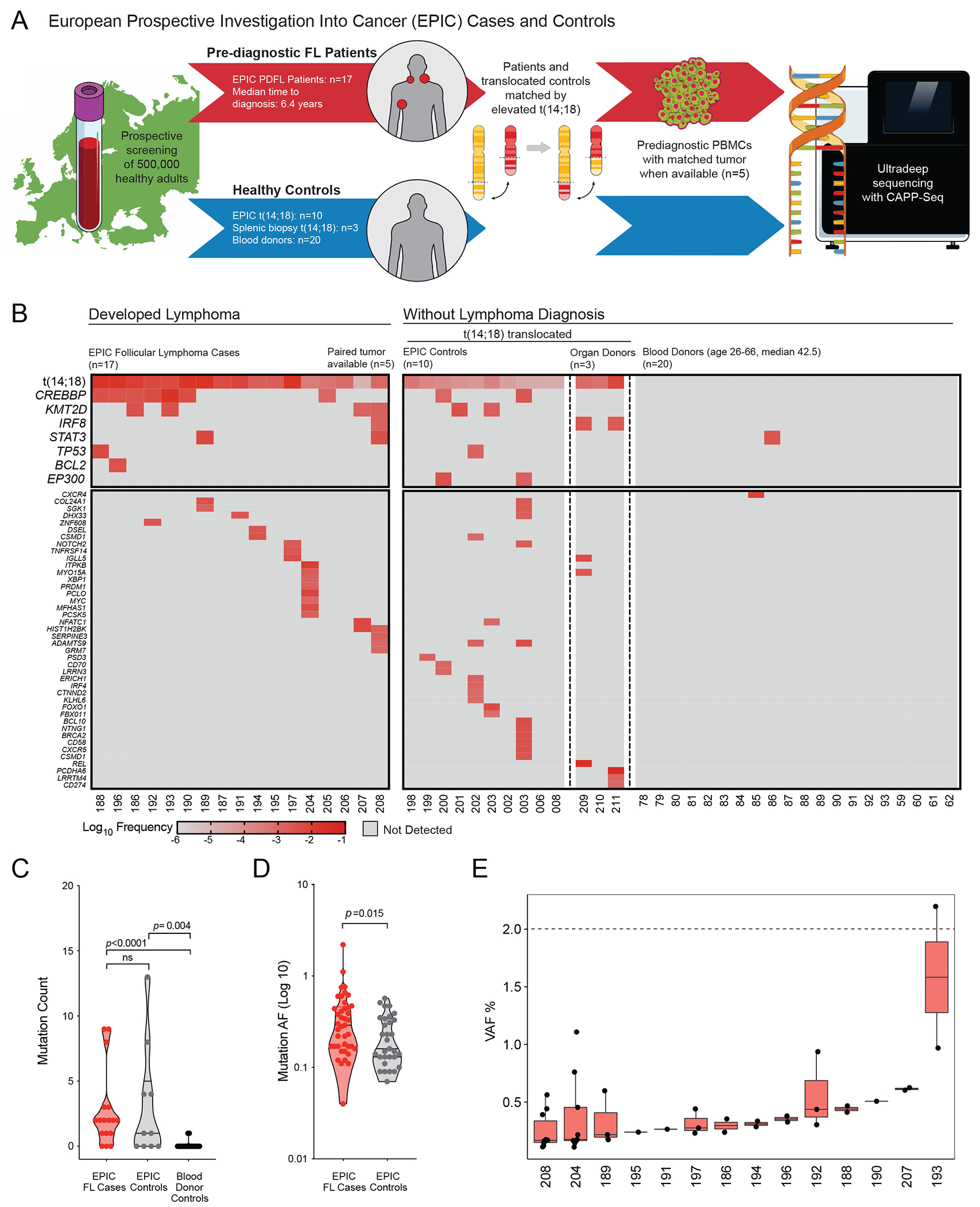
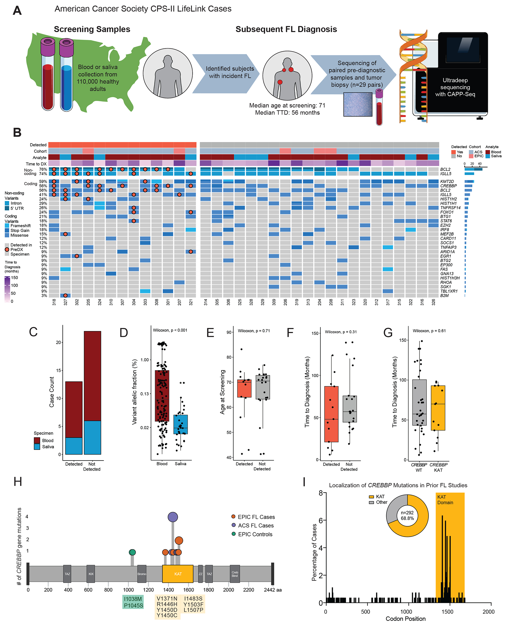
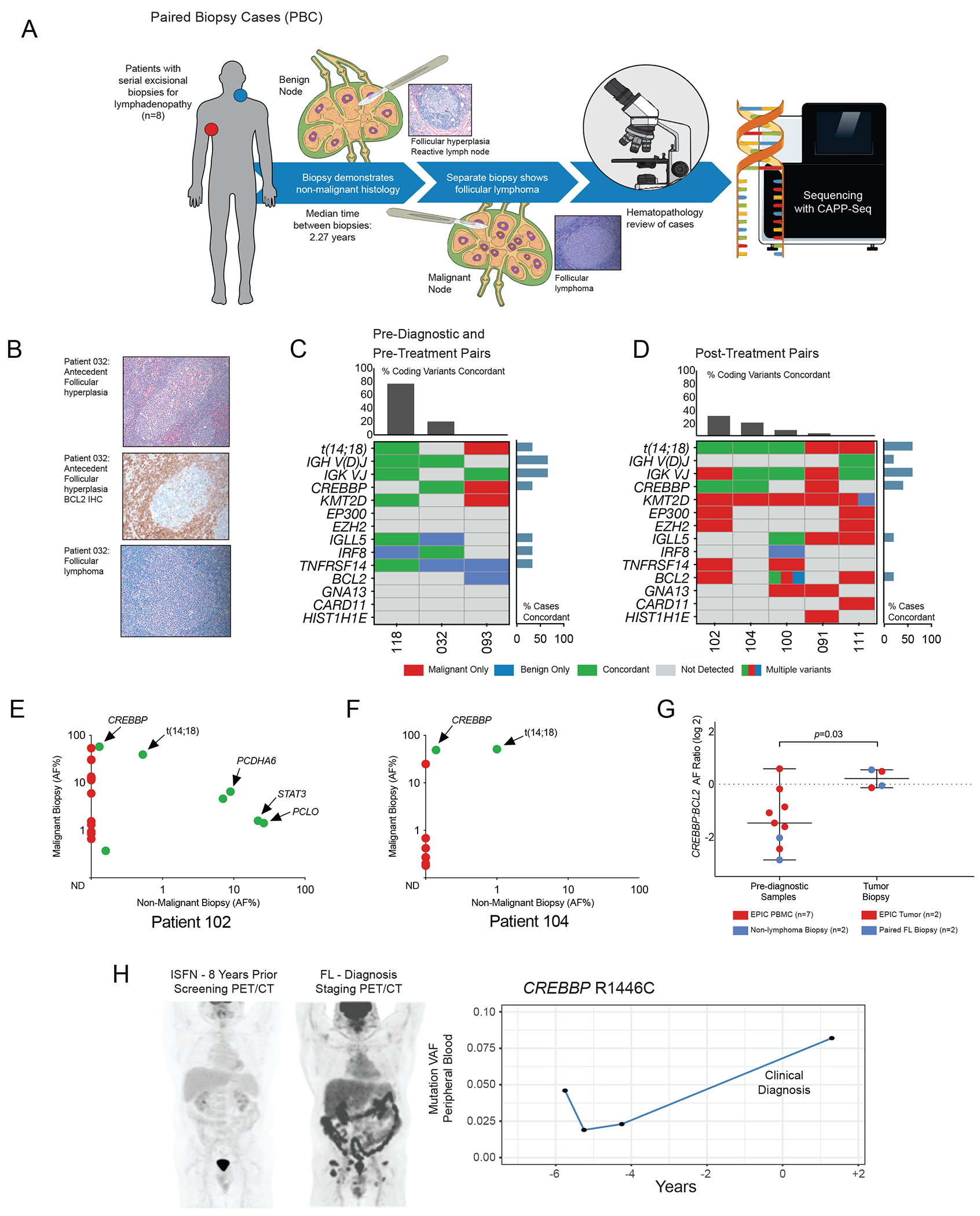
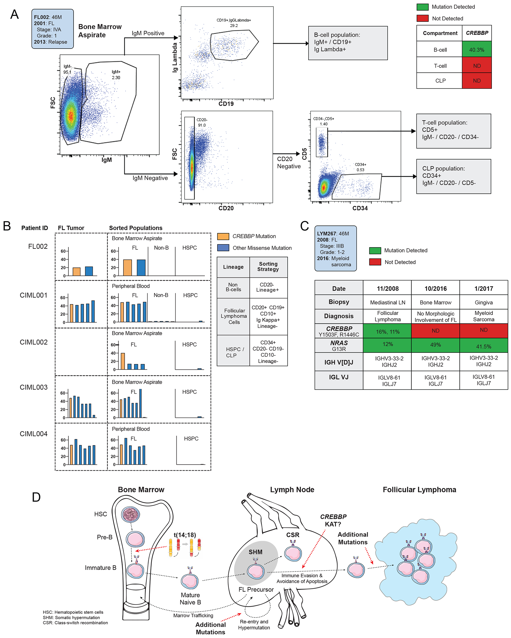
Follicular lymphomas (FL) are characterized by BCL2 translocations, often detectable in blood years before FL diagnosis, but also observed in aging healthy individuals, suggesting additional lesions are required for lymphomagenesis. We directly characterized early cooperating mutations by ultradeep sequencing of prediagnostic blood and tissue specimens from 48 subjects who ultimately developed FL. Strikingly, CREBBP lysine acetyltransferase (KAT) domain mutations were the most commonly observed precursor lesions, and largely distinguished patients developing FL (14/48, 29%) from healthy adults with or without detected BCL2 rearrangements (0/13, P = 0.03 and 0/20, P = 0.007, respectively). CREBBP variants were detectable a median of 5.8 years before FL diagnosis, were clonally selected in FL tumors, and appeared restricted to the committed B-cell lineage. These results suggest that mutations affecting the CREBBP KAT domain are common lesions in FL cancer precursor cells (CPC), with the potential for discriminating subjects at risk of developing FL or monitoring residual disease.

Significance: Our study provides direct evidence for recurrent genetic aberrations preceding FL diagnosis, revealing the combination of BCL2 translocation with CREBBP KAT domain mutations as characteristic committed lesions of FL CPCs. Such prediagnostic mutations are detectable years before clinical diagnosis and may help discriminate individuals at risk for lymphoma development. This article is highlighted in the In This Issue feature, p. 1275.


Conflict of interest statement

FS, JSM, JS, SR, GB, BN: no disclosures or conflict of interest

AAA reports ownership interest in CiberMed, FortySeven Inc., and Foresight Diagnostics, patent filings related to cancer biomarkers, research funding from Bristol Myers Squibb and Celgene, and paid consultancy from Genentech, Karyopharm, Roche, Chugai, Gilead, and Celgene.

Figures

Figure 1)
Figure 1). Recurrent CREBBP mutations are observed in screening blood samples years prior to FL diagnosis.
A. Schematic depiction of study design: from a large prospective European screening cohort, 17 patients with a subsequent follicular lymphoma (FL) diagnosis (PDFL patients) were paired with healthy participants with similar elevated circulating t(14;18) but no FL diagnosis at last follow-up. Healthy unrelated subjects (blood donors and normal spleen from organ donors) were included as controls. B. A heatmap illustrates the observed frequencies of FL-associated lesions in pre-diagnostic samples from EPIC PDFL patients (n=17) and controls (n=33), including recurrent t(14;18) and CREBBP mutations. C. A violin plot displays enrichment in total mutational burden among individuals bearing t(14;18) translocations as compared to blood donor controls. No significant difference in mutational burden was seen between t(14;18) translocated subjects based on eventual FL status. D. A violin plot demonstrates increased allelic fraction (AF) of somatic variants among PDFL patients as compared to t(14;18)-bearing controls. E. Box plots demonstrate the distribution of somatic variant AF in individual PDFL cases in relationship to the clinical CHIP threshold of 2% VAF.
Figure 2)
Figure 2). Pre-diagnostic CREBBP mutations are concordant in subsequent FL tumor biopsies.
A. Schematic depiction of study design: from a large prospective American screening cohort, 29 individuals were identified with subsequent FL diagnosis and available pre-diagnostic and tumor biopsy specimens. Pre-diagnostic and tumor specimens underwent parallel hybrid capture enrichment and sequencing, with ultra-deep sequencing of pre-diagnostic samples. B. An oncoprint depicts variants detected in 34 FL tumor biopsies. Cases are grouped by detection of tumor variants in prediagnostic specimens, with detected variants identified by a superimposed marker. C. A stacked bar graph demonstrates detection status of tumor-derived variants in pre-diagnostic blood (n=25) and saliva (n=9) specimens from FL patients. No difference in detection rate was observed by analyte (Fisher’s exact p=1). D. A bar plot depicts AF of tumor-confirmed variants depicted in pre-diagnostic specimens by analyte, with significantly lower AF seen in saliva specimens (p<0.001). E. A bar plot depicts the age at pre-diagnostic sample collection of participants developing FL in the EPIC and ACS CPS-II studies. Cases are grouped by detection status of tumor-confirmed variants, with no significant difference in age between patients with or without detected variants (p=0.71). F. A bar plot depicts the interval from pre-diagnostic sample collection to FL diagnosis of participants developing FL in the EPIC and ACS CPS-II studies. Cases are grouped by detection status of tumor-confirmed variants, with no significant difference in time to FL diagnosis in patients with or without detected variants (p=0.31) G. A bar plot depicts the interval from pre-diagnostic sample collection to FL diagnosis of participants developing FL in the EPIC and ACS CPS-II studies. Cases are grouped by detection of CREBBP KAT domain mutations, with no significant difference in time to diagnosis in patients with or without detected variants (p=0.61) H. A lollipop plot of the CREBBP protein illustrates the localization of observed pre-diagnostic mutations to the lysine acetyltransferase (KAT) domain. Two mutations in control samples (green) occurred outside of a characterized functional domain. I. A bar graph depicts the percentage of CREBBP mutations occurring at each codon in a pooled analysis of prior FL genotyping studies (15, 19, 36, 37, 38, 39). Mutations consistently concentrated within the KAT domain, with 68.8% (201/292 cases) occurring between codons 1300 and 1700.
Figure 3)
Figure 3). CREBBP KAT domain mutations are observed as persistent after cancer therapy and in association with histologic precursor lesions.
A. Schematic depiction of study design: Chronologically distant paired malignant and benign lymph node biopsies from 8 patients were re-reviewed by a blinded hematopathologist and sequenced in parallel. B. Representative images from pathology review of paired biopsy cases, including BCL2-negative antecedent follicular hyperplasia. C. A genotyping map demonstrates genetic concordance (indicated in green) between pre-diagnostic or pre-treatment and paired malignant biopsies. The concordance of selected recurrently mutated genes including chromatin modifying genes (CMGs) is included for comparison, with per-gene concordance displayed as a bar graph on the right and per-case concordance as a bar graph above. One patient (032) was t(14;18) negative by confirmatory PCR. D. A genotyping map demonstrates genetic concordance (indicated in green) between diagnostic FL biopsy and subsequent post-treatment histologically non-malignant specimens obtained for adenopathy. Annotation is as per the previous panel. E. A scatter plot demonstrates concordance of coding mutations in paired FL and post-treatment non-malignant biopsies for patient 102. The X-axis indicates log allelic fraction in the non-malignant sample and the Y-axis indicates log AF in the mature FL tumor, demonstrating persistence of t(14;18) and CREBBP KAT domain mutations. F. A scatter plot as in Fig 3E demonstrates post-treatment concordance of t(14 ;18) and CREBBP KAT domain mutation for patient 104. G. A scatter plot depicts the increase in CREBBP allelic fraction relative to t(14;18), with red points representing EPIC samples and blue points representing paired biopsies. At time of diagnostic biopsy these lesions were roughly equal (median CREBBP:BCL2 ratio 1.04, range 0.68-1.30), suggesting subclonal mutations in pre-diagnostic specimens proceeding to co-dominance of CREBBP mutations in mature FL tumors. H. PET/CT imaging of a representative patient at discovery of in-situ follicular neoplasia (ISFN, left) and eight years later at FL diagnosis (right). Serial sequencing of peripheral blood specimens collected during expectant management revealed a tumor-confirmed CREBBP KAT domain mutation (R1446C) detectable at first collected timepoint and increasing in AF to the time of diagnosis.
Figure 4)
Figure 4). Follicular lymphoma CREBBP mutations appear localized to mature B-cells.
A. Flow cytometry gating strategy for progenitor and mature lymphoid from human bone marrow aspirate of a representative FL patient at diagnosis (FL002). B. A table of bar graphs demonstrates variant allelic frequency of lymphoma-associated coding mutations in indicated cell subsets from 5 FL patients bearing CREBBP mutations. Each column represents a mutation initially detected in primary tumor or isotype-sorted marrow aspirate. C. A table demonstrates concordance and discordance of pathogentic mutations and VDJ rearrangements detected in serial biopsies from an FL patient (LYM267) with CREBBP and NRAS mutations who subsequently developed a myeloid neoplasm lacking CREBBP. D. Schematic figure for a proposed model of early follicular lymphomagenesis. The BCL2 translocation occurring in an early B-cell precursor provides evasion of programmed cell death, while the accrual of CREBBP KAT domain mutations in mature B-cells provides immune evasion and persistence of the GC program.

References

    1. Yunis JJ, Oken MM, Kaplan ME, Ensrud KM, Howe RR, Theologides A. Distinctive chromosomal abnormalities in histologic subtypes of non-Hodgkin’s lymphoma. New England Journal of Medicine. 1982;307(20):1231–6. - PubMed
    1. Tsujimoto Y, Cossman J, Jaffe E, Croce CM. Involvement of the bcl-2 gene in human follicular lymphoma. Science. 1985;228(4706):1440–3. - PubMed
    1. Roulland S, Navarro JM, Grenot P, Milili M, Agopian J, Montpellier B, et al. Follicular lymphoma-like B cells in healthy individuals: a novel intermediate step in early lymphomagenesis. The Journal of experimental medicine. 2006;203(11):2425–31. - PMC - PubMed
    1. Summers KE, Goff LK, Wilson AG, Gupta RK, Lister TA, Fitzgibbon J. Frequency of the Bcl-2/IgH rearrangement in normal individuals: implications for the monitoring of disease in patients with follicular lymphoma. Journal of clinical oncology : official journal of the American Society of Clinical Oncology. 2001;19(2):420–4. - PubMed
    1. Liu Y, Hernandez AM, Shibata D, Cortopassi GA. BCL2 translocation frequency rises with age in humans. Proceedings of the National Academy of Sciences of the United States of America. 1994;91(19):8910–4. - PMC - PubMed

Publication types

Substances