. 2023 Apr 27;97(4):e0016023.
doi: 10.1128/jvi.00160-23. Epub 2023 Mar 20.

Harnessing the Noncanonical Keap1-Nrf2 Pathway for Human Cytomegalovirus Control

Affiliations

Harnessing the Noncanonical Keap1-Nrf2 Pathway for Human Cytomegalovirus Control

Ayan K Ghosh et al. J Virol. .

Abstract

Host-derived cellular pathways can provide an unfavorable environment for virus replication. These pathways have been a subject of interest for herpesviruses, including the betaherpesvirus human cytomegalovirus (HCMV). Here, we demonstrate that a compound, ARP101, induces the noncanonical sequestosome 1 (SQSTM1)/p62-Keap1-Nrf2 pathway for HCMV suppression. ARP101 increased the levels of both LC3 II and SQSTM1/p62 and induced phosphorylation of p62 at the C-terminal domain, resulting in its increased affinity for Keap1. ARP101 treatment resulted in Nrf2 stabilization and translocation into the nucleus, binding to specific promoter sites and transcription of antioxidant enzymes under the antioxidant response element (ARE), and HCMV suppression. Knockdown of Nrf2 recovered HCMV replication following ARP101 treatment, indicating the role of the Keap1-Nrf2 axis in HCMV inhibition by ARP101. SQSTM1/p62 phosphorylation was not modulated by the mTOR kinase or casein kinase 1 or 2, indicating ARP101 engages other kinases. Together, the data uncover a novel antiviral strategy for SQSTM1/p62 through the noncanonical Keap1-Nrf2 axis. This pathway could be further exploited, including the identification of the responsible kinases, to define the biological events during HCMV replication. IMPORTANCE Antiviral treatment for human cytomegalovirus (HCMV) is limited and suffers from the selection of drug-resistant viruses. Several cellular pathways have been shown to modulate HCMV replication. The autophagy receptor sequestosome 1 (SQSTM1)/p62 has been reported to interact with several HCMV proteins, particularly with components of HCMV capsid, suggesting it plays a role in viral replication. Here, we report on a new and unexpected role for SQSTM1/p62, in HCMV suppression. Using a small-molecule probe, ARP101, we show SQSTM1/p62 phosphorylation at its C terminus domain initiates the noncanonical Keap1-Nrf2 axis, leading to transcription of genes under the antioxidant response element, resulting in HCMV inhibition in vitro. Our study highlights the dynamic nature of SQSTM1/p62 during HCMV infection and how its phosphorylation activates a new pathway that can be exploited for antiviral intervention.

Keywords: ARP101; SQSTM1/p62; antioxidant response element; human cytomegalovirus; noncanonical Keap1-Nrf2 pathway; p62-Keap1-Nrf2.


Conflict of interest statement

The authors declare no conflict of interest.

Figures
FIG 1
FIG 1
HCMV inhibition by ARP101. (A) A luciferase-based reporter assay was performed at 72 hpi on HFFs infected with the pp28-luciferase recombinant HCMV-Towne (MOI of 1 PFU/cell), followed by treatment with the indicated concentrations of ARP101. (B) HCMV-TB40 (200 PFU/well) was used to infect HFFs (1 × 106 cells in a 24-well plate), followed by treatment with the indicated concentrations of ARP101. On day 8 postinfection, plaques were enumerated. (C) HFFs were treated with the indicated concentrations of ARP101 for 72 h and 8 days, and a luminescence-based cell viability assay was performed to determine the 50% cellular viability (CC50). (D) The activity of ARP101 (10 μM) against pp28-luciferase ganciclovir (GCV)-resistant HCMV-Towne (GCVR HCMV) was measured by luciferase assay at 72 hpi. (E) HFFs were infected with HCMV-Towne (MOI of 0.1) for 24, 48, and 72 h and treated with ARP101 (10 μM) or GCV (5 μM). Infected cells were lysed, DNA was isolated, and viral load was measured by a US17 quantitative real-time PCR. (F) HFFs were infected with HCMV-Towne (MOI of 1) and treated with ARP101 (10 μM) or GCV (5 μM). Infected cells were lysed at 24, 48, and 72 hpi, lysates were run on 12% SDS-PAGE gels, and immunoblot assays were performed to detect IE1/2 (immediate early), UL84 (early late), and pp65 (late) antigens. Numbers presented below each panel of immunoblots represent the relative band intensities of each blot. Relative luciferase activity was calculated by dividing the luciferase units obtained at a given drug concentration by the luciferase units obtained following treatment with the vehicle control, dimethyl sulfoxide. All experiments were performed in triplicates, and represented values are mean ± SD.
FIG 2
FIG 2
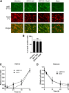
Timing of activity of ARP101. (A) An immunofluorescence assay was performed to test for HCMV entry. HFFs were pretreated with dimethyl sulfoxide (mock), ARP101, or GCV for 24 h and then infected with HCMV-Towne (MOI of 1). (B) Graph depicting percentage of pp65-positive cells in the microscopic data presented in panel A. (C and D) A luciferase-based reporter assay was performed to determine the timing of activity of ARP101. HFFs were infected with pp28-luciferase recombinant Towne, and ARP101 (10 μM) was either added to or removed from infected HFFs at 6, 24, 48, and 72 h postinfection. GCV (5 μM) was used as a control for timing of activity. Relative luciferase activity was calculated by dividing the luciferase units obtained at a given drug concentration by the luciferase units obtained following treatment with the vehicle control, dimethyl sulfoxide. All experiments were performed in triplicates, and represented values are mean ± SD.
FIG 3
FIG 3
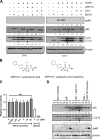
ARP101 induces both p62 and LC3 II in HCMV-infected HFFs. (A) HFFs were infected with HCMV-Towne (MOI of 1) and treated with ARP101 (10 μM) for 24 and 72 h. Bafilomycin A1 (Baf A1, 50 nM) was added to the respective conditions 3 h before harvesting. Following Baf A1 treatment, cells were lysed, and an immunoblot assay was performed for LC3 I/II and p62. Red arrows indicate HCMV-infected HFF samples, and black arrows indicate ARP101-treated HCMV-infected HFF samples. HCMV pp65 and β-actin were probed as infection and loading controls, respectively. (B) Structure of active ARP101 (having the hydroxamic acid functional group) and inactive ARP101 (carboxylic acid replaces the hydroxamic acid functional group). (C) HCMV inhibition by ARP101 hydroxamic acid and the carboxylic acid (inactive) was measured at 72 hpi with pp28-luciferase recombinant Towne (MOI of 1). (D) Whole-cell lysates from infected HFFs were analyzed by immunoblotting for p62, LC3 I/II, and viral pp65 following treatment with the active and inactive ARP101 (10 μM) at the indicated time points. Red arrows indicate HCMV-infected HFF samples, and black arrows indicate ARP101-treated HCMV-infected HFF samples at 72 h postinfection. The experiments were performed in triplicates, and the best representative data are presented. INF, infected with HCMV-Towne.
FIG 4
FIG 4
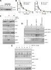
ARP101 induces phosphorylation of p62 at the KIR and UBA domains and increases expression of Nrf2 and heme oxygenase-1 (HO-1). (A) ATG5 knockdown (KD) by lentiviral transduction in HFFs. Cells expressing pLKO.1 (vector control) and plasmids containing ATG5 shRNA were grown to confluence following puromycin selection and lysed in cell lysis buffer. ATG5 expression in pLKO.1 control and ATG5 KD HFFs (#1, #2, and #3) was analyzed by immunoblotting. (B) A luciferase-based reporter assay was performed with ATG5 KD (#2 and #3) and pLKO.1 control HFFs infected with HCMV-Towne (MOI of 1) and treated with the indicated concentrations of ARP101 and GCV. The values are represented as the mean ± SD from two independent experiments. (C) HFFs were infected with HCMV-Towne (MOI of 1) and treated with ARP101 (10 μM) for 24, 48, and 72 h. Cells were lysed posttreatment, and equal amounts of proteins were analyzed on 12% SDS-PAGE gels. Immunoblot assays were performed for HO-1, Keap1, Nrf2, p62, and LC3 I/II. Viral pp65 and β-actin were probed for infection and loading controls, respectively. The red arrowhead indicates HCMV-infected HFF samples, and the black arrowhead indicates ARP101-treated HCMV-infected HFF samples at 72 hpi. (D) HCMV-infected cells (MOI of 1) were lysed posttreatment with active and inactive ARP101 (10 μM). Equal amounts of lysates were analyzed on 12% SDS-PAGE gels, and an immunoblot assay was performed for p-p62 (S349), p-p62 (S403), p62, viral UL84, and β-actin. The experiment was repeated in triplicate, and representative images are presented. (E) HFFs were treated with ARP101 (10 μM) and harvested at 24, 48, and 72 h posttreatment for analysis of levels of HO-1, Nrf2, Keap1, p-p62 (S349), p62, and LC3 I/II by Western blotting. β-Actin was probed as an internal control. NI, noninfected; INF, infected with HCMV-Towne.
FIG 5
FIG 5
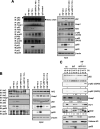
ARP101 releases Nrf2 from the p62-Keap1-Nrf2 complex, resulting in nuclear translocation. (A) Immunoprecipitation (IP) of p62 was performed in HCMV-infected (MOI of 1) ARP101 (10 μM)-treated cells at 48 and 72 hpi. Keap1, Nrf2, p-p62 (S349), and p-p62 (S403) were probed to measure the affinity of p62 for Keap1 and its effect on Nrf2 release from Keap1. Viral pp65 and pp28 were probed for analysis of the interaction of p62 with viral proteins. The right panel shows the profile of different proteins in the soluble fraction of the lysates (input), which was used for IP. IB, immunoblot. (B) Reverse IP of Nrf2 was performed in HCMV-infected ARP101-treated cells at 48 and 72 hpi. Keap1 and p62 were probed to measure the status of the p62-Keap1-Nrf2 complex. The right panel shows the input used for the IP. (C) HFFs were infected with HCMV-Towne (MOI of 1) and treated with ARP101 (10 μM) for 48 and 72 h. Cells were fractionated into cytoplasmic (C) and nuclear (N) fractions, concentrated by acetone precipitation, and quantified. Equal amounts of proteins were analyzed on 12% SDS-PAGE gels, and immunoblot assays were performed to measure the level of p62, p-p62 (Ser349), p-p62 (S403), Keap1, Nrf2, and HO-1. Histone H3 and GAPDH were probed as nuclear (N) and cytoplasmic (C) controls, respectively. Viral pp65 was probed as an infection control. The experiments were performed thrice, and representative images are presented. NI, noninfected; INF, infected with HCMV-Towne. IgG control experiments were conducted with INF cell lysates.
FIG 6
FIG 6
ARP101 induces binding of Nrf2 to specific promoters and activation of the antioxidant response element (ARE). (A to D) Binding of Nrf2 to several ARE promoters was analyzed by ChIP assay. HFFs were infected with HCMV-Towne (MOI of1) and treated with ARP101 or inactive ARP101 (10 μM) for 48 and 72 h. (E to H) Quantitative PCRs were performed for SQSTM1/p62 (E), HMOX1 (F), NQO1 (G), and GCLC (H) transcribed upon ARE activation in infected HFFs treated/untreated with ARP101 at 48 and 72 hpi. (I) Viral IE1 and IE2 expression was analyzed by quantitative PCR for infection control in the same samples from panels E to H. The experiments were performed thrice, and data from a single experiment with three technical replicates are presented. NI, noninfected; INF, infected with HCMV-Towne.
FIG 7
FIG 7
ARP101 fails to activate the ARE in Nrf2 KD cells and does not inhibit HCMV replication. (A) Nrf2 knockdown efficiency was analyzed by immunoblotting in the lysates of lentivirus-transduced HFF cells following puromycin selection. GAPDH was probed as a loading control. (B) A luciferase-based reporter assay was performed with infected (MOI of 1) Nrf2 KD and pLKO.1 control cells treated with the indicated concentrations of ARP101 and GCV. (C) Lysates from panel B were analyzed by Western blotting for Nrf2, viral pp65, and β-actin. (D) A plaque reduction assay was performed using Nrf2 KD and pLKO.1 control HFFs by infecting cells with HCMV-TB40 (200 PFU/well, used to infect 1 × 106 cells in a 24-well plate) and treating them with ARP101 (5 and 10 μM) and GCV (5 μM). (E) Nrf2 KD and pLKO.1 cells were infected with HCMV-TB40 (MOI of 1 PFU/cell), and supernatants were harvested after 120 h followed by titration using plaque assay. (F) Nrf2 KD and pLKO.1 control cells were infected with HCMV-Towne (MOI of 1) and treated with ARP101 (10 μM), and lysates were prepared after 72 hpi. Levels of Nrf2, Keap1, p-p62 (S349), p62, HO-1, and LC3 I/II were measured by Western blotting. Viral pp65 and GAPDH were probed as infection and internal controls, respectively. Experiments were performed thrice, and the best representative images are depicted. NI, noninfected; INF, infected with HCMV-Towne.
FIG 8
FIG 8
Keap1 is degraded through a p62-mediated process induced by ARP101 in HCMV-infected cells. (A) A cycloheximide chase assay was performed at 72 hpi, and expression of Keap1 was analyzed by immunoblotting. (B) Graphical representation of Keap1 levels measured at 72 hpi in panel A. The level of Keap1 was normalized to β-actin at the indicated time points after the addition of cycloheximide. (C) The levels of Keap1, along with several other proteins, at 72 hpi following ARP101 (10 μM) treatment were analyzed by Western blotting. Bafilomycin A1 (Baf A1; 50 nM) was added 3 h before harvesting at 72 hpi, whereas MG132 (10 μM) was added 8 h before harvesting at 72 hpi. β-Actin was probed as an internal control. (D) HFFs were treated with MG132 (10 μM), K67 (25 μM), and their combination for 18 h, and lysates were prepared for analysis of Keap1, Nrf2, p-p62 (S349), and p62 by immunoblotting. (E) HFFs were infected with HCMV-Towne (MOI of 1 PFU/cell) for 72 h and treated with ARP101 (10 μM), K67 (25 μM), and their combination. Lysates were analyzed by immunoblotting for Keap1, Nrf2, p-p62 (S349), viral pp65, and β-actin, respectively. (F) Immunoprecipitation of p-p62 (S349) followed by detection of Keap1 and LC3 II using noninfected, infected, and ARP101-treated cellular lysates harvested at 72 hpi. (G) IP of p-p62 (S349) followed by detection of Keap1 and LC3 II using HCMV-infected HFFs (MOI of 1 PFU/cell) treated with ARP101 or ARP101 plus K67 and harvested at 72 h postinfection. Experiments were performed thrice, and representative data are presented.
FIG 9
FIG 9
ARP101-induced p62 phosphorylation at S349/S403 is not modulated by the mTOR kinase or casein kinases 1 and 2 in HCMV-infected HFFs. HFFs were infected with HCMV-Towne (MOI of 1 PFU/cell) and treated with ARP101 (10 μM) for 24, 48, and 72 h. Torin-1 (250 nM) was added at 8 h (in panel B) or 24 h (in panel C) before harvesting the cells at 48 or 72 hpi, respectively. TBCA (30 μM), an inhibitor of casein kinase 2, and CK1-7 (50 μM), an inhibitor of casein kinase 1, were also added 8 h before harvesting at 72 hpi. (A) p-mTOR (S2448) and mTOR were analyzed by immunoblotting at the indicated time points. (B) Immunoblot analysis of indicated proteins at 72 hpi under various experimental conditions where kinase inhibitors including Torin-1 were added 8 h before harvesting and ARP101 was added immediately after infection. (C) p-p62 (S349), p-p62 (S403), p62, and p-4EBP1 were analyzed at 48 and 72 hpi following Torin-1 and ARP101 treatments, as mentioned above. Torin-1 was added 24 h before harvesting at indicated time points, and ARP101 was added immediately after infection. Viral pp65/UL84 and β-actin were probed for infection and loading controls, respectively. The experiments were repeated twice, and representative images are presented. NI, noninfected; INF, infected with HCMV-Towne.
FIG 10
FIG 10
Model depicting the mechanism of anti-HCMV activity of ARP101—induction of the noncanonical p62-Keap1-Nrf2 pathway, resulting in transcription of the antioxidant response element (ARE). (Left) HCMV does not interfere with p62-Keap-Nrf2 interaction and deactivates the ARE for successful replication. (Right) ARP101 induces p62 phosphorylation at Ser349 and Ser403 during infection, resulting in enhanced affinity of p62 for Keap1, and Nrf2 stabilization, which in turn translocates into the nucleus to bind to the specific promoter regions and activate the transcription of genes under the ARE. In the process, Keap1 associated with phosphorylated p62 undergoes degradation via association with LC3 II. Taken together, ARP101 generates a continuous supply of p62 through a feedback loop by the activation of ARE.
FIG 11
FIG 11
Synthesis of the inactive ARP101 analog compound 1. Reagents and conditions: (i) O-isopropylhydroxylamine hydrochloride, N-methylpyrrolidone, tetrahydrofuran, room temperature; (ii) t-butyl (2S)-2-hydroxy-3-methylbutanoate, diisopropyl-azodicarboxylate, PPh3, room temperature; (iii) trifluoroacetic acid, CH2Cl2, 0°C, 5 h.

References

    1. Staras SA, Dollard SC, Radford KW, Flanders WD, Pass RF, Cannon MJ. 2006. Seroprevalence of cytomegalovirus infection in the United States, 1988–1994. Clin Infect Dis 43:1143–1151. 10.1086/508173. - DOI - PubMed
    1. Griffiths PD, Clark DA, Emery VC. 2000. Betaherpesviruses in transplant recipients. J Antimicrob Chemother 45(Suppl T3):29–34. 10.1093/jac/45.suppl_4.29. - DOI - PubMed
    1. Kovacs A, Schluchter M, Easley K, Demmler G, Shearer W, La RP, Pitt J, Cooper E, Goldfarb J, Hodes D, Kattan M, McIntosh K. 1999. Cytomegalovirus infection and HIV-1 disease progression in infants born to HIV-1-infected women. Pediatric Pulmonary and Cardiovascular Complications of Vertically Transmitted HIV Infection Study Group. N Engl J Med 341:77–84. 10.1056/NEJM199907083410203. - DOI - PMC - PubMed
    1. Sabin CA, Phillips AN, Lee CA, Janossy G, Emery V, Griffiths PD. 1995. The effect of CMV infection on progression of human immunodeficiency virus disease is a cohort of haemophilic men followed for up to 13 years from seroconversion. Epidemiol Infect 114:361–372. 10.1017/s095026880005799x. - DOI - PMC - PubMed
    1. Sabin CA, Devereux HL, Clewley G, Emery VC, Phillips AN, Loveday C, Lee CA, Griffiths PD. 2000. Cytomegalovirus seropositivity and human immunodeficiency virus type 1 RNA levels in individuals with hemophilia. J Infect Dis 181:1800–1803. 10.1086/315476. - DOI - PubMed

Publication types

MeSH terms