Multicenter Study
. 2023 May 1;80(5):523-528.
doi: 10.1001/jamaneurol.2023.0265.

Quantification of Penumbral Volume in Association With Time From Stroke Onset in Acute Ischemic Stroke With Large Vessel Occlusion

Collaborators, Affiliations
Multicenter Study

Quantification of Penumbral Volume in Association With Time From Stroke Onset in Acute Ischemic Stroke With Large Vessel Occlusion

Pierre Seners et al. JAMA Neurol. .

Abstract

Importance: The benefit of reperfusion therapies for acute ischemic stroke decreases over time. This decreasing benefit is presumably due to the disappearance of salvageable ischemic brain tissue (ie, the penumbra).

Objective: To study the association between stroke onset-to-imaging time and penumbral volume in patients with acute ischemic stroke with a large vessel occlusion.

Design, setting, and participants: A retrospective, multicenter, cross-sectional study was conducted from January 1, 2015, to June 30, 2022. To limit selection bias, patients were selected from (1) the prospective registries of 2 comprehensive centers with systematic use of magnetic resonance imaging (MRI) with perfusion, including both thrombectomy-treated and untreated patients, and (2) 1 prospective thrombectomy study in which MRI with perfusion was acquired per protocol but treatment decisions were made with clinicians blinded to the results. Consecutive patients with acute stroke with intracranial internal carotid artery or first segment of middle cerebral artery occlusion and adequate quality MRI, including perfusion, performed within 24 hours from known symptoms onset were included in the analysis.

Exposures: Time from stroke symptom onset to baseline MRI.

Main outcomes and measures: Penumbral volume, measured using automated software, was defined as the volume of tissue with critical hypoperfusion (time to maximum >6 seconds) minus the volume of the ischemic core. Substantial penumbra was defined as greater than or equal to 15 mL and a mismatch ratio (time to maximum >6-second volume/core volume) greater than or equal to 1.8.

Results: Of 940 patients screened, 516 were excluded (no MRI, n = 19; no perfusion imaging, n = 59; technically inadequate perfusion imaging, n = 75; second segment of the middle cerebral artery occlusion, n = 156; unwitnessed stroke onset, n = 207). Of 424 included patients, 226 (53.3%) were men, and mean (SD) age was 68.9 (15.1) years. Median onset-to-imaging time was 3.8 (IQR, 2.4-5.5) hours. Only 16 patients were admitted beyond 10 hours from symptom onset. Median core volume was 24 (IQR, 8-76) mL and median penumbral volume was 58 (IQR, 29-91) mL. An increment in onset-to-imaging time by 1 hour resulted in a decrease of 3.1 mL of penumbral volume (β coefficient = -3.1; 95% CI, -4.6 to -1.5; P < .001) and an increase of 3.0 mL of core volume (β coefficient = 3.0; 95% CI, 1.3-4.7; P < .001) after adjustment for confounders. The presence of a substantial penumbra ranged from approximately 80% in patients imaged at 1 hour to 70% at 5 hours, 60% at 10 hours, and 40% at 15 hours.

Conclusions and relevance: Time is associated with increasing core and decreasing penumbral volumes. Despite this, a substantial percentage of patients have notable penumbra in extended time windows; the findings of this study suggest that a large proportion of patients with large vessel occlusion may benefit from therapeutic interventions.


Conflict of interest statement

Conflict of Interest Disclosures: Dr Heit reported receiving consultant fees from Medtronic and MicroVention and fees from iSchemaView for serving on the advisory board outside the submitted work. Dr Christensen reported holding equity in iSchemaView. Dr Cognard reported receiving consultant fees from MIVI, Medtronic, MicroVention, Cerenovus, and Stryker outside the submitted work. Dr Savatovsky reported receiving lecture fees from Canon, nonfinancial support from Bayer for travel expenses, personal fees from Biogen and Bayer for serving on the advisory boards, and holding stock in Guerbet and Chipiron. Dr Olivot reported receiving consultant fees from Abbvie, Acticor Biotech, and Bioxodes and speaker fees from Boehringer and Bristol Myers Squibb outside the submitted work. Dr Albers reported receiving personal fees from iSchemaView and Genentech and holding equity in iSchemaView outside the submitted work. No other disclosures were reported.

Figures

Figure 1.
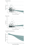
Figure 1.. Unadjusted Association Between Onset-to-Imaging Time and Penumbral Volume, Core Volume, and Substantial Penumbra Presence
A and B, The plain curve represents the unadjusted linear association between the volumes and onset-to-imaging time, and shaded areas the 95% CIs. C, The probability of substantial penumbra presence according to onset-to-imaging time. A substantial penumbra was defined as penumbral volume (time to maximum >6-second volume – core volume) greater than or equal to 15 mL and mismatch ratio (time to maximum >6-second volume/core volume) greater than or equal to 1.8. The regression curve estimates of the probability of substantial penumbra presence according to onset-to-imaging time for a typical patient aged 69 years with a National Institutes of Health Stroke Scale score of 17 (range, 0-42, with higher scores indicating greater stroke severity) and hypoperfusion intensity ratio of 0.43. The shaded area corresponds to the 95% CI (logistic regression model).
Figure 2.
Figure 2.. Representative Images of Patients at Increasing Times From Stroke Symptom Onset to Imaging Time
For each patient, the infarct core, critically hypoperfused, and penumbral volumes are automatically estimated using RAPID software, version 4.7 to 5.0 (iSchemaView). Infarct core is measured on diffusion-weighted imaging (apparent diffusion coefficient [ADC] <620 × 10−6 mm2/s), and the critically hypoperfused tissue on perfusion imaging (time-to-maximum [Tmax] >6.0 seconds). The volume of ischemic penumbra is defined as the difference between the critically hypoperfused and the core volumes (Tmax >6-second volume – core volume). Each patient has an acute occlusion of the intracranial internal carotid artery or the first segment of the middle cerebral artery. National Institutes of Health Stroke Scale score (range, 0-42, with higher scores indicating greater stroke severity) at the time of imaging was 11 in patient A, 11 in patient B, 13 in patient C, and 15 in patient D.

References

    1. Saver JL. Time is brain-quantified. Stroke. 2006;37(1):263-266. doi:10.1161/01.STR.0000196957.55928.ab - DOI - PubMed
    1. Jones TH, Morawetz RB, Crowell RM, et al. . Thresholds of focal cerebral ischemia in awake monkeys. J Neurosurg. 1981;54(6):773-782. doi:10.3171/jns.1981.54.6.0773 - DOI - PubMed
    1. Morawetz RB, DeGirolami U, Ojemann RG, Marcoux FW, Crowell RM. Cerebral blood flow determined by hydrogen clearance during middle cerebral artery occlusion in unanesthetized monkeys. Stroke. 1978;9(2):143-149. doi:10.1161/01.STR.9.2.143 - DOI - PubMed
    1. Touzani O, Young AR, Derlon JM, et al. . Sequential studies of severely hypometabolic tissue volumes after permanent middle cerebral artery occlusion: a positron emission tomographic investigation in anesthetized baboons. Stroke. 1995;26(11):2112-2119. doi:10.1161/01.STR.26.11.2112 - DOI - PubMed
    1. Baron JC. The core/penumbra model: implications for acute stroke treatment and patient selection in 2021. Eur J Neurol. 2021;28(8):2794-2803. doi:10.1111/ene.14916 - DOI - PubMed

Publication types