Review
. 2022 Feb 24;2(2):102-118.
doi: 10.1007/s43657-021-00042-x. eCollection 2022 Apr.

Positron Emission Tomography Molecular Imaging for Phenotyping and Management of Lymphoma

Affiliations
Review

Positron Emission Tomography Molecular Imaging for Phenotyping and Management of Lymphoma

Xiaohui Zhang et al. Phenomics. .

Abstract

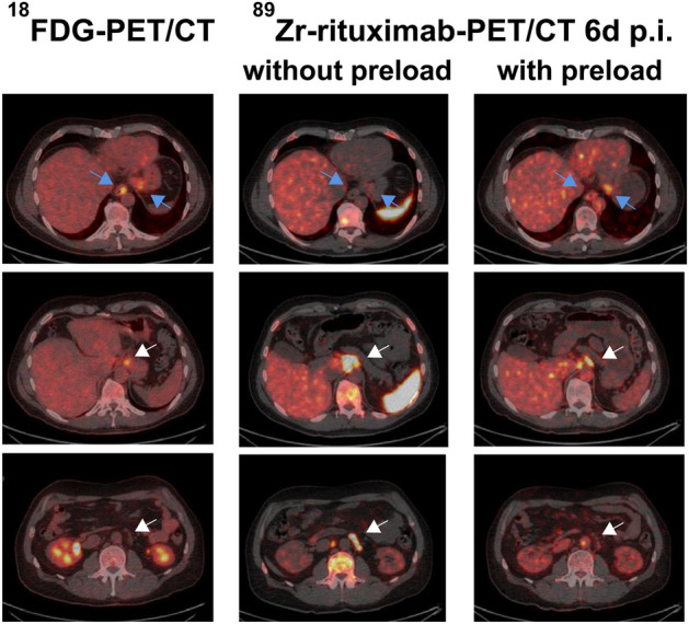
Positron emission tomography (PET) represents molecular imaging for non-invasive phenotyping of physiological and biochemical processes in various oncological diseases. PET imaging with 18F-fluorodeoxyglucose (18F-FDG) for glucose metabolism evaluation is the standard imaging modality for the clinical management of lymphoma. One of the 18F-FDG PET applications is the detection and pre-treatment staging of lymphoma, which is highly sensitive. 18F-FDG PET is also applied during treatment to evaluate the individual chemo-sensitivity and accordingly guide the response-adapted therapy. At the end of the therapy regiment, a negative PET scan is indicative of a good prognosis in patients with advanced Hodgkin's lymphoma and diffuse large B-cell lymphoma. Thus, adjuvant radiotherapy may be alleviated. Future PET studies using non-18F-FDG radiotracers, such as 68Ga-labeled pentixafor (a cyclic pentapeptide that enables sensitive and high-contrast imaging of C-X-C motif chemokine receptor 4), 68Ga-labeled fibroblast activation protein inhibitor (FAPI) that reflects the tumor microenvironment, and 89Zr-labeled atezolizumab that targets the programmed cell death-ligand 1 (PD-L1), may complement 18F-FDG and offer essential tools to decode lymphoma phenotypes further and identify the mechanisms of lymphoma therapy.

Keywords: Glucose metabolism; Lymphoma; Positron emission tomography (PET); Response assessment; Staging.


Conflict of interest statement

Conflicts of InterestThe authors declare that they have no conflicts of interest.

Figures

Fig. 1
Fig. 1
Pre-treatment 18F-FDG PET/CT for staging of lymphoma based on the Ann Arbor staging system. Stage I is defined as lymphomatous involvement confined to a single lymph node region or extranodal site. Stage II refers to lymphomatous involvement of more lymph node regions on one side of the diaphragm with or without limited contiguous extranodal involvement. Stage III is defined as lymph node involvement on both sides of the diaphragm. Stage IV refers to extensive extranodal involvement. White arrows denote bone marrow involvement. SUV indicates standardized uptake value
Fig. 2
Fig. 2
Coronal slices of pre-treatment and interim 18F-FDG PET images for response assessment in lymphoma, from left to right corresponding to Deauville score (DS) 1–5 with different FDG uptake (red arrows)
Fig. 3
Fig. 3
Serial 18F-FDG PET images of a 62-year-old woman with diffuse large B-cell lymphoma. a The initial staging PET image reveals lymphomatous involvement of para-aortic and iliac lymph nodes (defined as Stage II disease). b The PET image after six cycles of chemotherapy (R-CHOP) shows PET-positive residual uptake at the iliac lymph nodes (Deauville score 5). c The PET image after involved field radiotherapy indicates complete metabolic response
Fig. 4
Fig. 4
68Ga-FAPI PET/CT images of various extranodal sites in different subtypes of lymphoma patients. a Primary thyroid Burkitt lymphoma (arrow). b Hodgkin lymphoma with lung involvement (arrow). c Primary gastric diffuse large B-cell lymphoma (DLBCL, arrow). d Ileum DLBCL (arrow). e Left breast DLBCL (arrow). f Left testicle DLBCL (arrow). g Left temporal lobe DLBCL lesion (arrow) after left frontal lymphoma resection (thick arrow). Adapted from Jin et al. (2021). Reprint permission was obtained
Fig. 5
Fig. 5
18F-FDG PET/CT and 89Zr-rituximab immuno-PET/CT images of a 51-year-old man with follicular lymphoma. 89Zr-rituximab immuno-PET/CT images obtained 6 days after injection with and without a preload of unlabelled rituximab show lower tracer uptake in involved lymph nodes with the preload (white arrows), but higher uptake in less accessible visceral lesions (esophagus and stomach; blue arrows) resulting in better tumor targeting. Adapted from Muylle et al. (2015). Reprint permission was obtained
Fig. 6
Fig. 6
Pre-treatment and interim 18F-FDG PET/MRI images of a 14-year-old male with Hodgkin’s lymphoma. Coronal a short tau inversion recovery (STIR), b 18F-FDG PET, and c 18F-FDG PET/MRI fused images demonstrate lymphomatous involvement of right supraclavicular and right paratracheal lymph nodes, and a right lung nodule (arrows). Early response assessment following 2 cycles of chemotherapy shows d residual right supraclavicular lymph node on coronal STIR, with no uptake on coronal e 18F-FDG PET and f fused 18F-FDG PET/MRI (arrows). The other sites of disease have not been detected. Adapted from Verhagen et al. (2021). Reprint permission was obtained

References

    1. Adams HJ, Kwee TC, de Keizer B, Fijnheer R, de Klerk JM, Nievelstein RA. FDG PET/CT for the detection of bone marrow involvement in diffuse large B-cell lymphoma: systematic review and meta-analysis. Eur J Nucl Med Mol Imaging. 2014;41(3):565–574. doi: 10.1007/s00259-013-2623-4. - DOI - PubMed
    1. Adams HJ, Nievelstein RA, Kwee TC. Prognostic value of interim FDG-PET in Hodgkin lymphoma: systematic review and meta-analysis. Br J Haematol. 2015;170(3):356–366. doi: 10.1111/bjh.13441. - DOI - PubMed
    1. Afaq A, Fraioli F, Sidhu H, Wan S, Punwani S, Chen SH, et al. Comparison of PET/MRI with PET/CT in the evaluation of disease status in lymphoma. Clin Nucl Med. 2017;42(1):e1–e7. doi: 10.1097/RLU.0000000000001344. - DOI - PMC - PubMed
    1. Agostinelli C, Gallamini A, Stracqualursi L, Agati P, Tripodo C, Fuligni F, et al. The combined role of biomarkers and interim PET scan in prediction of treatment outcome in classical Hodgkin's lymphoma: a retrospective, European, multicentre cohort study. Lancet Haematol. 2016;3(10):e467–e479. doi: 10.1016/s2352-3026(16)30108-9. - DOI - PubMed
    1. André M, Girinsky T, Federico M, Reman O, Fortpied C, Gotti M, et al. Early positron emission tomography response-adapted treatment in stage I and II Hodgkin lymphoma: final results of the randomized EORTC/LYSA/FIL H10 trial. J Clin Oncol. 2017;35(16):1786–1794. doi: 10.1200/JCO.2016.68.6394. - DOI - PubMed

LinkOut - more resources