. 2023 Jun;23(6):324-330.
doi: 10.1089/vbz.2022.0087. Epub 2023 Mar 20.

Experimental Colonization of Sand Flies (Lutzomyia longipalpis; Diptera: Psychodidae) by Bartonella ancashensis

Affiliations

Experimental Colonization of Sand Flies (Lutzomyia longipalpis; Diptera: Psychodidae) by Bartonella ancashensis

Michael F Minnick et al. Vector Borne Zoonotic Dis. 2023 Jun.

Abstract

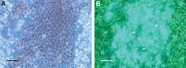
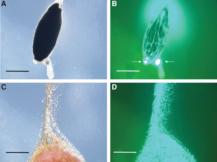
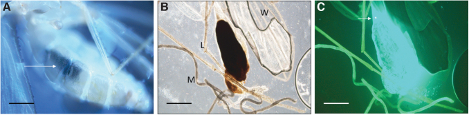
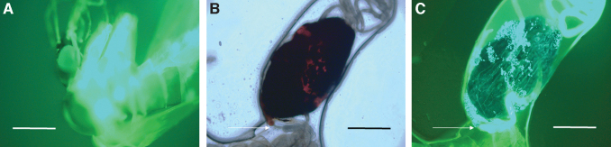
Background: Bartonella ancashensis is a recently described Bartonella species endemic to Peru, where it causes verruga peruana in humans. While the arthropod vector of B. ancashensis transmission is unknown, human coinfections with Bartonella bacilliformis suggest that phlebotomine sand flies are a vector. Materials and Methods: To address the hypothesis that sand flies are involved in the bacterium's transmission, Lutzomyia longipalpis sand flies were used as an infection model, together with green fluorescent protein-expressing B. ancashensis. Results: Results showed that bacterial infections were clearly established, limited to the anterior midgut of the female fly, and maintained for roughly 7 days. At 3-7 days postinfection, a prominent microcolony of aggregated bacteria was observed in the anterior midgut, immediately distal to the stomodeal valve of the esophagus. In contrast, eggs, diuretic fluid, feces, and other tissues were not infected. Conclusion: These results suggest that certain sand fly species within the endemic zone for B. ancashensis may play a role in the bacterium's maintenance and possibly in its transmission to humans.

Keywords: Bartonella ancashensis; Lutzomyia longipalpis; arthropod vector; infection model; sand fly.


Conflict of interest statement

The authors declare that they have no known competing financial interests or personal relationships that could constitute a conflict of interest or to have influenced the work reported in this paper.

Figures

FIG. 1.
FIG. 1.
Micrographs of sand fly anterior midgut contents at 2 days postfeeding. (A) Bloodmeal showing human erythrocytes by phase-contrast microscopy. (B) Corresponding fluorescence micrograph revealing GFP+ Bartonella ancashensis cells (examples are arrowed) in the dark-colored, central area of erythrocytes (1000 × magnification; scale bars = 35 μm). GFP, green fluorescent protein.
FIG. 2.
FIG. 2.
Micrographs of an isolated sand fly anterior midgut at 3 days postfeeding. (A) The bloodmeal is surrounded by a well-formed PM and is a dark, rusty color by phase-contrast microscopy. (B) Corresponding fluorescence micrograph revealing GFP+ B. ancashensis cells, forming a microcolony at the anterior end of the anterior midgut below the SV (arrowed). Some bacteria appear to be external to the PM but still within the anterior midgut. (C) Posterior end of anterior midgut leaking erythrocytes to the external milieu after applying a coverslip. (D) Corresponding fluorescence micrograph (C), revealing GFP+ B. ancashensis cells mixed in with the bloodmeal (400 × magnification; scale bars = 160 μm in (A, B), and 53 μm in (C, D). PM, peritrophic membrane; SV, stomodeal valve.
FIG. 3.
FIG. 3.
Micrographs of infected sand flies at 4 days postfeeding. (A) Intact sand fly with visible bloodmeal in the anterior midgut (arrowed). At this time the bloodmeal was dark brown when observed by light microscopy (10 × magnification; scale bar = 200 μm). (B) Isolated anterior midgut containing the bloodmeal. Other structures shown include a wing (W), malpighian tubules (M), and legs (L). (C) Corresponding fluorescence micrograph revealing GFP+ B. ancashensis throughout the anterior midgut with an apparent microcolony at the anterior end (arrowed) (400 × magnification; scale bars = 160 μm).
FIG. 4.
FIG. 4.
Micrographs of an isolated sand fly anterior midgut at 5 days postfeeding. The anterior portion of the anterior midgut is above the wing. (A) The blood meal was a dark, rusty color by phase-contrast microscopy (100 × magnification; scale bars = 160 μm). The SV is indicated. (B) Corresponding fluorescence micrograph revealing GFP+ B. ancashensis cells in the lumen of the anterior midgut with a microcolony at the anterior end of the anterior midgut just below the SV (arrowed). (C) Closeup image of (A) phase-contrast microscopy (400 × magnification; scale bars = 50 μm). (D) Corresponding fluorescence micrograph to (C), revealing GFP+ B. ancashensis cells just below the SV.
FIG. 5.
FIG. 5.
Micrographs of an infected sand fly at 6 days postfeeding. (A) UV fluorescence micrograph showing absence of apparent infection in head, thorax, and thoracic midgut (100 × magnification; scale bar = 3000 μm). (B) Anterior midgut showing rusty-brown bloodmeal by phase-contrast microscopy. The SV at the anterior is arrowed. (C) Corresponding fluorescence micrograph showing heavy colonization by GFP+ B. ancashensis below the SV and outside the PM but still within the confines of the anterior midgut (400 × magnification; scale bars = 100 μm in B, C). UV, ultraviolet.
FIG. 6.
FIG. 6.
Micrographs of an infected sand fly anterior midgut at 7 days postfeeding. (A) Intact anterior midgut showing a brownish-black bloodmeal by phase-contrast microscopy (100 × magnification; scale bar = 140 μm). In certain flies, the bloodmeal was completely absent at this time point. Fluorescence microscopy revealed relatively low densities of GFP+ B. ancashensis cells at 7 days, especially in the (B) posterior portion of the anterior midgut. (C) A microcolony was still apparent at the anterior end of the anterior midgut (400 × magnification; scale bar = 35 μm in B, C).

References

    1. Alexander B. A review of bartonellosis in Ecuador and Colombia. Am J Trop Med Hyg 1995;52(4):354–359; doi: 10.4269/ajtmh.1995.52.354 - DOI - PubMed
    1. Amano Y, Rumbea J, Knobloch J, et al. Bartonellosis in Ecuador: Serosurvey and current status of cutaneous verrucous disease. Am J Trop Med Hyg 1997;57(2):174–179; doi: 10.4269/ajtmh.1997.57.174 - DOI - PubMed
    1. Battisti JM, Lawyer PG, Minnick MF. Colonization of Lutzomyia verrucarum and Lutzomyia longipalpis sand flies (Diptera: Psychodidae) by Bartonella bacilliformis, the etiologic agent of Carrión's disease. PLoS Negl Trop Dis 2015;9(10):e0004128; doi: 10.1371/journal.pntd.0004128 - DOI - PMC - PubMed
    1. Battisti JM, Minnick MF. Development of a system for genetic manipulation of Bartonella bacilliformis. Appl Environ Microbiol 1999;65(8):3441–3448; doi: 10.1128/AEM.65.8.3441-3448.1999 - DOI - PMC - PubMed
    1. Battisti JM, Minnick MF. Laboratory maintenance of Bartonella quintana. Curr Protoc Microbiol 2008; Chapter 3:Unit 3C.1.1–3C.1.13; doi: 10.1002/9780471729259.mc03c01s10 - DOI - PubMed

Publication types

Supplementary concepts

LinkOut - more resources