. 2023 Sep;17(3):915-924.
doi: 10.1007/s12079-023-00738-x. Epub 2023 Mar 20.

Adipocytes reprogram prostate cancer stem cell machinery

Affiliations

Adipocytes reprogram prostate cancer stem cell machinery

Fabrizio Fontana et al. J Cell Commun Signal. 2023 Sep.

Abstract

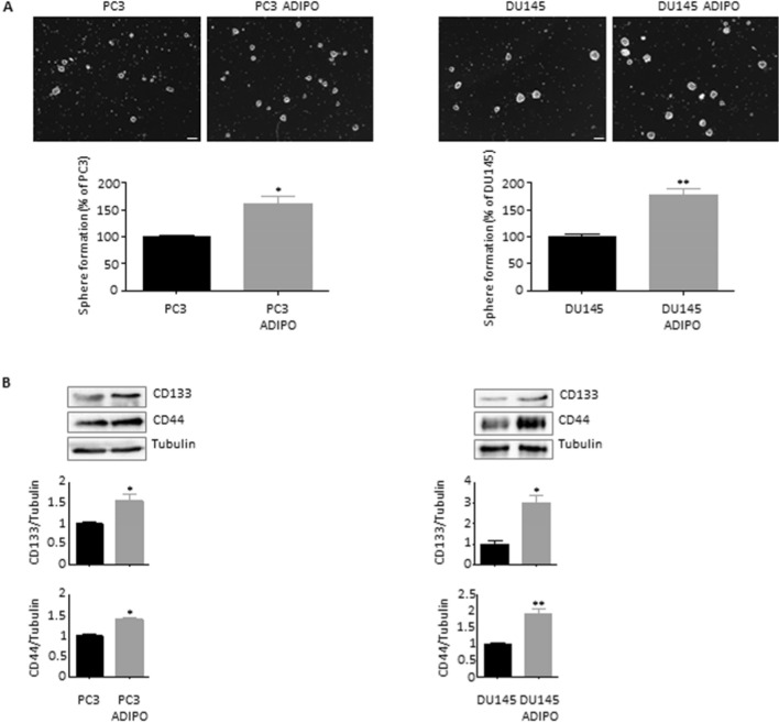
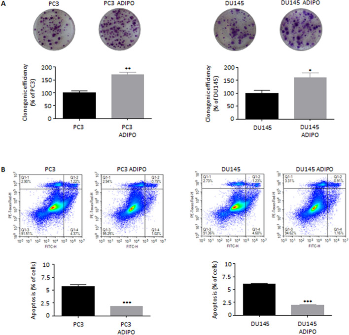
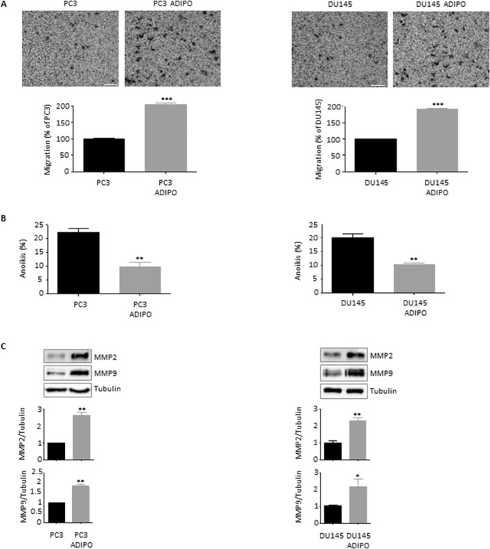
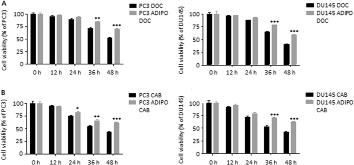
It is now well-established that an obese condition correlates with a higher risk of prostate cancer (PCa). A crosstalk between adipose tissue and PCa has been observed but is still poorly characterized. Herein, we demonstrated that 3T3-L1 adipocyte conditioned media (CM) could endow PC3 and DU145 PCa cells with stemness properties, by stimulating their sphere formation ability and promoting CD133 and CD44 expression. Moreover, after exposure to adipocyte CM both PCa cell lines underwent partial epithelial-to-mesenchymal transition (EMT), with E-/N-cadherin switch and Snail upregulation. Specifically, these changes in PC3 and DU145 cell phenotype were accompanied by increased tumor clonogenic activity and survival, as well as by enhanced invasion, anoikis resistance and matrix metalloproteinase (MMP) production. Finally, adipocyte CM-treated PCa cells exhibited reduced responsiveness to both docetaxel and cabazitaxel, demonstrating greater chemoresistance. Overall, these data indicate that adipose tissue can effectively contribute to PCa aggressiveness by reprogramming the cancer stem cell (CSC) machinery. Adipocytes endow prostate cancer cells with stem-like properties and mesenchymal traits, increasing their tumorigenicity, invasion and chemoresistance.

Keywords: Adipocytes; Cancer stem cells; Chemoresistance; EMT; Metastasis; Obesity; Prostate cancer.


Conflict of interest statement

The authors declare no conflict of interest.

Figures

Fig. 1
Fig. 1
Adipocytes promote prostate cancer cell stemness. (A) PC3 and DU145 cells were pre-treated with adipocyte CM for 24 h and then cultured in CSC media for 7 days. Sphere formation assay was performed to determine the spheroidogenic ability of cancer cells. Each experiment was repeated three times. Scale bars are 75 µm. Data represent mean values ± SEM and were analyzed by t-test. *P < 0.05 versus PC3 or DU145 (control). **P < 0.01 versus PC3 or DU145 (control). (B) Cells were pre-treated with adipocyte CM for 24 h and then cultured in CSC media for 7 days. Western blot analysis was performed to investigate the expression levels of CD133 and CD44 in PC3 and DU145 cells. Tubulin expression was evaluated as a loading control. Data represent mean values ± SEM and were analyzed by t-test. *P < 0.05 versus PC3 or DU145 (control). **P < 0.01 versus PC3 or DU145 (control)
Fig. 2
Fig. 2
Adipocytes stimulate prostate cancer cell tumorigenicity and survival. (A) PC3 and DU145 cells were pre-treated with adipocyte CM for 72 h and then cultured in proper media for 7–10 days. Colony formation assay was performed to determine the clonogenic activity of cancer cells. Each experiment was repeated three times. Data represent mean values ± SEM and were analyzed by t-test. *P < 0.05 versus PC3 or DU145 (control). **P < 0.01 versus PC3 or DU145 (control). (B) Cells were incubated in adipocyte CM for 96 h. Apoptotic rates were then evaluated by cytofluorimetric analysis after staining with eBioscience™ Annexin V-FITC Apoptosis Detection Kit (according to the manufacturer’s protocol). Each experiment was repeated three times. Data represent mean values ± SEM and were analyzed by t-test. ***P < 0.001 versus PC3 or DU145 (control)
Fig. 3
Fig. 3
Adipocytes endow prostate cancer cells with mesenchymal properties. (A) PC3 and DU145 cells were incubated in adipocyte CM for 96 h. Cell morphology was then evaluated by light microscopy. Each experiment was repeated three times. Scale bars are 40 µm. (B) After adipocyte CM treatment (24 h), Western blot analysis was performed to investigate the expression levels of E-cadherin, N-cadherin and Snail in PC3 and DU145 cells. Tubulin expression was evaluated as a loading control. Data represent mean values ± SEM and were analyzed by t-test. *P < 0.05 versus PC3 or DU145 (control). **P < 0.01 versus PC3 or DU145 (control)
Fig. 4
Fig. 4
Adipocytes drive prostate cancer cell invasion. (A) PC3 and DU145 cells were incubated in adipocyte CM for 24 h. Cell invasion was then evaluated by Boyden chamber assay. Each experiment was repeated three times. Scale bars are 100 µm. Data represent mean values ± SEM and were analyzed by t-test. ***P < 0.001 versus PC3 or DU145 (control). (B) Cells were incubated in adipocyte CM for 48 h. Anoikis was then evaluated by Trypan blue exclusion assay. Each experiment was repeated three times. Data represent mean values ± SEM and were analyzed by t-test. **P < 0.01 versus PC3 or DU145 (control). (C) After adipocyte CM treatment (24 h), Western blot analysis was performed to investigate the expression levels of MMP2 and MMP9 in PC3 and DU145 cells. Tubulin expression was evaluated as a loading control. Data represent mean values ± SEM and were analyzed by t-test. *P < 0.05 versus PC3 or DU145 (control). **P < 0.01 versus PC3 or DU145 (control)
Fig. 5
Fig. 5
Adipocytes confer chemoresistance to prostate cancer cells. (A) PC3 and DU145 cells were incubated in adipocyte CM for 24 h and then treated with docetaxel (10 nM) for 48 h. Cell viability was then evaluated by MTT assay. Each experiment was repeated three times. Data represent mean values ± SEM and were analyzed by two-way ANOVA followed by Bonferroni's test. **P < 0.01 versus PC3 DOC or DU145 DOC (control). ***P < 0.001 versus PC3 DOC or DU145 DOC (control). (B) PC3 and DU145 cells were incubated in adipocyte CM for 24 h and then treated with cabazitaxel (5 nM) for 48 h. Cell viability was then evaluated by MTT assay. Each experiment was repeated three times. Data represent mean values ± SEM and were analyzed by two-way ANOVA followed by Bonferroni’s test. *P < 0.05 versus PC3 CAB or DU145 CAB (control). **P < 0.01 versus PC3 CAB or DU145 CAB (control). ***P < 0.001 versus PC3 CAB or DU145 CAB (control)

References

    1. Bakir B, Chiarella AM, Pitarresi JR, Rustgi AK. EMT, MET, plasticity, and tumor metastasis. Trends Cell Biol. 2020;30:764–776. doi: 10.1016/j.tcb.2020.07.003. - DOI - PMC - PubMed
    1. Cao Y, Giovannucci E. Obesity and prostate cancer. In: Pischon T, Nimptsch K, editors. Obesity and cancer. Cham: Springer International Publishing; 2016. pp. 137–153.
    1. Chooi YC, Ding C, Magkos F. The epidemiology of obesity. Metabolism. 2019;92:6–10. doi: 10.1016/j.metabol.2018.09.005. - DOI - PubMed
    1. Di Franco S, Bianca P, Sardina DS, et al. Adipose stem cell niche reprograms the colorectal cancer stem cell metastatic machinery. Nat Commun. 2021;12:5006. doi: 10.1038/s41467-021-25333-9. - DOI - PMC - PubMed
    1. Dirat B, Bochet L, Dabek M, et al. Cancer-associated adipocytes exhibit an activated phenotype and contribute to breast cancer invasion. Cancer Res. 2011;71:2455–2465. doi: 10.1158/0008-5472.CAN-10-3323. - DOI - PubMed