. 2023 Mar 28;120(13):e2218847120.
doi: 10.1073/pnas.2218847120. Epub 2023 Mar 20.

Nonspherical ultrasound microbubbles

Affiliations

Nonspherical ultrasound microbubbles

Anshuman Dasgupta et al. Proc Natl Acad Sci U S A. .

Abstract

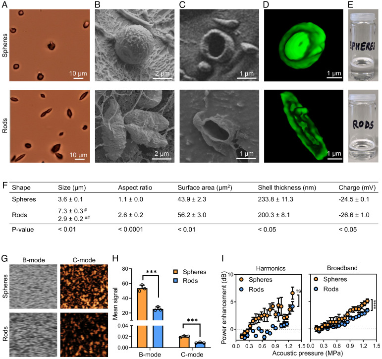
Surface tension provides microbubbles (MB) with a perfect spherical shape. Here, we demonstrate that MB can be engineered to be nonspherical, endowing them with unique features for biomedical applications. Anisotropic MB were generated via one-dimensionally stretching spherical poly(butyl cyanoacrylate) MB above their glass transition temperature. Compared to their spherical counterparts, nonspherical polymeric MB displayed superior performance in multiple ways, including i) increased margination behavior in blood vessel-like flow chambers, ii) reduced macrophage uptake in vitro, iii) prolonged circulation time in vivo, and iv) enhanced blood-brain barrier (BBB) permeation in vivo upon combination with transcranial focused ultrasound (FUS). Our studies identify shape as a design parameter in the MB landscape, and they provide a rational and robust framework for further exploring the application of anisotropic MB for ultrasound-enhanced drug delivery and imaging applications.

Keywords: microbubbles; nonspherical; shape; sonoporation; ultrasound.


Conflict of interest statement

The authors declare no competing interest.

Figures

Fig. 1.
Fig. 1.
Study setup. (A) Spherical MB were synthesized by anionic polymerization of butyl cyanoacrylate (BCA) in 1% (w/v) Triton™ X-100 at pH 2.5. Rod-shaped MB were generated by one-dimensionally stretching spherical MB at a temperature exceeding the glass transition temperature of PBCA, followed by cooling down to room temperature. (B) MB flow dynamics were studied by assessing their margination propensity, tumbling behavior, phagocytosis, and circulation kinetics. (C) Spherical and rod-shaped MB were intravenously administered, and their ability to permeate the BBB upon exposure to transcranial focused US was compared.
Fig. 2.
Fig. 2.
Generation and characterization of anisotropic MB. (A) Bright-field, (B and C) cryogenic scanning electron, and (D) 3D confocal laser scanning microscopy of spherical and rod-shaped MB. (E) Both spherical and anisotropic rod-shaped MB float in aqueous dispersions due to buoyancy. (F) Physicochemical characteristics of spherical and rod-shaped MB. # and ## represent the major and minor axis, respectively. (G and H) US imaging at 18 MHz center frequency and 0.03 mechanical index revealed that anisotropic MB produce less intense brightness-mode (B-mode) and contrast-mode (C-mode) US signals as compared to spherical MB. (I) Cavitation response analysis demonstrating that anisotropic MB emit lower levels of harmonic and broadband signals. Data represent mean ± SD of n = 3 independent MB batches. Statistical comparison between spheres and rods in panel F was performed using the unpaired Student t test. In panels H and I, unpaired t tests for multiple groups were used. ***P < 0.001 and ****P < 0.0001.
Fig. 3.
Fig. 3.
Anisotropic MB demonstrate increased margination and tumbling. (A) Coumarin 6–labeled spherical and rod-shaped MB were injected into a microfluidic channel in the presence of red blood cells (16% hematocrit). Margination was assessed by analyzing MB distribution at physiological flow conditions in the margin, intermediate, and central zones of the channel. (B) Fluorescence microscopy images exemplifying that anisotropic MB flow more toward the marginal zones than spherical MB. (C and D) Quantification of MB distribution in the microfluidic channels showing that the percentage of rod-shaped MB flowing in the marginal zones of the microfluidic channel was more than twofold higher than that for spherical MB. (E and F) Schematic and time-lapse fluorescence microscopy analysis of the tumbling motion of spherical and anisotropic MB, illustrating tumbling of the latter near the channel wall (gray lines). (Scale bar, 1 µm.) Statistical comparisons were performed using unpaired t tests for multiple groups. ****P < 0.0001.
Fig. 4.
Fig. 4.
Anisotropic shape reduces MB phagocytosis. J774A.1 murine macrophages were incubated with coumarin-labeled spherical and rod-shaped MB, and phagocytic uptake was analyzed. (AD) At 1 and 10 min after the initiation of incubation, rod-shaped MB showed less macrophage uptake as compared to spherical MB. Cell nuclei and cellular membranes were stained with DAPI (blue) and wheat germ agglutinin (red), respectively. (Scale bar, 20 µm.) Data represent mean ± SD. Statistical comparisons were performed using the unpaired Student t test. *P < 0.05.
Fig. 5.
Fig. 5.
Anisotropic MB display prolonged in vivo circulation times. (A) Time-lapse B-mode and C-mode US images at 18 MHz center frequency and 0.07 mechanical index, exemplifying that the US contrast of rod-shaped MB in the aorta decreases slower than that of spherical MB. Vessel borders outlined in yellow. (B) Continuous CEUS monitoring of MB signal up until 10 min after i.v. administration demonstrates prolonged circulation kinetics for anisotropic vs. spherical MB. (CE) Pharmacokinetic analysis of elimination rate, area under the curve (AUC), and circulation half-life for spherical vs. rod-shaped MB. (Scale bar, 1 mm.) Data represent mean ± SD (n = 5). Statistical comparison is based on an unpaired Student t test. *P < 0.05.
Fig. 6.
Fig. 6.
Anisotropic MB potentiate FUS-induced BBB permeation. (A) BBB permeation was induced by administering 1 × 109 MB and transcranially applying FUS to the brain. Trypan blue was used as a fluorescent model drug to capture the extent of BBB permeation. (B and C) Quantification of harmonic and broadband US signals in the brain indicates that in our experimental setup, spherical and anisotropic MB produced similar levels of cavitation. (D) Schematic showing how the enhanced margination and prolonged circulation time of rod-shaped MB may contribute to enhanced BBB permeation. (E and F) Ex vivo fluorescence imaging of a brain section and quantification (in n = 12 mice per group) showing that anisotropic rod-shaped MB more efficiently promote BBB sonopermeation than spherical MB. (G and H) Correlation of BBB permeation with acoustic emission signals, exemplifying a higher positive correlation and slope for anisotropic vs. spherical MB. Data represent mean ± SD. Statistical comparisons for panels B, C, and F were performed using unpaired Student t tests, while in panels G and H, comparisons were analyzed using the Spearman correlation. *P < 0.05.

References

    1. Stride E., et al. , Microbubble agents: New directions. Ultrasound Med. Biol. 46, 1326–1343 (2020). - PubMed
    1. Opacic T., et al. , Motion model ultrasound localization microscopy for preclinical and clinical multiparametric tumor characterization. Nat. Commun. 9, 1–13 (2018). - PMC - PubMed
    1. Sirsi S. R., Borden M. A., Microbubble compositions, properties and biomedical applications. Bubble Sci. Eng. Technol. 1, 3–17 (2009). - PMC - PubMed
    1. Sun T., et al. , Closed-loop control of targeted ultrasound drug delivery across the blood–brain/tumor barriers in a rat glioma model. Proc. Natl. Acad. Sci. U.S.A. 114, E10281–E10290 (2017). - PMC - PubMed
    1. Lipsman N., et al. , Blood–brain barrier opening in Alzheimer’s disease using MR-guided focused ultrasound. Nat. Commun. 9, 1–8 (2018). - PMC - PubMed

Publication types

LinkOut - more resources