. 2023 May 15:228:115202.
doi: 10.1016/j.bios.2023.115202. Epub 2023 Mar 8.

Ultra-sensitive label-free SERS biosensor with high-throughput screened DNA aptamer for universal detection of SARS-CoV-2 variants from clinical samples

Affiliations

Ultra-sensitive label-free SERS biosensor with high-throughput screened DNA aptamer for universal detection of SARS-CoV-2 variants from clinical samples

Ki Sung Park et al. Biosens Bioelectron. .

Abstract

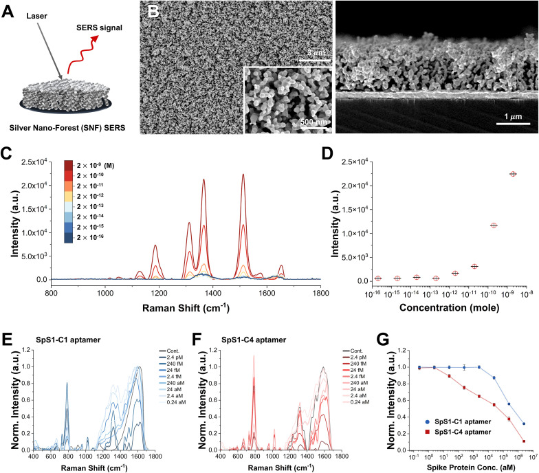
COVID-19, severe acute respiratory syndrome coronavirus 2 (SARS-CoV-2), has caused an ongoing global pandemic with economic and social disruption. Moreover, the virus has persistently and rapidly evolved into novel lineages with mutations. The most effective strategy to control the pandemic is suppressing virus spread through early detection of infections. Therefore, developing a rapid, accurate, easy-to-use diagnostic platform against SARS-CoV-2 variants of concern remains necessary. Here, we developed an ultra-sensitive label-free surface-enhanced Raman scattering-based aptasensor as a countermeasure for the universal detection of SARS-CoV-2 variants of concern. In this aptasensor platform, we discovered two DNA aptamers that enable binding to SARS-CoV-2 spike protein via the Particle Display, a high-throughput screening approach. These showed high affinity that exhibited dissociation constants of 1.47 ± 0.30 nM and 1.81 ± 0.39 nM. We designed a combination with the aptamers and silver nanoforest for developing an ultra-sensitive SERS platform and achieved an attomolar (10-18 M) level detection limit with a recombinant trimeric spike protein. Furthermore, using the intrinsic properties of the aptamer signal, we demonstrated a label-free aptasensor approach, enabling use without the Raman tag. Finally, our label-free SERS-combined aptasensor succeeded in detecting SARS-CoV-2 with excellent accuracy, even in clinical samples with variants of concern, including the wild-type, delta, and omicron variants.

Keywords: Label-free SERS biosensor; Particle display; SARS-CoV-2 spike binding aptamer; SERS-based aptasensor; Silver nanoforest.


Conflict of interest statement

Declaration of competing interest The authors declare that they have no known competing financial interests or personal relationships that could have appeared to influence the work reported in this paper.

Figures

Image 1
Graphical abstract
Fig. 1
Fig. 1
Design strategy for our label-free SERS-based aptasensor platform for SARS-CoV-2. (A) Targeted aptamer screening against spike (S) protein for detecting SARS-CoV-2 from clinical samples. Side and top view of the trimeric S protein on the surface of SARS-CoV-2 (PDB: 6VXX). (B) The particle display aptamer discovery process, in which solution-phase aptamer library molecules are converted to monoclonal aptamer particles, incubated with fluorescently-labeled S protein, and then subjected to fluorescence-activated cell sorting (FACS) to enrich library molecules with strong affinity for this target. (C) The aptamer is then conjugated onto a silver nanoforest (SNF) substrate for the detection of SARS-CoV-2. The intrinsic aptamer peaks shift in response to the conformational changes triggered by S protein binding to the aptamer.
Fig. 2
Fig. 2
Round by round assessment of the particle display screening process. (A) FACS dot plots from all four rounds of screening at the stated concentrations ([T]) of recombinant trimeric S protein. (B) Mean fluorescence intensity (MFI) within the sort gate at different target concentrations in each of the four rounds. (C) Additional sorting experiments in the final round of screening with another sort gate at different target concentrations. Overall, 5% of the top binders at 2 nM, 0.15% of the top binders at 2 nM, and 0.1% of the top binders at 0.5 nM of S protein were sorted using FACS.
Fig. 3
Fig. 3
Characterization of the isolated S protein-binding aptamers. (A) Candidate aptamers from each of the four consensus sequence families were chosen for further analysis. ‘FP bead’ indicates the forward primer-coated bead used as a negative control. (B) Specificity measurements were also conducted against the trimeric S protein, subunit 1, subunit 2, the receptor-binding domain (RBD), and bovine serum albumin (BSA). (C, D) Dissociation constant (Kd) measurements of the best aptamers using a bead-based fluorescence assay. Kd was calculated from the fluorescence intensity of binding in a 1:1 binding model using GraphPad software.
Fig. 4
Fig. 4
Design of the label-free SERS-based aptasensor. (A) Schematic illustration of SERS analysis using the SNF substrate. (B) Top and side view scanning electron microscopy (SEM) images of the SNF substrate. (C) Raman spectra with varying concentrations of rhodamine 6G on the SNF substrate. (D) Concentration-dependent Raman intensity of rhodamine 6G at 1,511 cm−1. (E–G) Raman shifts of intrinsic signals for (E) SpS1-C1 and (F) SpS1-C4 at varying concentrations of the recombinant trimeric S protein. (G) The data show the detection limit of SpS1-C1 and -C4 on the SNF substrate based on Raman signal intensity at 1,587 cm−1.
Fig. 5
Fig. 5
Clinical validation of our SERS-based aptasensor. (A–D) SARS-CoV-2 detection in 80 nasopharyngeal specimens representing (A) negative controls or patients with (B) wild-type, (C) Delta, and (D) Omicron variants of SARS-CoV-2. (E) Sensitivity and specificity of detection of SARS-CoV-2 variants based on a threshold (dashed lines) calculated from the receiver operating characteristic (ROC) curve. The box indicates the standard deviation, and the white circle indicates the mean value. (F–H) ROC curves of diagnosis for (F) wild-type, (G) Delta, and (H) Omicron variants of SARS-CoV-2.

References

    1. Abdelhamid M.A., Son R.G., Park K.S., Pack S.P. Colloids Surf., B. 2022;219 - PubMed
    1. Adamson B., Sikka R., Wyllie A., Premsrirut P. Medrxiv. 2022;5 doi: 10.1101/2022.01.04.22268770. Preprint posted online, 2022-01. - DOI
    1. Ahmad K.M., Oh S.S., Kim S., McClellen F.M., Xiao Y., Soh H.T. PLoS One. 2011;6(11) - PMC - PubMed
    1. Ambartsumyan O., Gribanyov D., Kukushkin V., Kopylov A., Zavyalova E. Int. J. Mol. Sci. 2020;21(9):3373. - PMC - PubMed
    1. Banerji A., Wickner P.G., Saff R., Stone C.A., Jr., Robinson L.B., Long A.A., Wolfson A.R., Williams P., Khan D.A., Phillips E. J. Allergy Clin. Immunol. Pract. 2021;9(4):1423–1437. - PMC - PubMed

Substances

Supplementary concepts