. 2023 Mar 20;14(1):1532.
doi: 10.1038/s41467-023-36996-x.

Digital nanoreactors to control absolute stoichiometry and spatiotemporal behavior of DNA receptors within lipid bilayers

Affiliations

Digital nanoreactors to control absolute stoichiometry and spatiotemporal behavior of DNA receptors within lipid bilayers

Vishal Maingi et al. Nat Commun. .

Abstract

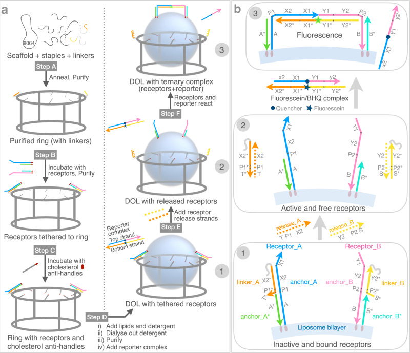
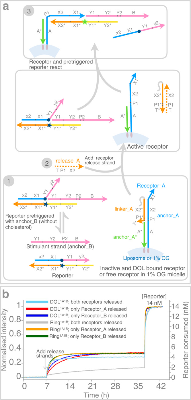
Interactions between membrane proteins are essential for cell survival but are often poorly understood. Even the biologically functional ratio of components within a multi-subunit membrane complex-the native stoichiometry-is difficult to establish. Here we demonstrate digital nanoreactors that can control interactions between lipid-bound molecular receptors along three key dimensions: stoichiometric, spatial, and temporal. Each nanoreactor is based on a DNA origami ring, which both templates the synthesis of a liposome and provides tethering sites for DNA-based receptors (modelling membrane proteins). Receptors are released into the liposomal membrane using strand displacement and a DNA logic gate measures receptor heterodimer formation. High-efficiency tethering of receptors enables the kinetics of receptors in 1:1 and 2:2 absolute stoichiometries to be observed by bulk fluorescence, which in principle is generalizable to any ratio. Similar single-molecule-in-bulk experiments using DNA-linked membrane proteins could determine native stoichiometry and the kinetics of membrane protein interactions for applications ranging from signalling research to drug discovery.


Conflict of interest statement

Certain aspects related to the internal immobilization and the release of membrane receptors employing DNA origami are covered in a pending US provisional patent application (63/415,546) owned by the California Institute of Technology with inventor V.M. The other authors declare no competing interests.

Figures

Fig. 1
Fig. 1. DOL synthesis and DNA circuit logic.
a DOL assembly (left column) and DNA receptor interaction (right column). Step A: DNA scaffold (8064 nucleotides; gray loop), regular staple strands (gray segments), and linker-extended staples (having orange and yellow sections) were annealed; excess staples were removed. Step B: Two types of DNA receptors modified with cholesterol (red ovals) were tethered to the ring and rings were repurified. Step C: Rings were incubated with cholesterol-modified anti-handles (gray lines with red ovals). Step D: Lipids and detergent were added; subsequent dialysis removed detergent and seeded liposome formation (blue spheres) on rings to create DOL. b Stepwise operation of a DNA circuit for the receptor release and interaction measurement. Step labels 1–3 correspond to labels in the right column of a. A zoomed segment of the liposome bilayer is shown. Initially (Step 1) both receptors are inactive and bound to the ring (not shown) via linker_A and linker_B (themselves attached to the ring via a short section of gray polyT). The inter-receptor distance (~45 nm) is not shown to scale. Receptors were detethered (Step 2) by adding release strands complementary to the linkers; domains T* and S* provided toeholds for this reaction. Released receptors diffuse freely within the bilayer but do not interact. Receptor interaction (Step 3) is mediated by a reporter complex consisting of a top strand with internal quencher (dark blue circle) and a bottom strand with an internal fluorophore (star; dark blue when quenched or green when fluorescent). Table 1 gives domains and sequences for all circuit components. DNA domains shown in different colors in b have a specific role as explained in Supplementary Table 1. DOL; DNA origami liposome, BHQ; black hole quencher.
Fig. 2
Fig. 2. Receptor reactions on three DOL variants.
ac Different platforms studied by varying the number of receptors or rings. Right side of arrow shows the ternary complex depending on the initial number of receptors tethered on a DOL platform (left side of arrow). The DNA circuit logic (Fig. 1b) is same in ac. Labels α and β represent the states corresponding to fluorescence intensity curves in d. d Kinetics curves acquired from plate reader experiments shown for receptor interaction event on the same surface of DOL (intra-DOL): DOL1A1B (cyan curve, pooled fraction 3 + 4, two repeats averaged), DOL2A2B (orange curve, fraction 5, single repeat), and dimer_DOL1A1B (green curve, fraction 6, two repeats averaged). Initial 7 h has DOL with reporter complex (4.7 nM). After 7 h release strands (100 nM) were added. DOL concentrations are the saturation endpoints, with single standard deviation (in square brackets) for two repeats where performed. Unreacted reporter was unquenched by adding excess of stimulant strands at 36 h evident as a quick spike in fluorescence. e Kinetics curves shown for receptor interaction between two different DOLs (inter-DOL) each containing only one receptor type: interDOL1A1B and interDOL2A2B. In all cases pooled fraction 3 + 4 were used and two repeats were performed (averaged curves shown). Concentrations were estimated from TEM data (see Supplementary Note 4). f Kinetics curves (averaged, three repeats each) shown for receptor interaction in solution. Receptors were activated by adding release strands. Plate reader experiment details in e and f are similar to d. Supplementary Figure 3 shows standard deviations for df. gi TEM images for the samples taken after completion of plate reader experiment (after 36 h) for the DOL cases in d. Similar TEM images observed in three different experiments for g and h and two different experiments for i. TEM images for inter-DOL cases in e are shown in Supplementary Figure 9. DOL; DNA origami liposome, 1 A/2 A; one or two Receptor_A; 1B/2B, one or two Receptor_B. Specific role of DNA domains shown in different colors in a explained in Supplementary Table  1. df data provided as a Source Data file.
Fig. 3
Fig. 3. Determining tethering efficiency.
Tethering efficiency of receptors to the DOL1A1B platform was determined by comparing the extent of receptor reaction when one or both the receptors were released and reacted normally within the DOL, and when one or both the receptors reacted with a receptor complex in the presence of a stimulant strand in solution. a shows a modified logic circuit in which only receptor Receptor_A was released; a stimulant strand (anchor_B without a cholesterol modification) was supplied in excess to make up for any missing Receptor_B. A reciprocal experiment using anchor_A without a cholesterol modification is not shown. b Fluorescence curves (as in Fig. 2) where either both the receptors were released with a normal reporter complex (cyan), only Receptor_A was released (red), or only Receptor_B was released (blue). Analogous curves are shown for a ring-only system (without a liposome), in which both the receptors were released (gray), only Receptor_A was released (orange, two repeats averaged), or only Receptor_B was released (green, two repeats averaged). As in Fig. 2, reporter complexes were quenched after ~36 hours with an excess of both stimulant strands, or whichever was missing. Supplementary Figure 3 shows standard deviations for Ring1A1B cases. DOL; DNA origami liposome, OG; n-octyl-β-D-glucoside. DNA domains shown in different colors in a have a specific role as explained in Supplementary Table 1. Source data for b provided as a Source Data file.

References

    1. Cho W, Stahelin RV. Membrane-protein interactions in cell signaling and membrane trafficking. Annu. Rev. Biophys. Biomol. Struct. 2005;34:119–151. doi: 10.1146/annurev.biophys.33.110502.133337. - DOI - PubMed
    1. Khoubza L, Chatelain FC, Feliciangeli S, Lesage F, Bichet D. Physiological roles of heteromerization: focus on the two-pore domain potassium channels. J. Physiol. 2021;599:1041–1055. doi: 10.1113/JP279870. - DOI - PubMed
    1. Kasai RS, Kusumi A. Single-molecule imaging revealed dynamic GPCR dimerization. Curr. Opin. Cell Biol. 2014;27:78–86. doi: 10.1016/j.ceb.2013.11.008. - DOI - PubMed
    1. Milligan G, Ward RJ, Marsango S. GPCR homo-oligomerization. Curr. Opin. Cell Biol. 2019;57:40–47. doi: 10.1016/j.ceb.2018.10.007. - DOI - PMC - PubMed
    1. Singh DR, King C, Salotto M, Hristova K. Revisiting a controversy: the effect of EGF on EGFR dimer stability. Biochim. Biophys. Acta Biomembr. 2020;1862:183015. doi: 10.1016/j.bbamem.2019.07.003. - DOI - PMC - PubMed

Publication types