Review
. 2023 May;24(5):271-298.
doi: 10.1038/s41583-023-00684-y. Epub 2023 Mar 20.

Shaping the brain vasculature in development and disease in the single-cell era

Affiliations
Review

Shaping the brain vasculature in development and disease in the single-cell era

Thomas Wälchli et al. Nat Rev Neurosci. 2023 May.

Abstract

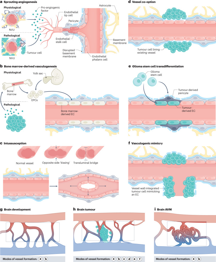
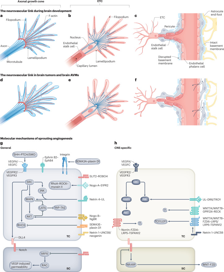
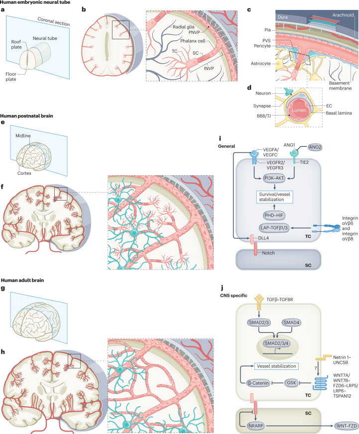
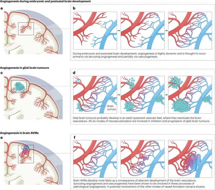
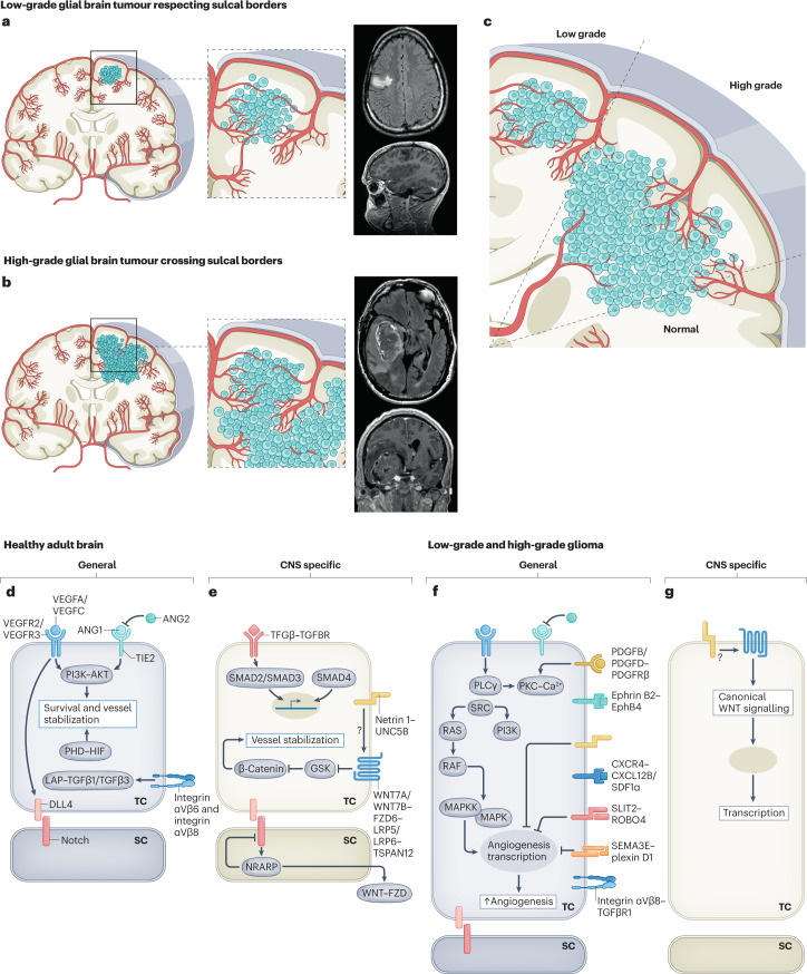
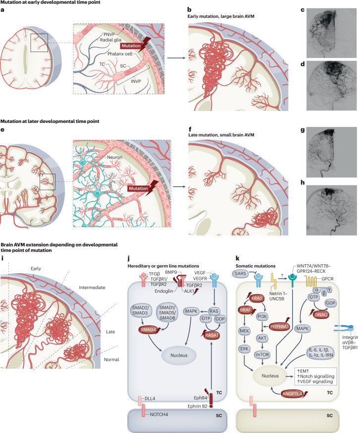
The CNS critically relies on the formation and proper function of its vasculature during development, adult homeostasis and disease. Angiogenesis - the formation of new blood vessels - is highly active during brain development, enters almost complete quiescence in the healthy adult brain and is reactivated in vascular-dependent brain pathologies such as brain vascular malformations and brain tumours. Despite major advances in the understanding of the cellular and molecular mechanisms driving angiogenesis in peripheral tissues, developmental signalling pathways orchestrating angiogenic processes in the healthy and the diseased CNS remain incompletely understood. Molecular signalling pathways of the 'neurovascular link' defining common mechanisms of nerve and vessel wiring have emerged as crucial regulators of peripheral vascular growth, but their relevance for angiogenesis in brain development and disease remains largely unexplored. Here we review the current knowledge of general and CNS-specific mechanisms of angiogenesis during brain development and in brain vascular malformations and brain tumours, including how key molecular signalling pathways are reactivated in vascular-dependent diseases. We also discuss how these topics can be studied in the single-cell multi-omics era.


Conflict of interest statement

The authors declare no competing interests.

Figures

Fig. 1
Fig. 1. Modes of vessel formation during brain development, in brain tumours and in brain AVMs.
Vascularization during brain development, in brain tumours and in brain arteriovenous malformations (AVMs) can occur via different modes of neovascularization. a, Neovascularization is possible via the formation of new blood vessels from pre-existing ones in response to pro-angiogenic signalling molecules secreted by components of the neurovascular unit (NVU) (defined as physiological sprouting angiogenesis) or by tumour cells (defined as pathological sprouting angiogenesis). For simplicity, the NVU (in physiological conditions) and tumour cells (in pathological conditions) are illustrated as sources of pro-angiogenic molecules for this mode of neovascularization. Note that the secretion of pro-angiogenic molecules is not limited to these sources but can also occur from brain vascular malformations and other vascular-dependent brain pathologies as well as from components of the extracellular matrix. New vessel sprouts are guided by specialized endothelial tip cells extending multiple filopodial protrusions sensing and reacting to pro-angiogenic, anti-angiogenic and hypoxia-related cues in the microenvironment. At the back of the leading tip cell, proliferating endothelial stalk cells elongate the growing blood vessel and initiate the formation of a functional lumen. Phalanx cells are the most quiescent of the endothelial cell (EC) subtypes, extend few filopodia and migrate and divide poorly in response to VEGF. Endothelial phalanx cells line vessels once the new vessel branches have been consolidated. b, Physiological vasculogenesis is defined as the de novo generation of blood vessels from either yolk sac-derived endothelial progenitor cells (EPCs) or bone marrow-derived EPCs, depending on the developmental time point. Pathological vasculogenesis occurs upon secretion of pro-angiogenic molecules by tumour cells that activate bone marrow to produce EPCs. Both indirect paracrine secretion of pro-angiogenic growth factors and direct luminal incorporation into sprouting nascent vessels contribute to vasculogenesis. Note that the secretion of pro-angiogenic molecules is not limited to these sources but can also occur from brain vascular malformations and other vascular-dependent brain pathologies as well as from components of the extracellular matrix. c, The splitting of existing blood vessels — vascular intussusception — allows the reorganization of existing cells without a corresponding increase in EC number. During this process, the opposite capillary walls invaginate into the vessel lumen in consecutive steps with the formation of a transluminal bridge of pericytes, myofibroblasts and extracellular matrix. df, Pathological conditions such as tumours or regenerative processes can exhibit the aforementioned modes of vessel formation and three additional ones, namely vessel co-option, glioma stem cell to EC transdifferentiation or glioma stem cell to pericyte transdifferentiation, and vasculogenic mimicry. Vessel co-option occurs when tumour cells co-opt existing vessels in response to angiopoietin 2 (ANG2) expression gradients (part d). In glioma stem cell transdifferentiation, glioma stem-like cells differentiate into either tumour-derived ECs or tumour-derived pericytes, induced predominantly by the TGFβ and NOTCH1 pathways in hypoxic conditions (part e). In vasculogenic mimicry, tumour cells (instead of ECs) are incorporated into the inner vessel wall, forming functional vessel-like structures and thereby mimicking ECs (part f). gi, Modes of vessel formation involved in angiogenesis during brain development (part g), in brain tumours (part h) and in brain AVMs (part i).
Fig. 2
Fig. 2. Neurovascular link molecules affecting endothelial tip cell sprouting during vascular brain development, in brain tumours and in brain AVMs.
a, The axonal growth cone at the leading edge of a growing axon is a specialized, subcellular ‘hand-like’ structure at the tip of an extending neuron. In the axonal growth cone, lamellipodia and filopodia sense and integrate attractive and repulsive guidance cues in the local tissue microenvironment, thereby guiding the extending axon to its target. The central domain of an axonal growth cone is rich in microtubules, whereas the peripheral domain predominantly contains filopodia (composed of F-actin bundles) and lamellipodia (composed of an actin meshwork). Some microtubules extend into the peripheral domain and rarely into filopodia. b, The endothelial tip cell (ETC) is a specialized vascular endothelial cell type at the tip of the newly forming blood vessel, followed by proliferating endothelial stalk cells. Similarly to axonal growth cones, ETCs are specialized, ‘hand-like’ structures at the forefront of growing blood vessels that sense environmental cues using lamellipodia and ‘finger-like’ filopodia, thereby guiding the growing blood vessels to their respective targets. Endothelial phalanx cells comprise a third, mostly silent vascular endothelial cell type, lining the border of functional, established blood vessels (not shown). ETCs use actin-based lamellipodia and filopodia sensing attractive and repulsive guidance cues in the local tissue microenvironment to reach their target. Microtubules have not been detected in filopodia so far. c, A newly forming blood vessel sprout including a migrating ETC extending multiple filopodia, followed by proliferating endothelial stalk cells creating a newly formed capillary lumen, and quiescent endothelial phalanx cells lining an established vascular blood vessel. Pericytes, astrocytes and the basement membrane are also depicted. df, Schematic illustrations showing the characteristics of the axonal growth cone (part d), ETC (part e) and vessel sprouting (part f) in pathological conditions. Newly formed vessels often show a disrupted basement membrane, vascular leakage and a reduced pericyte coverage (part f). g,h, Molecularly, sprouting angiogenesis into the CNS is regulated by neurovascular link molecules that act in a non-CNS-specific way (part g), such as VEGFA–VEGFR2, SEMA3A/SEMA3E–plexin D1, ephrin B2–EphB4 and SLIT2–ROBO4, or a CNS-specific manner (part h), such as WNT7A/WNT7B–GPR124–FZD6–RECK and DR6–TROY. Of note, the VEGFA/VEGFC–VEGFR2/VEGFR3 and netrin 1–UNC5B signalling axes are shown in part h because even though they represent non-CNS-specific mechanisms, multiple CNS-specific mechanisms interact with these pathways downstream. AVM, arteriovenous malformation; SC, stalk cell; TC, tip cell; UL, unknown ligand.
Fig. 3
Fig. 3. Structural and molecular mechanisms of angiogenesis at the embryonic, postnatal and adult stages of vascular brain development.
a, A human neural tube at the embryonic stage with the roof and floorplate illustrated on the coronal cutting plane. b, Sprouting angiogenesis into the neural tube during embryogenesis. The perineural vascular plexus (PNVP) is formed by vasculogenesis from mesodermal-derived angioblasts at around 7 weeks of gestational age in humans (embryonic day 8.5 (E8.5) in mice). Subsequently, at around 8 weeks of gestational age in humans (E9.5 in mice), angiogenic sprouts of the intraneural vascular plexus (INVP) are formed along radial glia via sprouting angiogenesis using endothelial tip cell (ETC) filopodia, invading the CNS parenchyma and migrating towards the ventricle, where pro-angiogenic and anti-angiogenic factors such as VEGFA and WNT proteins are produced. At the forefront of these angiogenic sprouts, ETCs guide the CNS-invading blood vessels using ETC filopodia. c, The anatomical organization of the meningeal layers, including dura, arachnoid and pia mater with intradural lymphatic vessels (blue) and blood vessels (red). An angiogenic vascular sprout emanating from the extraparenchymal PNVP composed of ETCs, endothelial stalk cells and endothelial phalanx cells invading the intraparenchymal INVP is shown. A perivascular space (PVS) surrounds the base of the vascular sprout. d, The neurovascular unit (NVU) for established blood vessels that is composed of a variety of cell types, including endothelial cells (ECs), pericytes, astrocytes and neurons. ECs and pericytes are ensheathed by a common basal lamina, the endothelial basement membrane. The blood–brain barrier (BBB) is composed of microvascular ECs that are mutually connected via complex tight junctions (TJs), thereby regulating or inhibiting paracellular diffusion of water-soluble molecules. ECs regulate the transport of molecules between the blood and the brain parenchyma via the expression of influx and efflux transporters. e, A coronal section of a human brain during postnatal development. f, At the postnatal stage, sprouting angiogenesis is the main mode of neovascularization, and vascular sprouting occurs in all directions throughout cortical layers 1–6. Endothelial sprouts invading the CNS parenchyma from week 8 of gestational age (E9.5 in mice) onwards grow along radial glia fibres towards the ventricle. g,h, In the healthy adult brain, the vasculature is almost quiescent, with only very few ECs proliferating. i,j, Molecularly, numerous pathways have been implicated in EC quiescence, survival and maintained inhibition of paracellular permeability, and the molecular cues can be either non-CNS specific or CNS specific. The TGFβ–TGFβR signalling axis is shown here because even though it is a non-CNS-specific mechanism of angiogenesis, it interacts downstream with the CNS-specific WNT7A/WNT7B–GPR124–FZD6–RECK pathway. ANG1, angiopoietin 1; ANG2, angiopoietin 2; SC, stalk cell; TC, tip cell.
Fig. 4
Fig. 4. Angiogenesis during brain development, in glial brain tumours and in brain AVMs.
a,b, Angiogenesis during embryonic and postnatal brain development is initiated by bone marrow-derived de novo vasculogenesis followed by sprouting angiogenesis with the formation and elongation of new vessel sprouts from pre-existing vessels. Newly formed vessels fuse with other vascular sprouts in a process called ‘anastomosis’, thereby forming a healthy capillary bed within a three-dimensional network of perfused, functional vasculature. c,d, Glial brain tumours develop in a vascular bed where they reactivate the surrounding quiescent brain vasculature but also form their own blood vessels within the tumour mass. All six modes of neovascularization are active in glial brain tumours. e,f, Brain arteriovenous malformations (AVMs) develop as a consequence of aberrant vascular development of a healthy capillary bed in which the initial formation of arteriovenous shunts leads to further progression towards brain AVMs. Sprouting angiogenesis and bone marrow-derived vasculogenesis (in the AVM nidus) play an important role during the initiation and progression of brain AVMs.
Fig. 5
Fig. 5. Molecular mechanisms regulating the vasculature during initiation and progression of glial brain tumours.
Figure illustrating the hypothetical concept postulating the gyral confinement and respect of sulcal borders during progression from low-grade glial brain tumours to high grade glial brain tumours based on radiological observations and the concept of sprouting angiogenesis and recruitment of blood vessels from the adjacent brain parenchyma. ac, Cross sections of the adult human brain in the coronal plane showing pathological angiogenesis in glial brain tumours. Illustrations and T1-weighted coronal and sagittal MRI scans with gadolinium show that low-grade gliomas are often confined to one gyrus, thereby respecting sulcal borders (parts a,c). Illustrations and T1-weighted coronal and sagittal MRI scans with gadolinium show that invasive high-grade gliomas do often not respect gyral confinement and cross sulcal borders (parts b,c). d,e, Molecularly, numerous signalling pathways have been implicated in the adult healthy brain, regulating endothelial cell quiescence, survival and maintained inhibition of paracellular permeability. Molecular cues can be either non-CNS specific (part d) or CNS specific (part e). These signalling pathways are thought to be of importance during both embryonic and postnatal vascular brain development, as well as to contribute to the maintenance of the quiescent healthy adult brain vasculature. f,g, Molecularly, different non-CNS-specific and CNS-specific angiogenic molecular mechanisms have been implicated in glioma initiation and progression, and they include the reactivation of developmentally active ligand–receptor pairs. ANG1, angiopoietin 1; ANG2, angiopoietin 2; SC, stalk cell; TC, tip cell. Images in parts a,b courtesy of P. Nicholson.
Fig. 6
Fig. 6. Molecular mechanisms regulating the vasculature during initiation and progression of brain AVMs.
Figure illustrating the hypothesis stating that the timing of mutation influences the size and location of the arteriovenous malformation (AVM). a,b, Cross section of the human brain in the coronal plane illustrating that mutations occurring in progenitor endothelial cells (ECs) at an early developmental time point will ‘trace’ the future developmental territory of their daughter cells, resulting in a large lesion spreading along a radial axis from the pial cortical surface to the ventricles. c,d, Anterior–posterior (c) and lateral (d) digital subtraction angiography of the right intracarotid artery showing a large AVM. e,f, Cross section of the human brain in the coronal plane illustrating that mutations at later developmental time point result in smaller lesions restricted to a local vascular territory. Note that these smaller AVMs are located around the pial, sulcal and cortical areas or alternatively in the ventricular, ependymal and subependymal zones (that is, choroidal AVMs) but do not occur isolated midway in the white matter without reaching either the cortical surface or the ventricular surface. g,h, Anterior–posterior and lateral digital subtraction angiography of the right intracarotid artery showing a smaller AVM. i, AVM extension as result of early, intermediate and late time points of mutation. j,k, Various molecular pathways have been implicated in AVM initiation and progression. The mutations shown belong to either hereditary or germ line mutations (part j) or somatic mutations in genes in the endothelial tip and stalk cells (part k). The proteins encoded by mutated genes are indicated with a flash symbol. Additional molecules and ligand–receptor pairs involved in regulating the vasculature during initiation and progression of brain AVMs can be found in Supplementary Table 2. BMP9, bone morphogenetic protein 9; EMT, endothelial-to-mesenchymal transition; GPCR, G protein-coupled receptor; INVP, intraneural vascular plexus; SARS, seryl-tRNA synthetase 1; SC, stalk cell; TC, tip cell. Images in parts c,d,g,h courtesy of P. Nicholson.

References

    1. Mink JW, Blumenschine RJ, Adams DB. Ratio of central nervous system to body metabolism in vertebrates: its constancy and functional basis. Am. J. Physiol. 1981;241:R203–R212. - PubMed
    1. Zlokovic BV, Apuzzo ML. Strategies to circumvent vascular barriers of the central nervous system. Neurosurgery. 1998;43:877–878. doi: 10.1097/00006123-199810000-00089. - DOI - PubMed
    1. Wälchli T, et al. Quantitative assessment of angiogenesis, perfused blood vessels and endothelial tip cells in the postnatal mouse brain. Nat. Protoc. 2015;10:53–74. doi: 10.1038/nprot.2015.002. - DOI - PubMed
    1. Zlokovic BV. The blood-brain barrier in health and chronic neurodegenerative disorders. Neuron. 2008;57:178–201. doi: 10.1016/j.neuron.2008.01.003. - DOI - PubMed
    1. Stewart PA, Wiley MJ. Developing nervous tissue induces formation of blood-brain barrier characteristics in invading endothelial cells: a study using quail–chick transplantation chimeras. Dev. Biol. 1981;84:183–192. doi: 10.1016/0012-1606(81)90382-1. - DOI - PubMed

Publication types