. 2023 Mar 20;19(1):5.
doi: 10.1186/s12993-023-00208-9.

Whole-body vibration ameliorates glial pathological changes in the hippocampus of hAPP transgenic mice, but does not affect plaque load

Affiliations

Whole-body vibration ameliorates glial pathological changes in the hippocampus of hAPP transgenic mice, but does not affect plaque load

Tamas Oroszi et al. Behav Brain Funct. .

Abstract

Background: Alzheimer's disease (AD) is the core cause of dementia in elderly populations. One of the main hallmarks of AD is extracellular amyloid beta (Aβ) accumulation (APP-pathology) associated with glial-mediated neuroinflammation. Whole-Body Vibration (WBV) is a passive form of exercise, but its effects on AD pathology are still unknown.

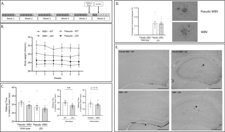
Methods: Five months old male J20 mice (n = 26) and their wild type (WT) littermates (n = 24) were used to investigate the effect of WBV on amyloid pathology and the healthy brain. Both J20 and WT mice underwent WBV on a vibration platform or pseudo vibration treatment. The vibration intervention consisted of 2 WBV sessions of 10 min per day, five days per week for five consecutive weeks. After five weeks of WBV, the balance beam test was used to assess motor performance. Brain tissue was collected to quantify Aβ deposition and immunomarkers of astrocytes and microglia.

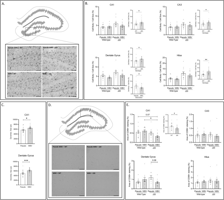
Results: J20 mice have a limited number of plaques at this relatively young age. Amyloid plaque load was not affected by WBV. Microglia activation based on IBA1-immunostaining was significantly increased in the J20 animals compared to the WT littermates, whereas CD68 expression was not significantly altered. WBV treatment was effective to ameliorate microglia activation based on morphology in both J20 and WT animals in the Dentate Gyrus, but not so in the other subregions. Furthermore, GFAP expression based on coverage was reduced in J20 pseudo-treated mice compared to the WT littermates and it was significantly reserved in the J20 WBV vs. pseudo-treated animals. Further, only for the WT animals a tendency of improved motor performance was observed in the WBV group compared to the pseudo vibration group.

Conclusion: In accordance with the literature, we detected an early plaque load, reduced GFAP expression and increased microglia activity in J20 mice at the age of ~ 6 months. Our findings indicate that WBV has beneficial effects on the early progression of brain pathology. WBV restored, above all, the morphology of GFAP positive astrocytes to the WT level that could be considered the non-pathological and hence "healthy" level. Next experiments need to be performed to determine whether WBV is also affective in J20 mice of older age or other AD mouse models.

Keywords: Amyloid beta; J20 mice; Motor coordination; Neuroinflammation; Passive exercise.


Conflict of interest statement

The authors declare that they have no competing interests.

Figures

Fig. 1
Fig. 1
Experimental design (A): 5 months old male hAPP-J20 mice and their wild (WT) littermates underwent 5 weeks of whole body vibration intervention (WBV) with twice daily session of 10 min exposure, five times per week (gray color marks the days of treatment). After 5 weeks, balance beam test was performed to assess motor coordination. Animals were terminated on week 6 at the age of ~ 6.5 months and brain tissue was collected for immunohistological analyses. Effects of intervention (pseudo vs. WBV) and genotype (J20 vs. WT) on body weight (B), motor performance (C) and plaque load (D). WT animals showed significatly higher body weight during the intervention compared to the J20 animals (B). Walking distance in the balance beam test was only significantly improved in the J20 animals compared to the WT (C). Amyloid plaque deposition in the hippocampus was not significantly affected by WBV intervention (D). Images of 6e10 were taken about the whole hippocampus at 50 × magnifications to visualze total amyloid plaque distribution (E), representative images of areas marked by + are depicted in D. Data are depicted as mean ± SEM. ** indicates: P < .01. Scale bars in D are 50 um and in E are 500 um
Fig. 2
Fig. 2
Microglia visualized by IBA1 staining in the Hilus subregions is depicted in (A). Effects of intervention (pseudo vs. WBV) and genotype (WT vs. J20) on microglia activation in the CA1, CA3, DG and Hilus subregions are depicted in (B). Significant increase of microglia activation was observed in the J20 animals compared to the WT controls in all subregions (B, CA1, CA3, Dentate Gyrus and Hilus). Microglia activation was only significantly decreased by WBV in the DG (B, Dentate Gyrus). Furthermore, WBV treatment significantly increased the size of microglia dendritic processes in the CA1 and DG subregions (C, CA1 and Dentate Gyrus). Expression of CD68 coverage in the CA1 subregion is visualized in D. Significant decrease of CD68 expression in the CA1 area was detected in the J20 animals compared to the WT controls (E, CA1). In addition, a tendency (p = 0.06) of interaction effect (intervention vs. genotype) on CD68 expression was also observed in the CA1 subregion; additional post-hoc analysis showed a significant decrease for the J20—WBV group compared to the WT—WBV group, as well as the same tendency (p = 0.07) compared to the WT—pseudo WBV group (E, CA1). A significant effect of interaction (intervention vs. genotype) in CD68 expression was observed in the DG area; additional post hoc revealed a strong tendency (p = 0.08) of decrease CD68 expression in the J20—WBV treated animals compared to the J20—pseudo WBV controls (E, Dentate Gyrus). CD68 expression was not significantly altered in the CA3 and Hilus areas (E, CA3 and Hilus). Representative Images of IBA1 and CD68 were taken about the Hilus and CA1 subregions at 200 × magnifications to visualze microglia (A and C). Relevant statistical differences are marked in B, C and D. Data are depicted as mean ± SEM. * indicates: P = .05. Scale bars in A and D are 100 um
Fig. 3
Fig. 3
GFAP + astrocytes in the CA3 hipocampal subregion are visualized in (A). Effects of intervention (pseudo vs. WBV) and genotype (J20 vs. WT) on expression of GFAP positive cells in the CA1, CA3, DG and Hilus regions are depicted in B. Significantly decreased coverage of GFAP positive cells in the J20 animals was detected in the CA1 and Hilus regions (B, CA1 and Hilus). The same trend was observed in the CA3 and Hilus subregions (B, CA3 and Hilus). Significant effect of interaction (intervention vs. genotype) was detected in the CA1 and CA3 subregions (B, CA1 and CA3). Additional posthoc analysis revealed that WT—WBV and pseudo WBV groups showed significantly higher coverage compared to the J20 – pseudo WBV group in the CA1 region (B, CA1). Significantly increased GFAP coverage was detected in the J20—WBV and WT—pseudo WBV groups compared to the J20—pseudo WBV group in the CA3 region (B, CA3). In addition, significant increase of GFAP coverage was detected by WBV in the Hilus region (Panel B, Hilus). Representative images of GFAP + astrocytes were taken about the CA3 hippocampal subregion at 200 × magnifications (A). Data are depicted as mean ± SEM. * indicates: P = .05. ** P < .01. *** P < .001. Scale bars in A are 100 um

References

    1. Breijyeh Z, Karaman R. Comprehensive review on Alzheimer’s disease: causes and treatment. Molecules. 2020;25:5789. doi: 10.3390/molecules25245789. - DOI - PMC - PubMed
    1. Fakhoury M. Microglia and astrocytes in Alzheimer’s disease: implications for therapy. Curr Neuropharmacol. 2018;16:508–518. doi: 10.2174/1570159X15666170720095240. - DOI - PMC - PubMed
    1. Hansen DV, Hanson JE, Sheng M. Microglia in Alzheimer’s disease. J Cell Biol. 2018;217:459–472. doi: 10.1083/jcb.201709069. - DOI - PMC - PubMed
    1. Matsuoka Y, Picciano M, Malester B, LaFrancois J, Zehr C, Daeschner JM, et al. Inflammatory responses to amyloidosis in a transgenic mouse model of Alzheimer’s disease. Am J Pathol. 2001;158:1345–1354. doi: 10.1016/S0002-9440(10)64085-0. - DOI - PMC - PubMed
    1. Ries M, Sastre M. Mechanisms of Aβ clearance and degradation by glial cells. Front Aging Neurosci. 2016 doi: 10.3389/fnagi.2016.00160. - DOI - PMC - PubMed

Substances