

Therapeutic effects of kartogenin on temporomandibular joint injury by activating the TGF-β/SMAD pathway in rats
- PMID: 36941805
- PMCID: PMC10666730
- DOI: 10.1177/15353702231157945
Therapeutic effects of kartogenin on temporomandibular joint injury by activating the TGF-β/SMAD pathway in rats
Abstract
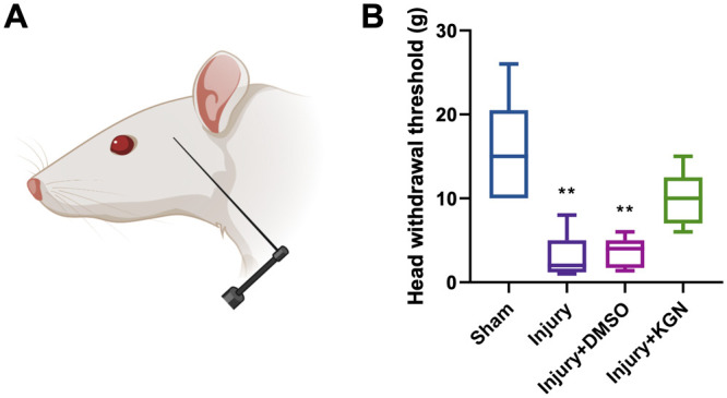
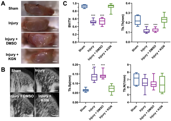
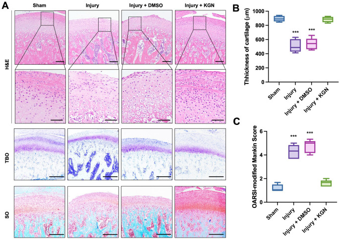
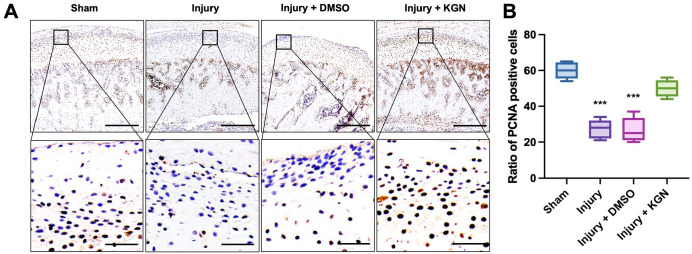
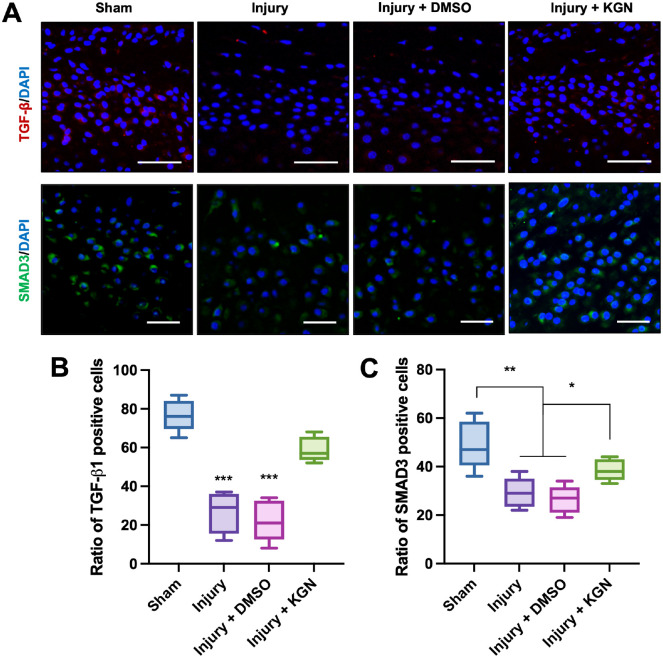
Patients with temporomandibular dysfunction (TMD) usually suffer from pathology or malpositioning of the temporomandibular joint (TMJ) disk, leading to the degenerative lesion of condyles. Kartogenin can promote the repair of damaged cartilage. This study aimed to explore whether intra-articular injection of kartogenin could alleviate the TMJ injury induced by type II collagenase. We measured the head withdrawal threshold and found that kartogenin alleviated the pain around TMD induced by type II collagenase. We observed the morphology of the condylar surface and found that kartogenin protected the integration of the condylar surface. We analyzed the density of the subchondral bone and found that kartogenin minimized the damage of TMJ injury to the subchondral bone. We next explored the histological changes and found that kartogenin increased the thickness of the proliferative layer and more collagen formation in the superficial layer. Then, to further ensure whether kartogenin promotes cell proliferation in the condyle, we performed immunohistochemistry of proliferating cell nuclear antigen (PCNA). The ratio of PCNA-positive cells was significantly increased in the kartogenin group. Next, immunofluorescence of TGF-β1 and SMAD3 was performed to reveal that kartogenin activated the TGF-β/SMAD pathway in the proliferative layer. In conclusion, kartogenin may have a therapeutic effect on TMJ injury by promoting cell proliferation in cartilage and subchondral bone. Kartogenin may be promising as an intra-articular injection agent to treat TMD.
Keywords: Temporomandibular joint; cartilage; cell proliferation; collagenase; dysfunction; kartogenin.
Conflict of interest statement
Declaration of Conflicting InterestsThe author(s) declared no potential conflicts of interest with respect to the research, authorship, and/or publication of this article.
Figures
Similar articles
-
Installing and thereafter removing an aberrant prosthesis elicited opposite remodelling responses in growing mouse temporomandibular joints.J Oral Rehabil. 2015 Sep;42(9):685-92. doi: 10.1111/joor.12304. Epub 2015 May 1. J Oral Rehabil. 2015. PMID: 25940877
-
Kartogenin potentially protects temporomandibular joints from collagenase-induced osteoarthritis via core binding factor β and runt-related transcription factor 1 binding - A rat model study.J Dent Sci. 2023 Oct;18(4):1553-1560. doi: 10.1016/j.jds.2023.03.002. Epub 2023 Mar 14. J Dent Sci. 2023. PMID: 37799879 Free PMC article.
-
Rebamipide Attenuates Mandibular Condylar Degeneration in a Murine Model of TMJ-OA by Mediating a Chondroprotective Effect and by Downregulating RANKL-Mediated Osteoclastogenesis.PLoS One. 2016 Apr 28;11(4):e0154107. doi: 10.1371/journal.pone.0154107. eCollection 2016. PLoS One. 2016. PMID: 27123995 Free PMC article.
-
Temporomandibular Joint Osteoarthritis: Pathogenic Mechanisms Involving the Cartilage and Subchondral Bone, and Potential Therapeutic Strategies for Joint Regeneration.Int J Mol Sci. 2022 Dec 22;24(1):171. doi: 10.3390/ijms24010171. Int J Mol Sci. 2022. PMID: 36613615 Free PMC article. Review.
-
Pathological mechanism of chondrocytes and the surrounding environment during osteoarthritis of temporomandibular joint.J Cell Mol Med. 2021 Jun;25(11):4902-4911. doi: 10.1111/jcmm.16514. Epub 2021 May 5. J Cell Mol Med. 2021. PMID: 33949768 Free PMC article. Review.
Cited by
-
TGF-β1 maintains the developmental potential of embryonic submandibular gland epithelia separated with mesenchyme.Heliyon. 2024 Jun 25;10(13):e33506. doi: 10.1016/j.heliyon.2024.e33506. eCollection 2024 Jul 15. Heliyon. 2024. PMID: 39040362 Free PMC article.
References
-
- Valesan LF, Da-Cas CD, Réus JC, Denardin ACS, Garanhani RR, Bonotto D, Januzzi E, de Souza BDM. Prevalence of temporomandibular joint disorders: a systematic review and meta-analysis. Clin Oral Investig 2021;25:441–53 - PubMed
-
- Lomas J, Gurgenci T, Jackson C, Campbell D. Temporomandibular dysfunction. Aust J Gen Pract 2018;47:212–5 - PubMed
-
- Gauer RL, Semidey MJ. Diagnosis and treatment of temporomandibular disorders. Am Fam Physician 2015;91:378–86 - PubMed
Publication types
MeSH terms
Substances
LinkOut - more resources
Full Text Sources
Medical
Miscellaneous