Review
. 2023 Jan 3;5(4):100667.
doi: 10.1016/j.jhepr.2022.100667. eCollection 2023 Apr.

Management of splanchnic vein thrombosis

Affiliations
Review

Management of splanchnic vein thrombosis

Laure Elkrief et al. JHEP Rep. .

Abstract

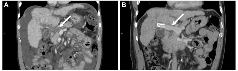
The expression splanchnic vein thrombosis encompasses Budd-Chiari syndrome and portal vein thrombosis. These disorders have common characteristics: they are both rare diseases which can cause portal hypertension and its complications. Budd-Chiari syndrome and portal vein thrombosis in the absence of underlying liver disease share many risk factors, among which myeloproliferative neoplasms represent the most common; a rapid comprehensive work-up for risk factors of thrombosis is needed in these patients. Long-term anticoagulation is indicated in most patients. Portal vein thrombosis can also develop in patients with cirrhosis and in those with porto-sinusoidal vascular liver disease. The presence and nature of underlying liver disease impacts the management of portal vein thrombosis. Indications for anticoagulation in patients with cirrhosis are growing, while transjugular intrahepatic portosystemic shunt is now a second-line option. Due to the rarity of these diseases, studies yielding high-grade evidence are scarce. However, collaborative studies have provided new insight into the management of these patients. This article focuses on the causes, diagnosis, and management of patients with Budd-Chiari syndrome, portal vein thrombosis without underlying liver disease, or cirrhosis with non-malignant portal vein thrombosis.

Keywords: BCS, Budd-Chiari syndrome; CALR, calreticulin; Cavernoma; DOACs, direct-acting oral anticoagulants; Direct oral anticoagulants; EHPVO, extrahepatic portal vein obstruction; GFR, glomerular filtration rate; JAK2, Janus kinase 2; LMWH, low-molecular-weight heparin; MPN, myeloproliferative neoplasm; MTHFR, methylene-tetrahydrofolate reductase; PNH, paroxysmal nocturnal hemoglobinuria; PVT, portal vein thrombosis; Portal biliopathy; Portal vein recanalisation; SVT, splanchnic vein thrombosis; TIPS, transjugular intrahepatic portosystemic shunt; VKAs, vitamin K antagonists; Vascular liver diseases.


Conflict of interest statement

The authors declare no conflicts of interest that pertain to this work. Please refer to the accompanying ICMJE disclosure forms for further details.

Figures

Fig. 1
Fig. 1
Management of liver nodules in patients with Budd-Chiari syndrome. AFP, alpha-fetoprotein.
Fig. 2
Fig. 2
Example of acute portal vein thrombosis with progressive development of a cavernous transformation. A female patient presented with abdominal pain. Contrast-enhanced CT (all portal venous phase, axial view) showed a complete occlusion of an enlarged portal trunk and intrahepatic portal branches by non-enhancing and hypoattenuating material (arrow) consistent with an acute portal vein thrombosis. Note the heterogeneous enhancement of the hepatic parenchyma with central hypoenhancement relative to the liver periphery (zonal perfusion, ∗). Over time, CT shows the progressive development of numerous tortuous veins in the hepatic pedicle and the hepatic hilum corresponding to a cavernous transformation of the portal vein (dashed arrows). Note the absence of recanalization of the portal veins and the progressive enlargement of the cavernoma, with progressive extension to the pancreas. No bile duct dilatation was noted.
Fig. 3
Fig. 3
Proposed algorithm for anticoagulant therapy in patients with portal vein thrombosis with and without cirrhosis. DOACs, direct-acting oral anticoagulants; EHPVO, extrahepatic portal vein obstruction; LMWH, low-molecular-weight heparin; PVT, portal vein thrombosis; TIPS, transjugular intrahepatic portosystemic shunt; VKAs, vitamin K antagonists.
Fig. 4
Fig. 4
Correlation between radiological findings and pathology. (A) Recent portal vein thrombosis in a 59-year-old man with viral cirrhosis. Contrast-enhanced CT (coronal view, portal venous phase) shows partially occlusive non-enhancing material corresponding to the clot in the lumen of the portal trunk (arrows). The downstream venous branches are patent. (B) HES staining. Low magnification. Recent portal vein partial thrombosis. (C) HES staining. Higher magnification. Recent portal vein thrombi composed of fibrinous deposits associated with red blood cells. (D) Chronic portal vein thrombosis in a 52-year-old man with alcohol-related cirrhosis. Contrast-enhanced CT (coronal view, portal venous phase) shows minimally occlusive non-enhancing material corresponding to the incorporation of the clot in the wall of the portal trunk and the superior mesenteric vein (arrows). (E) HES staining. Low magnification. Non-malignant chronic portal vein thrombi. (F) HES staining. Higher magnification. Chronic portal vein thrombi with fibro-oedematous intimal thickening with numerous siderophagous and neovessels partially repermeabilizing the lumen.
Fig. 5
Fig. 5
CT scan showing a patient with decompensated cirrhosis and occlusive portal vein thrombosis in whom portal vein recanalization was achieved after TIPS insertion. Patient with (A) occlusive portal vein thrombosis (arrow) despite anticoagulation for 3 months, in whom (B) portal vein recanalization was achieved after TIPS insertion. TIPS, transjugular intrahepatic portosystemic shunt.

References

    1. Jones D.E.J., Sturm E., Lohse A.W. Access to care in rare liver diseases: new challenges and new opportunities. J Hepatol. 2018;68:577–585. doi: 10.1016/j.jhep.2017.11.004. - DOI - PubMed
    1. De Gottardi A., Sempoux C., Berzigotti A. Porto-sinusoidal vascular disorder. J Hepatol. 2022;77:1124–1135. doi: 10.1016/j.jhep.2022.05.033. - DOI - PubMed
    1. Denninger M.H., Chaït Y., Casadevall N., Hillaire S., Guillin M.C., Bezeaud A., et al. Cause of portal or hepatic venous thrombosis in adults: the role of multiple concurrent factors. Hepatology. 2000;31:587–591. doi: 10.1002/hep.510310307. - DOI - PubMed
    1. Darwish Murad S., Plessier A., Hernandez-Guerra M., Fabris F., Eapen C.E., Bahr M.J., et al. Etiology, management, and outcome of the Budd-Chiari syndrome. Ann Intern Med. 2009;151:167–175. - PubMed
    1. Plessier A., Darwish-Murad S., Hernandez-Guerra M., Consigny Y., Fabris F., Trebicka J., et al. Acute portal vein thrombosis unrelated to cirrhosis: a prospective multicenter follow-up study. Hepatology. 2010;51:210–218. doi: 10.1002/hep.23259. - DOI - PubMed