. 2023 Feb;35(2):147-153.
doi: 10.1016/j.sdentj.2023.01.003. Epub 2023 Jan 5.

Human umbilical cord mesenchymal stem cells induction in peri-implantitis Rattus norvegicus accelerates and enhances osteogenesis activity and implant osseointegration

Affiliations

Human umbilical cord mesenchymal stem cells induction in peri-implantitis Rattus norvegicus accelerates and enhances osteogenesis activity and implant osseointegration

Nike Hendrijantini et al. Saudi Dent J. 2023 Feb.

Abstract

Peri-implantitis additional treatment generally aims to repair damaged tissue through a regenerative approach. Human umbilical cord mesenchymal stem cells (hUCMSCs) produce a high osteogenic effect and are capable of modulating the immune system by suppressing inflammatory response, modulating bone resorption, and inducing endogenous osteogenesis.

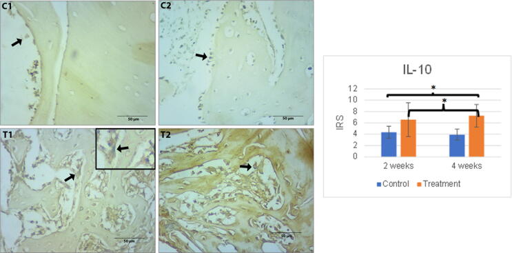
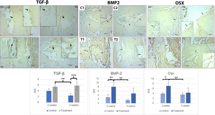
Aim: This study was intended to discover the effect of hUCMSCs on an implant osseointegration process in peri-implantitis rat subjects as assessed by several markers including interleukin-10 (IL-10), transforming growth factor-β (TGF-β), receptor activator of nuclear factor kappa- β ligand (RANKL), bone morphogenic protein (BMP-2), osterix (Osx), and osteoprotegerin (OPG).

Material and methods: The research design implemented during this study represented a true experimental design incorporating the use of Rattus norvegicus (Wistar strain) as subjects.

Results: Data analysed by means of a Brown Forsythe test indicated differences between the increase in BMP-2 expression (p < 0.000) and Osx expression (p < 0.001) and between RANKL expression (p < 0.001, Tukey HSD) and OPG expression (p < 0.000, Games Howell).

Conclusion: According to the findings of this research, hUCMSCs induction is successful in accelerating and enhancing osteogenic activity and implant osseointegration in peri-implantitis rat subjects.

Keywords: Human umbilical cord mesenchymal stem cells; Implant; Medicine; Osseointegration; Peri-implantitis.


Figures

Fig. 1
Fig. 1
Left: The immunohistochemical staining of IL-10 expression in all peri-implantitis groups contained in osteoblast cells around the implant site. The black arrow indicates osteoblast cells with an overexpression of IL-10. Scale bar = 50 µm. Each box is presented at 1000x magnification. C1 (implant 2 weeks), C2 (implant 4 weeks), T1 (implant + hUCMSCs 2 weeks), T2 (implant + hUCMSCs 4 weeks). Right: Mean of IRS of IL-10 expressions together with its standard deviation. Single asterisk (*) above the bars represents p value < 0.05, double asterisk (**) represents p value < 0.01, and triple asterisk (***) represents p value < 0.001.
Fig. 2
Fig. 2
(A) The immunohistochemical staining of BMP-2 A in all peri-implantitis groups in the osteoblast cells around the implant site. (B). The immunohistochemical staining of Osx expression in all peri-implantitis groups in osteoblast cells around the implant site. The black arrow indicates the osteoblast cells with an overexpression of BMP-2 and Osx. Scale bar = 50 µm. Each box represented positive cells at 1000x magnification. C1 (implant 2 weeks), C2 (implant 4 weeks), T1 (implant + hUCMSCs 2 weeks), T2 (implant + hUCMSCs 4 weeks). (C) Mean of IRS of BMP-2 and Osx expressions together with its standard deviation. The single asterisk (*) above the bars represents p value < 0.05, double asterisk (**) represents p value < 0.01, and triple asterisk (***) represents p value < 0.001.
Fig. 3
Fig. 3
(A) The immunohistochemical staining of RANKL expression of all peri-implantitis groups in the osteoblast cells around the implant site. (B). The immunohistochemical staining of OPG expression in all peri-implantitis groups in osteoblast cells around the implant site. The black arrow indicates the osteoblast cells with an overexpression of RANKL and OPG. Scale bar = 50 µm. Each box represents cells at 1000x magnification. C1 (implant 2 weeks), C2 (implant 4 weeks), T1 (implant + hUCMSCs 2 weeks), T2 (implant + hUCMSCs 4 weeks). (C) Mean of IRS of RANKL and OPG expressions together with its standard deviation. Single asterisk (*) above bars represents p value < 0.05, double asterisk (**) represents p value < 0.01, and triple asterisk (***) represents p value < 0.001.

References

    1. Alghamdi H.S. Methods to improve osseointegration of dental implants in low quality (Type-IV) bone: an overview. J. Funct. Biomater. 2018;9 doi: 10.3390/JFB9010007. - DOI - PMC - PubMed
    1. Amengual-Peñafiel L., Córdova L.A., Constanza Jara-Sepúlveda M., Brañes-Aroca M., Marchesani-Carrasco F., Cartes-Velásquez R. Osteoimmunology drives dental implant osseointegration: a new paradigm for implant dentistry. Jpn Dent. Sci. Rev. 2021;57:12. doi: 10.1016/J.JDSR.2021.01.001. - DOI - PMC - PubMed
    1. Atkins G.J., Kostakis P., Pan B., Farrugia A., Gronthos S., Evdokiou A., Harrison K., Findlay D.M., Zannettino A.C.W. RANKL expression is related to the differentiation state of human osteoblasts. J. Bone Mineral Res. 2003;18:1088–1098. doi: 10.1359/JBMR.2003.18.6.1088. - DOI - PubMed
    1. Fernandes M.H., de Gomes P. Bone cells dynamics during Peri-Implantitis: a theoretical analysis. J Oral Maxillofac Res. 2016;7 doi: 10.5037/JOMR.2016.7306. - DOI - PMC - PubMed
    1. Hendrijantini N., Kusumaningsih T., Rostiny R., Mulawardhana P., Danudiningrat C.P., Rantam F.A. A potential therapy of human umbilical cord mesenchymal stem cells for bone regeneration on osteoporotic mandibular bone. Eur. J. Dent. 2018;12:358–362. doi: 10.4103/EJD.EJD_342_17. - DOI - PMC - PubMed

LinkOut - more resources