. 2023 May 8;51(8):3770-3792.
doi: 10.1093/nar/gkad171.

CAF-1 deposits newly synthesized histones during DNA replication using distinct mechanisms on the leading and lagging strands

Affiliations

CAF-1 deposits newly synthesized histones during DNA replication using distinct mechanisms on the leading and lagging strands

Clément Rouillon et al. Nucleic Acids Res. .

Abstract

During every cell cycle, both the genome and the associated chromatin must be accurately replicated. Chromatin Assembly Factor-1 (CAF-1) is a key regulator of chromatin replication, but how CAF-1 functions in relation to the DNA replication machinery is unknown. Here, we reveal that this crosstalk differs between the leading and lagging strand at replication forks. Using biochemical reconstitutions, we show that DNA and histones promote CAF-1 recruitment to its binding partner PCNA and reveal that two CAF-1 complexes are required for efficient nucleosome assembly under these conditions. Remarkably, in the context of the replisome, CAF-1 competes with the leading strand DNA polymerase epsilon (Polϵ) for PCNA binding. However, CAF-1 does not affect the activity of the lagging strand DNA polymerase Delta (Polδ). Yet, in cells, CAF-1 deposits newly synthesized histones equally on both daughter strands. Thus, on the leading strand, chromatin assembly by CAF-1 cannot occur simultaneously to DNA synthesis, while on the lagging strand these processes may be coupled. We propose that these differences may facilitate distinct parental histone recycling mechanisms and accommodate the inherent asymmetry of DNA replication.


Figures

Figure 1.
Figure 1.
DNA controls CAF-1 recruitment to DNA-loaded PCNA. (A–C) SDS PAGE following separation on SEC of a CAF-1–PCNA binding reaction on DNA plasmids using WT CAF-1 (A), a CAF-1_PIP** mutant (B), or WT CAF-1 in absence of ATP (C). The grey arrow indicates the elution volume of the plasmid DNA. Chromatograms are shown in Supplementary Figure S1C. (D) SDS PAGE of glutaraldehyde crosslinking reactions of fluorescent PCNA (3 μM), CAF-1 (1.5 μM), RFC (150 nM) and nicked pUC19 plasmid (300 nM) after nuclease digestion. Fluorescence scan for PCNA (546 nm) and Coomassie staining are shown. The CAF-1 and PCNA interaction is dependent on PCNA loading onto DNA. (E, F) Coomassie-stained SDS PAGE following SEC of a CAF-1–PCNA binding reaction on DNA plasmids using CAF-1_KER* (E) and CAF-1_WHD* (F) mutants. (G) Crosslinking experiment between CAF-1 (3 μM) and labeled PCNA (4.5 μM) on DNA fragments (1.5 μM) of various sizes. RFC and ATP were not added to actively load PCNA and DNA was not digested in these reactions. Full gels are shown in Supplementary Figure S1I.
Figure 2.
Figure 2.
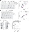
The WHD of CAF-1 controls PCNA-dependent nucleosome assembly. (A) (Left) Native agarose gel of PCNA-NAQ assay reactions. A reaction containing all components, and reactions where we removed either ATP, RFC, PCNA or CAF-1 are shown. Fluorescence signal for H2B-T112C labeled with AF647 (H2B-AF647) or DNA (SybrGOLD), and their overlay are shown. H2B fluorescence on the nicked plasmid (top panel) represents PCNA-dependent histone deposition. (Right) Native PAGE stained with SybrGOLD to detect protected DNA fragments following MNase digestion of samples in A. 150bp DNA fragments are characteristic of nucleosomal DNA, a 621bp loading control is used to monitor DNA retrieval during the purification procedure. Bands around 120bp represent hexasomes. (B) Quantification of the H2B fluorescence signal on the nicked plasmid relative to the total H2B signal in each lane in panel A as a measure of PCNA-dependent nucleosome assembly. C) Quantification of the PCNA-dependent nucleosome assembly activity for CAF-1_PIP**, CAF-1 KER* and CAF-1_WHD*. Mean ± SD is shown, * P < 0.05, ** P < 0.01, *** P < 0.001, **** P < 0.0001 (one-way ANOVA comparing WT CAF-1 to control conditions (B) or each mutant (C)). Gels are shown in Supplementary Figure S2H.
Figure 3.
Figure 3.
Two CAF-1 complexes bind to DNA-loaded PCNA and histones regulate this interaction. (A) (Left) SEC of crosslinked CAF-1–PCNA complexes after DNA digestion using 1 μM PCNA, 0.15 μM RFC, 1.5 μM WT CAF-1 and 0.3 μM nicked pUC19. After crosslinking with 0.2% glutaraldehyde and quenching, the samples were treated with nuclease to digest the DNA plasmid. (Right) Coomassie SDS-PAGE of the collected fractions. The fractions that were used to prepare mass photometry samples are shown as Peak1 and Peak2. (B) Mass photometry data of pooled fractions of Peak1 (left) and Peak2 (right) from experiment in panel A. Theoretical masses are listed and calculated masses from the fitted data are shown in each graph. Normalized counts are shown. (C) SDS PAGE of protein-protein crosslinking reactions after DNA digestion. These reactions contain 50 nM fluorescently labeled PCNA, 15 nM full-length RFC, 15 nM pUC19 and increasing CAF-1 concentrations. PCNA fluorescence signal is shown. Full gels are shown in Supplementary Figure S3A. (D) Quantification of the fluorescence intensity of bands in C. Data are shown as mean ± SD of three independent experiments. (E) SEC and SDS-PAGE of crosslinked CAF-1–PCNA complexes after DNA digestion with CAF-1_WHD*, as in panel A. WT curved is shown in dashed gray line for comparison. (F) SDS PAGE of crosslinking reactions containing fluorescent PCNA (5.5 μM) and H3–H4 (H4-E63C, 1.5 μM dimer concentration), CAF-1 or tCAF-1 (1.5 μM). DNA or RFC are not present in these reactions. (G) SDS PAGE of crosslinking reactions containing fluorescent PCNA (5.5 μM) and H3–H4 (H4-E63C, 1.5 μM dimer concentration), CAF-1 or CAF-1_ΔAD (1.5 μM). DNA or RFC are not present in these reactions.
Figure 4.
Figure 4.
CAF-1 competes with Polϵ, not with Polδ, for PCNA binding. (A, B) (Top) Fluorescence scan of a denaturing alkaline agarose gel of primer extension reactions with Polδ (A) or Polϵ (B). The fluorescently labelled primer signal is shown. The polymerases were at 120 nM, PCNA 480 nM and CAF-1 concentrations as shown. (Bottom) Quantification of the full-length product band relative to the total fluorescence in each lane (expressed as percentages) from the top panels. Mean ± SD are shown for independent experiments (Polδ n = 2 – Polϵ n = 4). (C) Fluorescence scan of denaturing alkaline agarose gel of primer extension reactions with Polϵ with CAF-1_PIP** and CAF-1_KER* mutants (300 nM). (D) Quantification of primer extension by Polϵ in the presence of CAF-1 mutants. Mean ± SD are shown for three independent experiments. (E) Fluorescence scan of denaturing alkaline agarose gel of primer extension reactions with Polϵ with FEN1_DA, FEN1_DA PIP* and CAF-1 (300 nM). (F) Quantification of primer extension by Polϵ in the presence of FEN1_DA and its PIP* mutant version. Mean ± SD are shown for three independent experiments.
Figure 5.
Figure 5.
Polϵ function and interplay with CAF-1 are independent of histone binding. (A, B) (Left) Fluorescence scan of denaturing alkaline agarose gel of primer extension reactions with Polϵ (A) or Polδ (B) in the presence of H3–H4. H3–H4 were either preincubated with the DNA polymerase or with CAF-1, as indicated by the •. The fluorescently labelled primer signal is shown. (Right) Quantification of primer extension by Polϵ (A) and Polδ (B) in presence of histones. Mean and SD is shown for three independent experiments. (C) (Left) Native PAGE stained with SybrGOLD to detect protected DNA fragments following MNase digestion during primer extension reactions with Polϵ or Polδ in presence of CAF-1. H3–H4 were co-incubated with the polymerase or with CAF-1 throughout the reaction, H2A–H2B were added before treatment with 80 units MNase. (Right) Bioanalyzer-based quantification of protected nucleosomal fragments from samples on the left, relative to the loading control band in each lane. Mean ± SD is shown for three independent experiments. * P < 0.05, ** P > 0.01 (unpaired t-test comparing Polϵ or Polδ to the condition containing CAF-1).
Figure 6.
Figure 6.
CAF-1 deposits newly synthesized H3–H4 on both leading and lagging strands. (A) Average SCAR-Seq profile of parental (H3K27me3) (left) or newly synthesized (H4K20me0) (right) histones across all replication initiation zones (N(IZ) = 2102) in control (DMSO) or dTAG treated samples. Partition is calculated as the proportion of forward (F) and reverse (R) read counts. Replication fork directionality (RFD) in WT cells measured by Okazaki fragment sequencing (OK-Seq) is shown for comparison. (B) Spike-in normalized values for parental (H3K27me3) and new (H4K20me0) histone modification shows a significant reduction in H4K20me0 samples when CAF-1 is depleted. n = 3 independent experiments. * P < 0.05, ** P < 0.01 (two-way ANOVA).
Figure 7.
Figure 7.
CAF-1 and Polϵ compete for PCNA binding within the replisome. (A) Autoradiography scan of a denaturing agarose gel of DNA replication products from a pulse-chase experiment in presence of the yeast replisome (Polδ and TopoII are omitted), to test the PCNA-dependent effect of CAF-1 on Polϵ. All proteins were present during the pulse step (3 min 20 seconds). After addition to the chase solution, reactions were stopped at the indicated time points (4, 5, 6 and 7 min). (B) Quantification of the maximum replication fork rate for pulse-chase experiments in A. Data are shown as mean ± SD of four independent experiments. (C) Graph of normalized replication rates in relation to the + RFC/PCNA sample for each repeat. n = 4 independent experiments. * P < 0.05, **** P < 0.0001, ns = not significant (one-way ANOVA). (D) Autoradiography scan of a denaturing agarose gel of DNA replication products from a pulse-chase experiment in presence of the yeast replisome (Polδ and TopoII are omitted), to test the PCNA-dependent effect of CAF-1 and FEN1_DA on Polϵ. All proteins were present during the pulse step (3 min 20 s). After addition to the chase solution, reactions were stopped at the indicated time points (4, 4.8 and 5.4 min). (E) Quantification of the maximum replication fork rate for pulse-chase experiments in (D). Data are shown as mean ± SD of three independent experiments. F) Graph of normalized replication rates in relation to the + RFC/PCNA sample for each repeat. n = 3 independent experiments. * P < 0.05, ns = not significant (unpaired t-test). (G) The crosstalk of CAF-1 mediated nucleosome assembly with the DNA replication machinery differs between the leading and lagging strand of replication forks. Two CAF-1 complexes associate with PCNA on DNA to assemble a nucleosome. CAF-1 competes with the leading strand DNA polymerase Polϵ for PCNA binding, but not with the lagging strand polymerase Polδ. Nevertheless, CAF-1 deposits newly synthesized histones on both daughter strands. This means that on the leading strand, chromatin assembly by CAF-1 cannot occur on the same PCNA that is occupied by Polϵ. On the lagging strand, CAF-1 may share PCNA with Polδ, but other scenarios could also be envisioned. A direct isolation of the CAF-1–PCNA-Polδ complex is required to prove this hypothesis. The model was created with BioRender.com.

References

    1. Bellelli R., Boulton S.J.. Spotlight on the replisome: aetiology of DNA replication-associated genetic diseases. Trends Genet. 2021; 37:317–336. - PubMed
    1. Escobar T.M., Loyola A., Reinberg D. Parental nucleosome segregation and the inheritance of cellular identity. Nat. Rev. Genet. 2021; 22:379–392. - PMC - PubMed
    1. Hills S.A., Diffley J.F.X.. DNA replication and oncogene-induced replicative stress. Curr. Biol. 2014; 24:R435–R444. - PubMed
    1. Stewart-Morgan K.R., Petryk N., Groth A.. Chromatin replication and epigenetic cell memory. Nat. Cell Biol. 2020; 22:361–371. - PubMed
    1. Bell S.P., Labib K.. Chromosome duplication in Saccharomyces cerevisiae. Genetics. 2016; 203:1027–1067. - PMC - PubMed

Publication types

MeSH terms