. 2023 Sep 1;78(3):803-819.
doi: 10.1097/HEP.0000000000000354. Epub 2023 Mar 22.

Sepsis-induced endothelial dysfunction drives acute-on-chronic liver failure through Angiopoietin-2-HGF-C/EBPβ pathway

Affiliations

Sepsis-induced endothelial dysfunction drives acute-on-chronic liver failure through Angiopoietin-2-HGF-C/EBPβ pathway

Grant Elias et al. Hepatology. .

Abstract

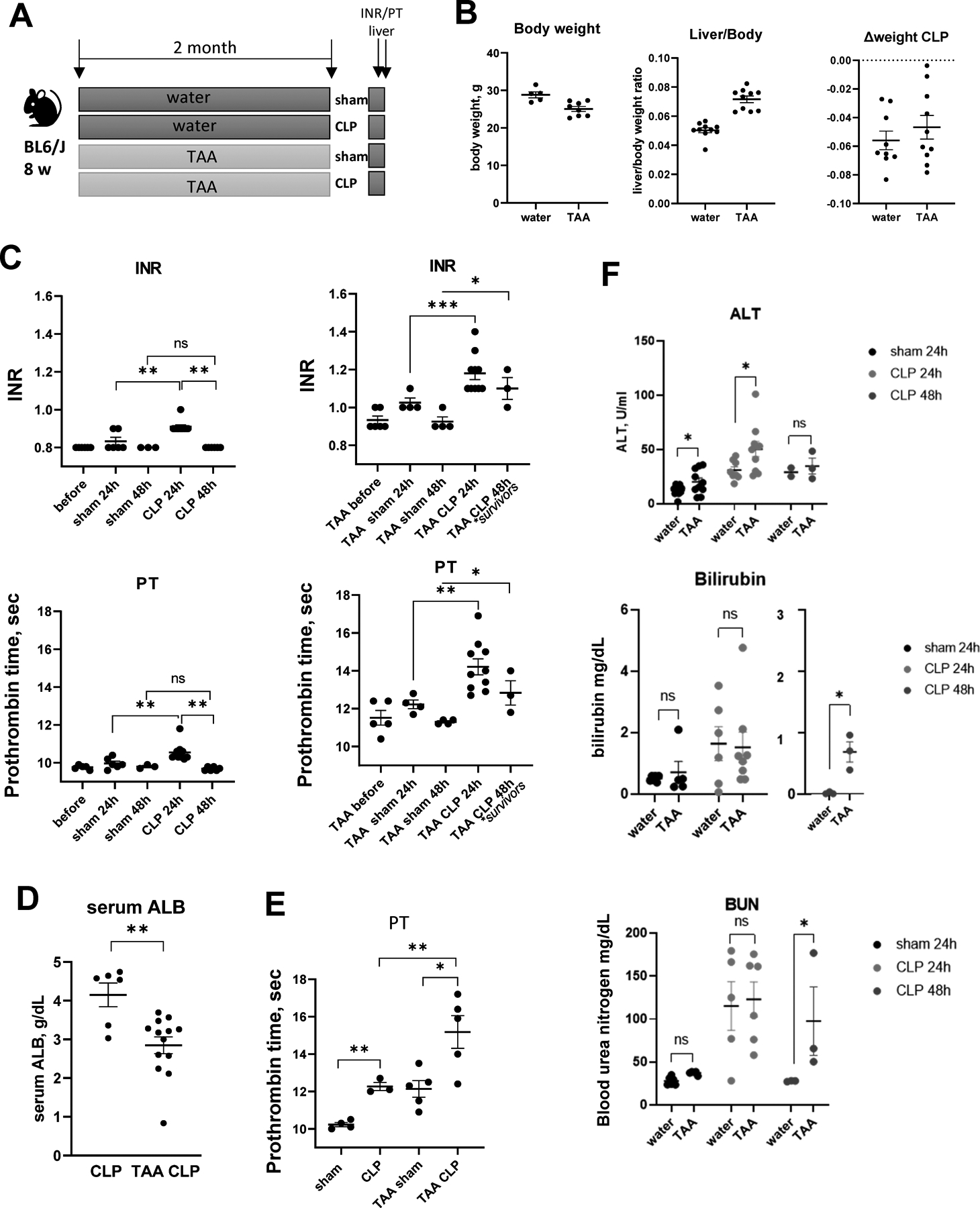
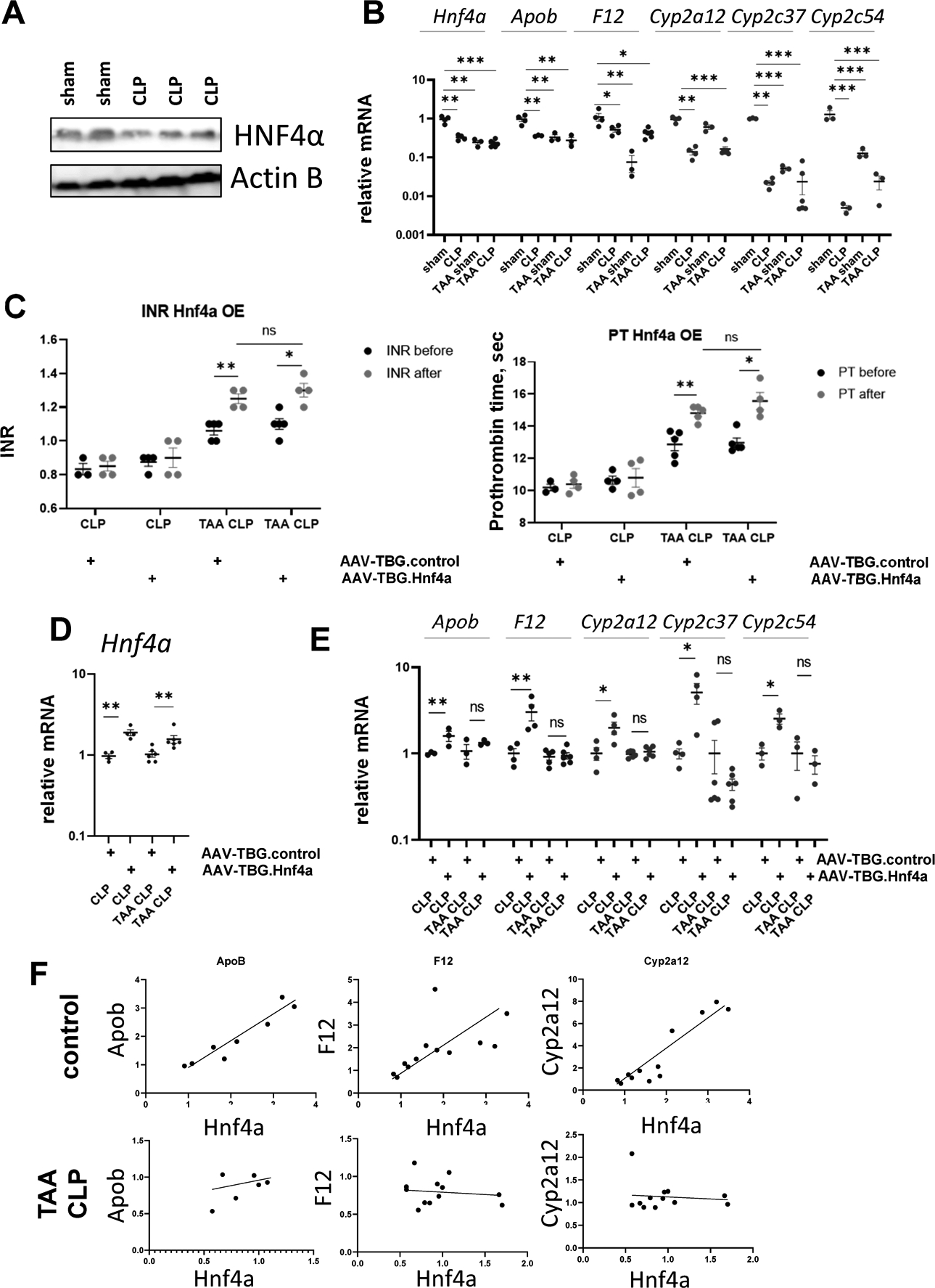
Background and aims: Acute-on-chronic liver failure (ACLF) is an acute liver and multisystem failure in patients with previously stable cirrhosis. A common cause of ACLF is sepsis secondary to bacterial infection. Sepsis-associated ACLF involves a loss of differentiated liver function in the absence of direct liver injury, and its mechanism is unknown. We aimed to study the mechanism of sepsis-associated ACLF using a novel mouse model.

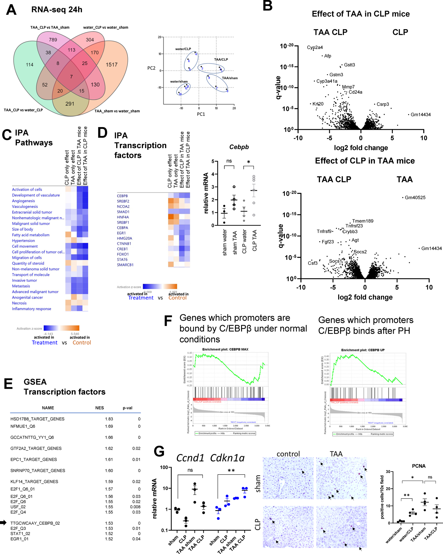
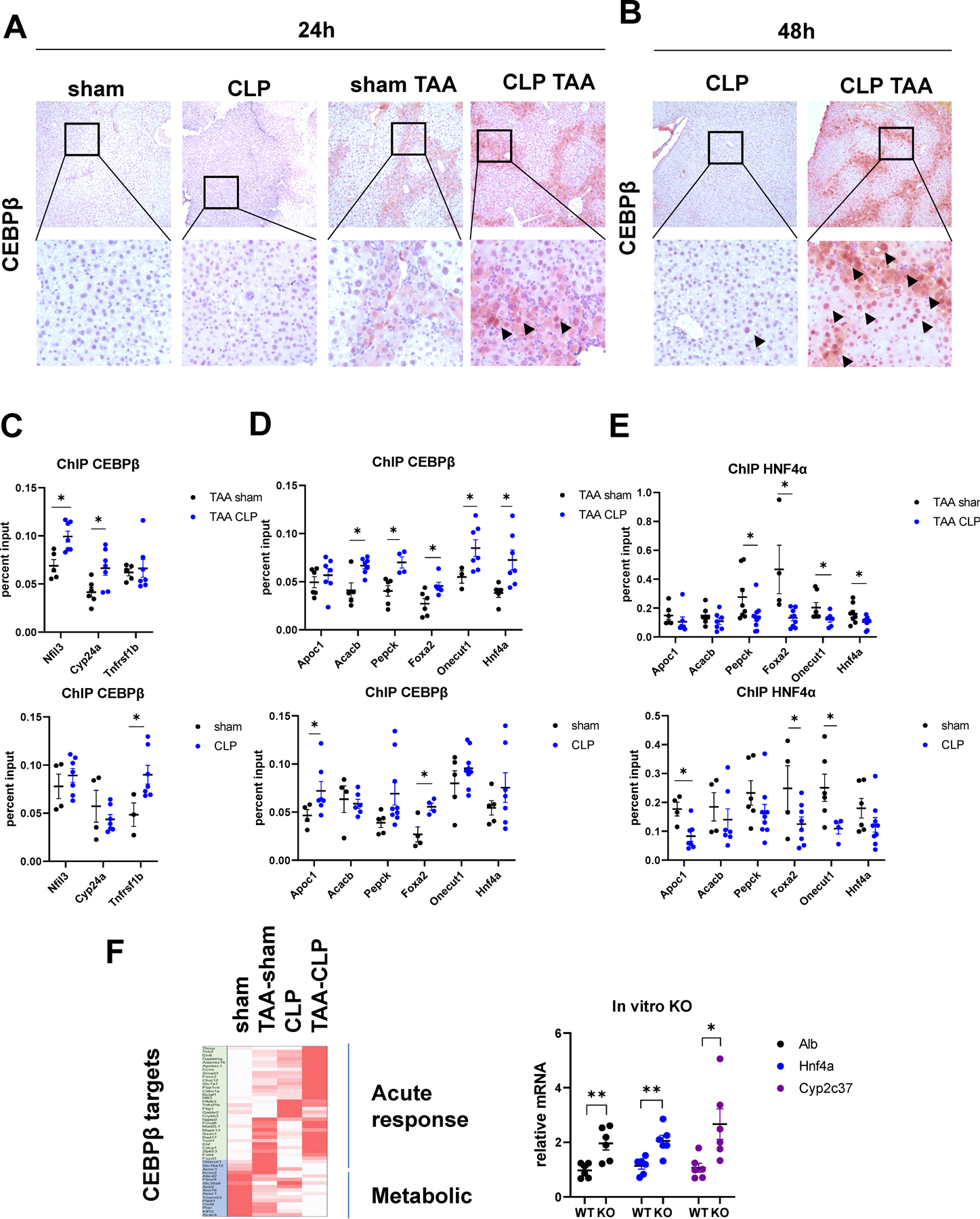
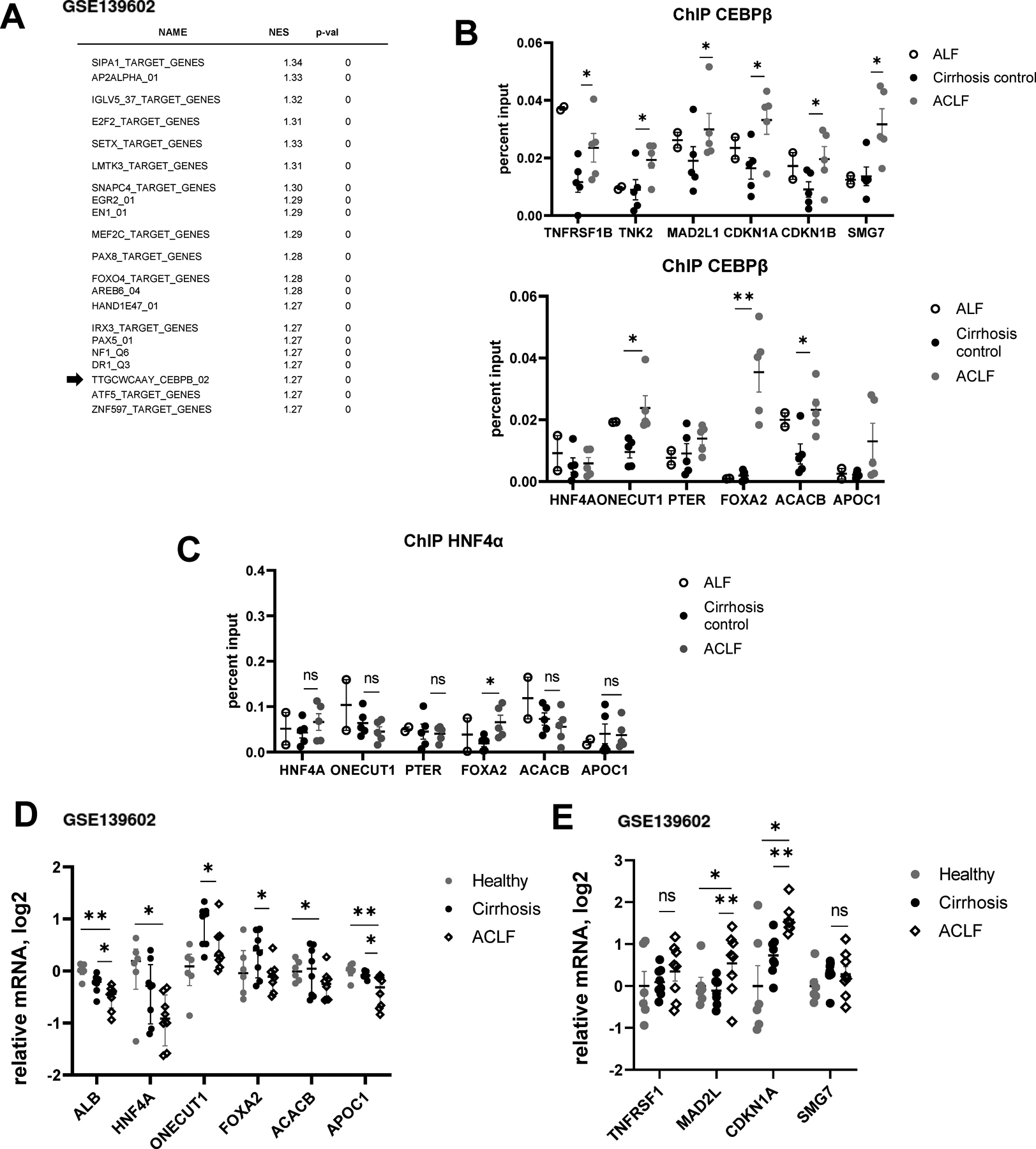
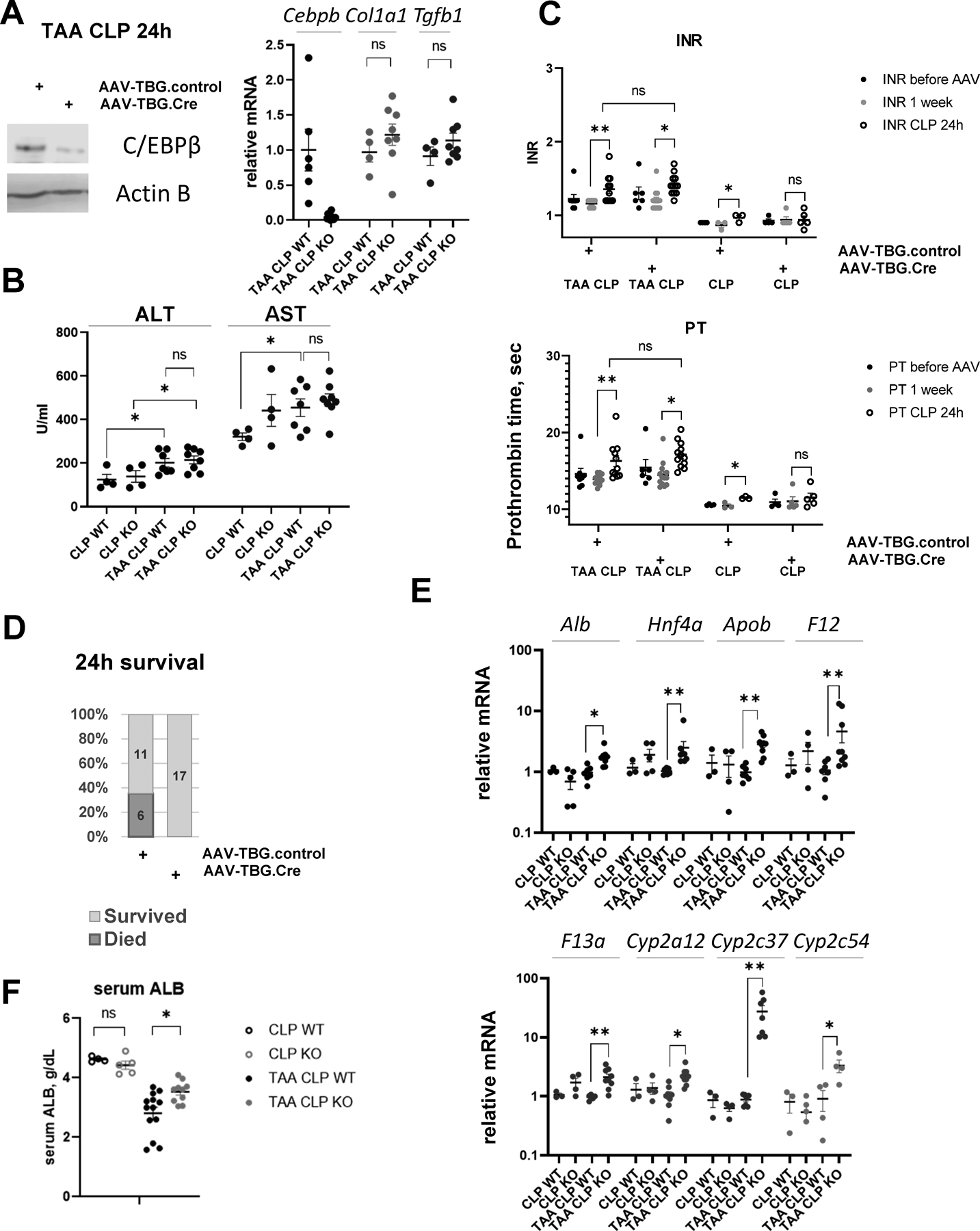
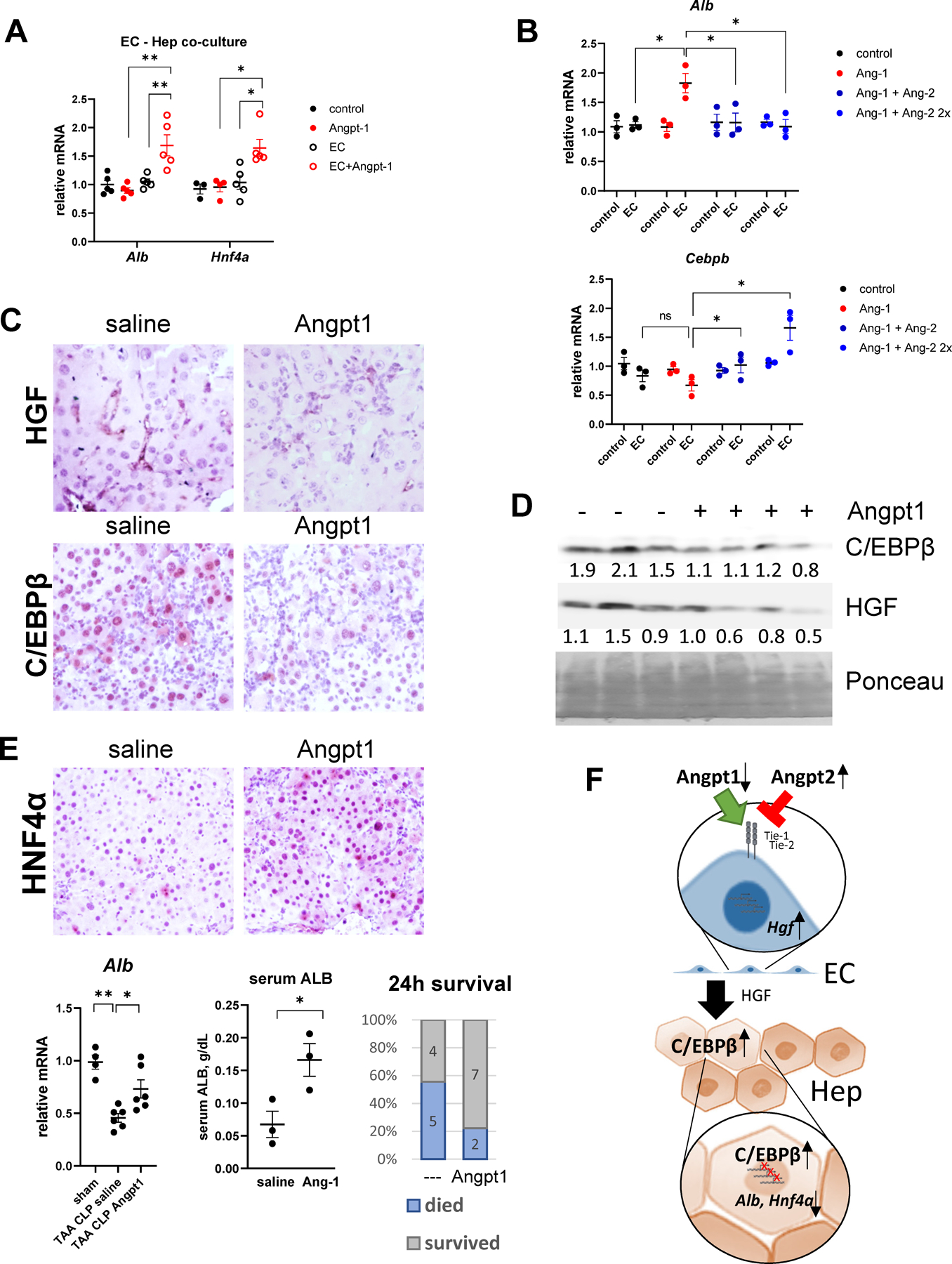
Approach and results: Sepsis-associated ACLF was induced by cecal ligation and puncture procedure (CLP) in mice treated with thioacetamide (TAA). The combination of TAA and CLP resulted in a significant decrease in liver synthetic function and high mortality. These changes were associated with reduced metabolic gene expression and increased CCAAT enhancer binding protein beta (C/EBPβ) transcriptional activity. We found that C/EBPβ binding to its target gene promoters was increased. In humans, C/EBPβ chromatin binding was similarly increased in the ACLF group compared with control cirrhosis. Hepatocyte-specific Cebpb knockout mice had reduced mortality and increased gene expression of hepatocyte differentiation markers in TAA/CLP mice, suggesting that C/EBPβ promotes liver failure in these mice. C/EBPβ activation was associated with endothelial dysfunction, characterized by reduced Angiopoietin-1/Angiopoietin-2 ratio and increased endothelial production of HGF. Angiopoietin-1 supplementation or Hgf knockdown reduced hepatocyte C/EBPβ accumulation, restored liver function, and reduced mortality, suggesting that endothelial dysfunction induced by sepsis drives ACLF through HGF-C/EBPβ pathway.

Conclusions: The transcription factor C/EBPβ is activated in both mouse and human ACLF and is a potential therapeutic target to prevent liver failure in patients with sepsis and cirrhosis.


Conflict of interest statement

Conflict of Interest statement: The authors declare that they have no conflicts of interest with the contents of this article.

Figures

Figure 1.
Figure 1.. TAA and CLP combination results in rapid and sustained loss of liver function.
Mice (male and female) were treated with TAA (200mg/L in the drinking water) and subjected to cecal ligation and puncture (CLP) or sham surgery. A. Treatment groups. B. Liver/body weight ratios and weight loss after CLP surgery. C-E. INR, prothrombin time (PT) (C) in male mice and serum albumin (D) and prothrombin time (PT) in female mice (E) as indicated. F. Serum ALT (top), Bilirubin (middle) and blood urea (BUN, bottom) in sham and CLP mice as indicated. *, P<0.05, **, P<0.01.
Figure 2.
Figure 2.. HNF4α is inactivated in TAA/CLP mice.
A. HNF4α protein levels in whole liver lysates from mice that were subjected to CLP or sham surgeries. B. HNF4α target gene expression in four groups of mice. C-E. Mice (male and female) were treated with TAA (200mg/L in the drinking water) or received water only and subjected to cecal ligation and puncture (CLP). One week before surgeries mice were treated with AAV.TBG.control or AAV.TBG.Hnf4a vectors at 10^11 gc/mouse. C. INR and PT in these mice one day before or one day after CLP. D-E. Relative gene expression in these mice. F. Correlation between gene expression of Hnf4a and HNF4α target genes in control (water/sham+water/CLP) groups and TAA/CLP group. *, P<0.05, **, P<0.01, ***, P<0.001.
Figure 3.
Figure 3.. TAA and CLP combination results in unique transcriptional changes.
Mice (male and female) were treated with TAA and subjected to cecal ligation and puncture or sham surgery. Whole liver mRNA from 4 groups (3 mice per group) at 24h post-CLP was analyzed using RNA-seq. A. Number of differentially expressed genes in pairwise comparisons. Right. Principal component analysis. B. Volcano plots comparing TAA/CLP combination to single treatments. C-D. Ingenuity pathway analysis for each of the pairwise comparisons. Pathway and transcription regulators that are activated in treatment group (blue) and in control group (orange). Right. Cebpb gene expression in four groups of mice. *, P<0.05. E. Gene set enrichment analysis. Top transcription factors predicted to be activated in TAA/CLP group compared to all other groups. F. C/EBPβ target genes enrichment in genes activated in TAA/CLP group compared to the rest. G. Cell cycle and proliferation associated gene expression in four groups of mice. *, P<0.05, **, P<0.01. Middle. PCNA staining in four groups. Right. Average number of PCNA potive cells per 10x field. *, P<0.05, **, P<0.01.
Figure 4.
Figure 4.. TAA and CLP combination results in C/EBPβ activation.
A-B. Mice (male and female) were treated with TAA and subjected to cecal ligation and puncture or sham surgery. Liver section from indicated groups of mice at 24 hours (A) and 48 hours (B) post-surgery were stained using C/EBPβ specific antibodies (total C/EBPβ). C-E Chromatin immunoprecipitation using C/EBPβ specific antibodies (total C/EBPβ) or HNF4α specific antibodies in four groups of mice. Data is presented as percent input. N= 4–8 mice per group. *, P<0.05. F. Relative gene expression changes in TAA, CLP and TAA/CLP mice of top genes bound by C/EBPβ at 3 hours after partial hepatectomy. Right. Relative gene expression in mouse primary hepatocytes isolated from Cebpb floxed mice treated with Ad-Cre vector or control for 48 hours. *, P<0.05, **, P<0.01.
Figure 5.
Figure 5.. C/EBPβ promoter binding is increased in human ACLF compared to control cirrhosis.
A. Gene set enrichment analysis. Top transcription factors predicted to be activated in ACLF group compared to healthy controls using published dataset GSE139602. B-C. Chromatin immunoprecipitation using C/EBPβ specific antibodies (total C/EBPβ) or HNF4α specific antibodies. Data are presented as percent input. N= 5 (ACLF and cirrhosis) N=2 (ALF). *, P<0.05, **, P<0.01. D-E. C/EBPβ target gene expression in ACLF, cirrhosis and healthy controls (GSE139602). *, P<0.05, **, P<0.01.
Figure 6.
Figure 6.. Hepatocyte specific Cebpb knockout protects from mortality and loss of liver function in TAA/CLP mice.
Cebpb floxed mice received AAV-TBG.CRE (KO) or AAV.TBG.control (WT) at 10^11 gc/mouse 1 week before surgeries. A. C/EBPβ protein levels in hepatocyte specific knockout mice. Right. Relative gene expression at one day after CLP. B. Serum ALT and AST in wild type (WT) and knockout mice (KO). C. INR and PT in mice before AAV injection, one day before CLP and one day after CLP. *, P<0.05, **, P<0.01. D. Survival in TAA/CLP mice. N=17 per group. E-F. Whole liver mRNA in these mice *, P<0.05, **, P<0.01.
Figure 7.
Figure 7.. TAA and CLP combination causes endothelial cell dysfunction and HGF upregulation.
A. Serum samples from TAA/CLP and CLP mice (pooled from N=3 mice each) were used for cytokine array. Relative abundance of cytokines and circulating molecules in two groups. B. Liver section from indicated groups of mice at 24 hours post-CLP were stained using HGFα specific antibodies. C-D. Mice were treated with AAV.shScrambled or AAV.shHgf at 10^11 gc/mouse one week before CLP. C/EBPβ expression and relative gene expression in the livers of TAA/CLP mice 24 hours after CLP. E. Left. Gene set enrichment analysis. Top pathways predicted to be activated in TAA/CLP group compared to all other groups. Right. Angiogenesis genes enrichment in genes activated in TAA/CLP group compared to the rest. F. CD146 positive liver endothelial cells were isolated from control and TAA treated mice or treated with 100ng/ml of LPS. Relative gene expression. N=4 mice per group. G. LSECs were treated with recombinant Angiopoietin-1 (10 ng/ml) in the presence or absence of LPS for 48 hours. N=3–5. *, P<0.05, **, P<0.01. Unpaired t-test. LPS vs LPS Ang-1:Hgf P=0.006, Icam1 P=0.032, Stab2 P=0.045. H. LSECs were treated with recombinant Angiopoietin-1 (10 ng/ml) and Angiopoietin-2 (10 and 20 ng/ml). N=3 independent experiments. *, P<0.05 using paired ttest. Ang-1 vs control: Hgf P=0.040, Vcam1 P=0.048, Stab2 P=0.0096. Ang2+Ang1 vs Ang-1 Hgf P=0.049, Icam P=0.0002, Vcam1 P=0.009, Stab2 P=0.0079.
Figure 8
Figure 8. Angiopoietin-1 supplementation prevents C/EBPβ activation in hepatocytes.
A. Primary mouse hepatocytes alone or in co-culture with LSECs were treated with recombinant Angiopoietin-1 (10 ng/ml). Relative gene expression. N=3–4 per group. *, P<0.05, **, P<0.01. B. Primary mouse hepatocytes alone or in co-culture with LSECs were treated with recombinant Angiopoietin-1 (10 ng/ml) and Angiopoietin-2 (10 and 20 ng/ml). Relative gene expression. N=3 independent experiments. *, P<0.05 using paired ttest. C-E. TAA/CLP mice received 5 μg of recombinant Angiopoietin-1 or saline control at 2 hours after CLP. C. Liver section from indicated groups of mice at 24 hours post-CLP were stained using HGFα or C/EBPβ specific antibodies (total C/EBPβ). D. Western blot analysis of C/EBPβ and HGF protein levels in the livers. D. Whole liver mRNA gene expression and mortality in TAA/CLP mice. E. Top. Liver section from indicated groups of mice at 24 hours post-CLP were stained using HNF4α specific antibodies. Bottom. Albumin liver mRNA and serum levels and mortality at 24h. F. Proposed model of ACLF pathogenesis.

References

    1. Moreau R, Gao B, Papp M, Bañares R, Kamath PS. Acute-on-chronic liver failure: A distinct clinical syndrome. J Hepatol 2021;75 Suppl 1:S27–s35. - PubMed
    1. Heron M Deaths: Leading Causes for 2019. Natl Vital Stat Rep 2021;70:1–114. - PubMed
    1. Solé C, Solà E. Update on acute-on-chronic liver failure. Gastroenterol Hepatol 2018;41:43–53. - PubMed
    1. Mucke MM, Rumyantseva T, Mucke VT, Schwarzkopf K, Joshi S, Kempf VAJ, Welsch C, et al. Bacterial infection-triggered acute-on-chronic liver failure is associated with increased mortality. Liver Int 2018;38:645–653. - PubMed
    1. Li X, Wang LK, Wang LW, Han XQ, Yang F, Gong ZJ. Blockade of high-mobility group box-1 ameliorates acute on chronic liver failure in rats. Inflamm Res 2013;62:703–709. - PubMed

Publication types