. 2023 Mar 21;11(2):e0528122.
doi: 10.1128/spectrum.05281-22. Online ahead of print.

Broad Host Tropism of Flaviviruses during the Entry Stage

Affiliations

Broad Host Tropism of Flaviviruses during the Entry Stage

Yanan Zhang et al. Microbiol Spectr. .

Abstract

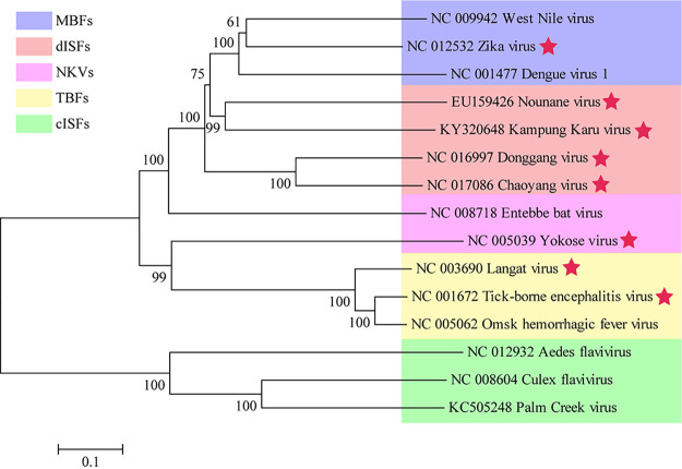
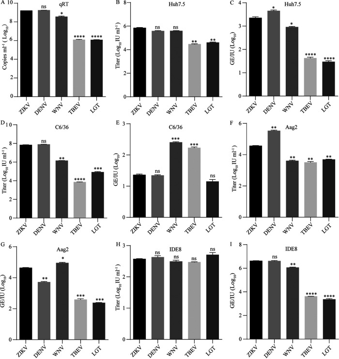
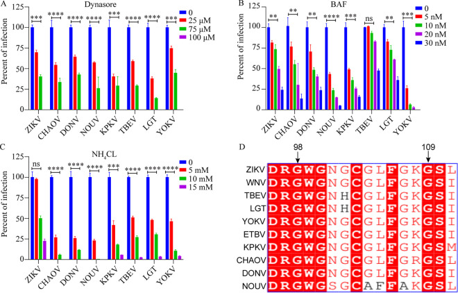
The genus Flavivirus consists of viruses with various hosts, including insect-specific flaviviruses (ISFs), mosquito-borne flaviviruses (MBFs), tick-borne flaviviruses (TBFs), and no-known vector (NKV) flaviviruses. Using the reporter viral particle (RVP) system, we found the efficient entry of ISFs into vertebrate cells, MBFs into tick cells, as well as NKVs and TBFs into mosquito cells with similar entry characteristics. By construction of reverse genetics, we found that Yokose virus (YOKV), an NKV, could enter and replicate in mosquito cells but failed to produce infectious particles. The complete removal of the glycosylation modification on the envelope proteins of flaviviruses had no obvious effect on the entry of all MBFs and TBFs. Our results demonstrate an entry-independent host-tropism mechanism and provide a new insight into the evolution of flaviviruses. IMPORTANCE Vector-borne flaviviruses, such as Zika virus, have extremely broad host and cell tropism, even though no critical entry receptors have yet been identified. Using an RVP system, we found the efficient entry of ISFs, MBFs, TBFs, and NKVs into their nonhost cells with similar characteristics. However, glycan-binding proteins cannot serve as universal entry receptors. Our results demonstrate an entry-independent host-tropism mechanism and give a new insight into the cross-species evolution of flaviviruses.

Keywords: Zika virus; entry independent; flaviviruses; host tropism.


Conflict of interest statement

The authors declare no conflict of interest.

Figures

FIG 1
FIG 1
Maximum-likelihood phylogenetic tree of different groups of flaviviruses. Complete polyprotein amino acid sequences were aligned, and a maximum likelihood phylogenetic tree was reconstructed in MEGA7 (41). The consensus tree inferred based on 500 replicates (42) was taken to represent the evolutionary history of the taxa. Stars indicate viruses tested in Fig. 2 and 4. Purple, mosquito-borne flaviviruses (MBFs); orange, dual-host affiliated insect-specific flaviviruses (dISFs); pink, no known vector flaviviruses (NKVs); yellow, tick-borne flaviviruses (TBFs); and green, classical insect-specific flaviviruses (cISFs).
FIG 2
FIG 2
RVPs of ISF and NKV can infect mosquito and human cells. (A to E) RVPs were produced by cotransfection of CprME-expression plasmid and WNV replicon encoding a GFP reporter in 293T cells. (A) RVP secretion was measured by real-time PCR with primers according to the WNV replicon. RVP titers in the supernatant were gauged on C6/36 (B) or Huh7.5 (D) cells 72 h post-transfection. (C and E) The infectivity of RVPs was further calculated as GE/IU. The data were analyzed by unpaired t test (n = 3). (F to H) cISF RVP was produced by cotransfection in C6/36 cells. (F) cISF RVP secretion was measured by real-time PCR with primers based on the WNV replicon. (G) RVP titer was gauged on Huh7.5 and C6/36 cells. (H) The infectivity of cISF RVP is shown as GE/IU. The data were analyzed by two-tailed unpaired t test (n = 3). Error bars indicate standard deviation (SD). The results show the average and standard deviation of three independent experiments; ns, not significant, P ≥ 0.05; *, P < 0.05; **, P < 0.01, ***; P < 0.001; ****, P < 0.0001.
FIG 3
FIG 3
YOKV can replicate in mosquito cells. (A) Schematic diagram of the YOKV infectious clone. The cDNA of YOKV was cloned into the pACYC177 vector with a CMV promoter and HDV RBZ termination sequence. (B) Viral titers in the supernatant were determined at the indicated time points by a focus-forming assay. YOKV titers peaked at 72 h post-infection at around 106 FFU/mL (n = 3). (C) Virus replication was detected by real-time PCR in transfected cells at indicated temperatures. The replication-deficient GAA mutations were negative controls (n = 3). Error bars indicate standard deviation (SD). (D) Immunofluorescence of viral double-stranded RNA by anti-dsRNA mAb and envelop protein E by 4G2 mAb in transfected vertebrate cells and insect cells under various temperatures. ND, not detected. The nuclei were stained with Hoechst 33342; scale bar, 10 μm. GAA is a replication-deficient mutation. These results show the average and standard deviation of three independent experiments. The data were analyzed by two-tailed multiple t test; ns, not significant, P ≥ 0.05; *, P < 0.05; **, P < 0.01; ***, P < 0.001; ****, P < 0.0001.
FIG 4
FIG 4
Cross-species barrier of YOKV occurs post-entry. (A) Virus replication was detected by real-time PCR in infected cells at different temperatures. Cells were infected at an MOI of 0.5 and cultured at the indicated temperatures. Viral RNAs were tested by real-time PCR 72 h postinfection. Error bars indicate standard deviation (SD). (B) Immunofluorescence of viral double-stranded RNA by anti-dsRNA mAb and envelop protein E by 4G2 mAb in infected vertebrate cells and insect cells under indicated temperatures. ND, not detection. The nuclei were stained with Hoechst 33342; scale bar, 10 μm; n = 3. These results show the average and standard deviation of three independent experiments. The data were analyzed by two-tailed multiple t test; ns, not significant, P ≥ 0.05; *, P < 0.05; **, P < 0.01; ***, P < 0.001; ****, P < 0.0001.
FIG 5
FIG 5
RVPs of TBFs can infect mosquito cells and MBFs are able to infect tick cells. RVPs of TBFs (TBEV and LGTV) and MBFs (ZIKV, DENV, and WNV) were produced as above. (A) RVP secretion was measured by real-time PCR with primers based on the WNV replicon. RVP titers in the supernatant were gauged on Huh7.5 (B), C6/36 (D), Aag2 (F) and IDE8 (H) cells at 72 h posttransfection. (C, E, G, and I) The infectivity of TBFs and MBFs is shown as GE/IU (n = 3). Error bars indicate standard deviation (SD). The results show the average and standard deviation of three independent experiments. The data were analyzed by two-tailed unpaired t test; ns, not significant, P ≥ 0.05; *, P < 0.05; **, P < 0.01; ***, P < 0.001; ****, P < 0.0001.
FIG 6
FIG 6
ISFs, NKVs, and MBFs are similar in the endocytosis and membrane fusion. Huh7.5 cells were pretreated with indicated concentrations of dynasore (A), bafilomycin A (BAF) (B), or NH4Cl (C). Then, 1 h later, indicated RVPs were added to Huh7.5 cells. The infectivity of all RVPs was inhibited by the compounds in a similar pattern. (D) Alignment of the fusion loop of dISFs, NKVs, TBFs, and MBFs (n = 3). Error bars indicate standard deviation (SD). The results show the average and standard deviation of three independent experiments. The data were analyzed by two-tailed multiple t test; ns, not significant, P ≥ 0.05; **, P < 0.01; ***, P < 0.001; ****, P < 0.0001.
FIG 7
FIG 7
N-glycosylation of flavivirus envelope proteins is not responsible for virus entry. (A) Alignment analysis of the N154 (according to the amino acid sequence of the E protein of ZIKV MR766 NIID strain) glycosylation site of flaviviruses. (B) The removal of the N-glycosylation site was confirmed by Western blot with an anti-E antibody. The mutant migrated faster than the WT. (C) The maturation activity of overexpressed furin was confirmed with a polyclonal antibody against ZIKV prM. prM was detected on ZIKV RVPs produced by naive 293T cells but not by cells with furin overexpression. WT and glycosylation-negative mutant RVPs were produced in naive 293T cells and 293T cells overexpressing a furin protease. RVP titers were gauged on Huh7.5, C6/36, and Aag2 cells. (D) RVP secretion was measured by real-time PCR with primers based on the WNV replicon. (E, G, and I) RVP titers in the supernatant were gauged on Huh7.5 (E), C6/36 (G), and Aag2 (I) cells 72 h post-transfection. (F, H, and J) The infectivity of TBFs and MBFs is shown as GE/IU (n = 3). Error bars indicate standard deviation (SD). The results show the average and standard deviation of three independent experiments. The data were analyzed by two-tailed multiple t test; ns, not significant, P ≥ 0.05; *, P < 0.05; **, P < 0.01; ***, P < 0.001; ****, P < 0.0001.

Similar articles

Cited by

References

    1. Blitvich BJ, Firth AE. 2015. Insect-specific flaviviruses: a systematic review of their discovery, host range, mode of transmission, superinfection exclusion potential and genomic organization. Viruses 7:1927–1959. doi:10.3390/v7041927. - DOI - PMC - PubMed
    1. Blitvich BJ, Firth AE. 2017. A review of flaviviruses that have no known arthropod vector. Viruses 9:v9060154. doi:10.3390/v9060154. - DOI - PMC - PubMed
    1. Gould EA, Solomon T. 2008. Pathogenic flaviviruses. Lancet 371:500–509. doi:10.1016/S0140-6736(08)60238-X. - DOI - PubMed
    1. Guzman H, Contreras-Gutierrez MA, Travassos da Rosa APA, Nunes MRT, Cardoso JF, Popov VL, Young KI, Savit C, Wood TG, Widen SG, Watts DM, Hanley KA, Perera D, Fish D, Vasilakis N, Tesh RB. 2018. Characterization of three new insect-specific flaviviruses: their relationship to the mosquito-borne flavivirus pathogens. Am J Trop Med Hyg 98:410–419. doi:10.4269/ajtmh.17-0350. - DOI - PMC - PubMed
    1. Lindenbach BD, Thiel H-J, Rice CM. 2007. Flaviviruses: the viruses and their replication, p 1101–1152. In Knipe DM, Howley PM (ed), Fields' virologv, 5th ed. Lippincott, Williams and Wilkins, Philadelphia, PA.

LinkOut - more resources