Review
. 2023 Aug;39(4-6):351-373.
doi: 10.1089/ars.2022.0123. Epub 2023 May 9.

Surviving and Adapting to Stress: Translational Control and the Integrated Stress Response

Affiliations
Review

Surviving and Adapting to Stress: Translational Control and the Integrated Stress Response

Ronald C Wek et al. Antioxid Redox Signal. 2023 Aug.

Abstract

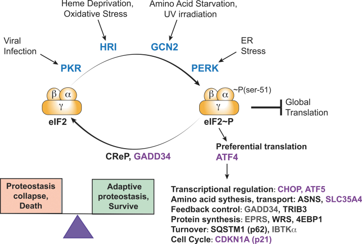
Significance: Organisms adapt to changing environments by engaging cellular stress response pathways that serve to restore proteostasis and enhance survival. A primary adaptive mechanism is the integrated stress response (ISR), which features phosphorylation of the α subunit of eukaryotic translation initiation factor 2 (eIF2). Four eIF2α kinases respond to different stresses, enabling cells to rapidly control translation to optimize management of resources and reprogram gene expression for stress adaptation. Phosphorylation of eIF2 blocks its guanine nucleotide exchange factor, eIF2B, thus lowering the levels of eIF2 bound to GTP that is required to deliver initiator transfer RNA (tRNA) to ribosomes. While bulk messenger RNA (mRNA) translation can be sharply lowered by heightened phosphorylation of eIF2α, there are other gene transcripts whose translation is unchanged or preferentially translated. Among the preferentially translated genes is ATF4, which directs transcription of adaptive genes in the ISR. Recent Advances and Critical Issues: This review focuses on how eIF2α kinases function as first responders of stress, the mechanisms by which eIF2α phosphorylation and other stress signals regulate the exchange activity of eIF2B, and the processes by which the ISR triggers differential mRNA translation. To illustrate the synergy between stress pathways, we describe the mechanisms and functional significance of communication between the ISR and another key regulator of translation, mammalian/mechanistic target of rapamycin complex 1 (mTORC1), during acute and chronic amino acid insufficiency. Finally, we discuss the pathological conditions that stem from aberrant regulation of the ISR, as well as therapeutic strategies targeting the ISR to alleviate disease. Future Directions: Important topics for future ISR research are strategies for modulating this stress pathway in disease conditions and drug development, molecular processes for differential translation and the coordinate regulation of GCN2 and other stress pathways during physiological and pathological conditions. Antioxid. Redox Signal. 39, 351-373.

Keywords: eIF2; eIF2 kinases; integrated stress response; translation initiation; translational control.


Conflict of interest statement

R.C.W. is a member of the advisory board in HiberCell, Inc. K.A.S. and T.G.A. are consultants for HiberCell, Inc., and K.A.S. receives research support from HiberCell, Inc.

Figures

FIG. 1.
FIG. 1.
p-eIF2α triggers global and gene specific translation during the ISR. Four protein kinases phosphorylate the α subunit of eIF2 at serine-51 in response to different stress conditions. Enhanced levels of p-eIF2α inhibit the GEF eIF2B, lowering eIF2•GTP levels and global translation. The constitutive expressed CReP and ISR-induced GADD34 combine with PP1 to dephosphorylate p-eIF2α, ensuring that the levels of p-eIF2α are appropriate for the stress condition. Enhanced levels of p-eIF2α also elicits preferential translation of select mRNAs, including that encoding transcriptional regulator ATF4. Increased levels of ATF4 induce the transcriptional expression of a collection of ISR genes, including those encoding additional transcription factors (CHOP and ATF5), amino acid synthesis and transport [ASNS (Chen et al., 2004) and SLC35A4 (Andreev et al., 2015)], feedback control [GADD34 and TRIB3 (Jousse et al., ; Nikonorova et al., ; Ohoka et al., 2005)], protein synthesis [EPRS and WRS (Baird et al., ; Harding et al., ; Young et al., 2016a) and 4EBP1], protein turnover [IBTKα (Baird et al., ; Willy et al., 2017) and SQSTM1 (p62) (B'chir et al., 2013)], and cell cycle [CDK1A (p21) (Collier et al., ; Lehman et al., 2015)]. Many of the ISR genes that are transcriptionally induced by ATF4 are also preferentially translated during enhanced p-eIF2α (indicated in purple), ensuring their enhanced expression during the ISR. The ISR is critical for maintenance of proteostasis during stress, ensuring survival. However, during chronic induction of the ISR can instead be maladaptive, which can lead to proteostatic collapse and cell death. Hence, the magnitude and duration of the ISR can determine the balance between cell survival and death. 4EBP1, 4E binding protein 1; ASNS, asparagine synthetase; ATF, activating transcription factor; CReP, constitutive repressor of eIF2α phosphorylation; eIF, eukaryotic initiation factor; GADD34, growth arrest and DNA damage-inducible gene 34; GEF, guanine nucleotide exchange factor; ISR, integrated stress response; mRNA, messenger RNA; p-eIF2α, phosphorylation of eIF2α; PP1, protein phosphatase 1; WRS, Wolcott-Rallison syndrome.
FIG. 2.
FIG. 2.
The eIF2α kinases monitor stress arrangements via different regulatory regions juxtaposed to their kinase catalytic domains. Each of the eIF2α kinases shares a related protein kinase domain (orange bars) that is juxtaposed to distinct regulatory and targeting regions that function to monitor ligands that are modulated by different stress conditions (green bars). The size of the eIF2α kinase domains varies among the family members due to the different lengths of insert sequences in the N-terminal lobe of the kinase domains. Regulatory regions include two dsRBMs in PKR and heme-binding regions in its N-terminus and kinase insert of HRI. PERK has a SS for entry into the ER, an IRE1-related region, and an ER TM segment. GCN2 regulatory regions include the RWD, partial kinase, HARS, and CTD that facilitates oligomerization and contributes to the eIF2α kinase association with ribosomes. For a review of the structures of eIF2α kinase catalytic and regulatory domains, see Rothenburg et al. (2016). CTD, carboxy terminal domain; dsRBM, double-stranded RNA-binding motif; ER, endoplasmic reticulum; GCN, general control nonderepressible; HARS, histidyl-tRNA synthetase; HRI, heme regulated inhibitor; IRE1, inositol requiring enzyme 1; PERK, PKR-like ER kinase; PKR, protein kinase RNA-activated; RNA, ribonucleic acid; SS, signal sequence; TM, transmembrane; tRNA, transfer RNA.
FIG. 3.
FIG. 3.
Scheme depicting the function and regulation of 2B GEF activity. (A) During the process of translation initiation, GTP bound to eIF2 is hydrolyzed to GDP, and the GEF eIF2B functions to recycle eIF2•GDP to eIF2•GTP to facilitate Met-tRNAiMet binding to ribosomes for further rounds of translation initiation. The GAP and GDI functions of bound eIF5 are also depicted and show that eIF2B must displace eIF5 to facilitate nucleotide exchange and form the new TC. p-eIF2α converts it from a substrate to an inhibitor of eIF2B exchange activity. eIF2B exchange activity is inhibited by both p-eIF2α and by direct phosphorylation by GSK-3 as indicated in red. The metabolite F6P acts as an allosteric activator of eIF2B GEF activity indicated by the green arrow. ISRIB and 2BAct (eIF2B activator) function as inhibitors to block p-eIF2 binding to eIF2B, rendering the GEF insensitive to its effects also indicated in red. F6P, fructose-6-phosphate; GAP, GTPase activator protein; GCP, guanosine diphosphate; GDI, guanine nucleotide dissociation inhibitor; GSK-3, glycogen synthase kinase 3; GTP, guanosine trisphosphate; ISRIB, ISR inhibitor; Met-tRNAiMet, methionyl initiator tRNA; TC, ternary complex.
FIG. 4.
FIG. 4.
The uORFs function in the bar code for translational control in the ISR. The mRNAs are indicated as a solid line with the 5′-cap. The CDS for each transcript are indicated as a blue bar, with uORFs that allow for reinitiation (green) and repressing uORFs (red) indicated by bars. The small and large ribosomal subunits (ovals) are shown to scan, elongate, and terminate and release during the processes of uORF-directed translational control. During translation initiation, the eIF2 is released from ribosomes (light gray oval) and reinitiating ribosomes resume scanning and reacquire TC (dark oval). Gene transcripts that have the indicated uORF configurations as listed and are further described in detail in the text. CDS, downstream coding sequence; uORF, upstream open reading frame.
FIG. 5.
FIG. 5.
Delayed reinitiation mechanism for preferential translation of ATF4 mRNA in response to p-eIF2α during stress. In absence of stress there are low levels of p-eIF2α and ample eIF2•GTP for delivery of initiator tRNA to ribosomes. Ribosomes (ovals represents large and small subunits) initiate translation at the 5′-proximal uORF1 (green bar), and following termination rapidly reacquire new TC and reinitiate at the inhibitory uORF2 (red bar). The uORF2 extends out-of-frame into the ATF4 CDS (blue bar) and after translation termination of the inhibitory uORF, there are low amounts of ATF4 and its target genes expressed in the absence of stress. An abbreviated uORF consisting of only start-stop codons can be situated upstream of uORF1, and the shortened uORF is suggested to also allow for ribosome reinitiation. During stress, induced p-eIF2α and lowered eIF2 TC is suggested to delay ribosome reinitiation after translation of uORF1, enabling 40S ribosomes (light gray oval) to scan through uORF2 before reacquiring a new eIF2 TC (dark gray oval). The translation competent 40S ribosome can instead initiate at the ATF4 CDS and the resulting increased synthesis of ATF4 protein directly enhances transcriptional expression of ISR-targeted genes.
FIG. 6.
FIG. 6.
Preferential translation of GADD34 occurs by a bypass mechanism while CReP translation is constitutive. The GADD34 and CReP mRNA encode targeting subunits that facilitates protein phosphatase 1 dephosphorylation of p-eIF2α. As illustrated in this figure, both gene transcripts contain two uORFs, with GADD34 being preferentially translated by ribosomes (ovals represents large and small subunits) in response to induced p-eIF2α, whereas translation of CrEP occurs independent of p-eIF2α status. (A) The GADD34 mRNA contains two upstream uORFs that overlap in part and are out-of-frame. The start codon context is shown for the uORF2. The uORF1 (gray bar) is nominally translated, and the inhibitory uORF2 (red bar) can be bypassed in response to induced p-eIF2α during stress. In the absence of stress and low p-eIF2α levels, there is abundant eIF2 TC and scanning ribosomes (dark gray oval) readily initiate translation at uORF2. During the initiation phase of translation, the eIF2 is released from ribosomes (light gray oval). Elongating ribosomes translate the uORF2, and the encoded Pro-Pro-Gly-Stop is suggested to lead to inefficient translation termination, facilitating release of ribosomes from the mRNA and lowering ribosome reinitiation at the downstream GADD34 CDS. During stress, induced p-eIF2α and the resulting lowered TC are suggested to allow for a portion of the scanning ribosomes to proceed through the uORF2 and instead initiate translation at the downstream CDS (blue bar). ISR-directed transcription of the GADD34 gene, along with preferential translation, potently increases GADD34 protein that facilitates feedback dephosphorylation of p-eIF2α, allowing for resumption of bulk translation. (B) The CReP transcript also has two uORFs, with only uORF2 (red bar) being well translated. However, uORF2 allows for ribosomes to resume scanning and reacquire TC (dark oval), enabling reinitiation at the downstream CReP CDS. Therefore, CReP is well translated independent of stress and serves to help maintain appropriate p-eIF2α levels during both nonstressed and stressed conditions.
FIG. 7.
FIG. 7.
Reciprocal coregulation of GCN2 and mTORC1 during amino acid insufficiency affects translational and transcriptional expression. (Upper green panel) During conditions of amino acid insufficiency, such as leukine deprivation or asparaginase exposure, there is reciprocal regulation of GCN2 and mTORC1 and their corresponding phosphorylation of target proteins. With wild-type GCN2 (GCN2 WT), p-eIF2α is increased alongside reduced mTORC1 phosphorylation of 4EBP1 (p-4EBP1) and S6K1 (p-S6K1), slowing translation initiation at the eIF2B and eIF4F steps. Diminished GEF activity of eIF2B lowers bulk protein synthesis and promotes preferential translation of ATF4 and other ISR target genes. Reduced S6K1 activity suppresses translation of gene transcripts with TOP tracts. Together, these changes slow anabolic capacity, facilitating proteostasis recovery. (Upper red panel) In cells or tissues lacking GCN2 (GCN2 KO), the activities of eIF2B, eIF4F, and S6K1 continue, resulting in a proteostasis mismatch, with sustained anabolic capacity in a time of nutrient scarcity. (Middle green panel) Maintenance of adaptive proteostasis during chronic amino acid insufficiency promotes ATF4 synthesis, which executes targeted changes to the transcriptome to resolve the nutrient stress and limit ISR activation (e.g., ASNS, GADD34). Other ATF4 target genes (e.g., SESTRIN2, REDD1) function to repress mTORC1, increasing protein breakdown and reducing anabolism. (Lower red panel) A disrupted GCN2-ATF4 axis during prolonged amino acid insufficiency alters or amplifies ISR target gene execution, which fails to appropriately trigger mTORC1 repression, leading to discordant ISR and mTORC1 regulation of protein synthesis, promoting ER stress and cell injury and premature death. KO, knockout; mTORC1, mammalian/mechanistic target of rapamycin complex 1; REDD1, regulated in development and DNA damage response 1; S6K1, ribosomal protein S6 kinase; TOP, 5′-terminal oligopyrimidine.
FIG. 8.
FIG. 8.
A dynamic range of the duration and magnitude of p-eIF2α and translational control ensures successful adaptation to stress. The ISR features stress-activation of eIF2α kinase(s), which induces bulk translation repression and attendant ISR gene expression that helps to ameliorate the stress, followed by feedback dephosphorylation of p-eIF2α by GADD34-directed PP1 and resumption of protein synthesis (A) The schematic illustrates the reciprocal regulation between increasing p-eIF2α (in black) and decreasing levels of mRNA translation (in blue) that are featured in the ISR. The adaptive zone (yellow) represents the dynamic range of the translational control response during successful adaptation to stress. Cells encounter stress, triggering p-eIF2α, which signals an adaptive response that alleviates cell damage and mitigates the stress. As indicated, the successful adaptation to stress by ISR largely functions in the adaptive zone. However, in situations of chronic stress or with genetic changes that dysregulate the ISR, there can be hypo- or hyperphosphorylation of eIF2α (orange to dark red zones), and these extremes in translational control can trigger cell damage and death. (B) In the example of hypoinduction of the ISR, mutations in PERK and GCN2 can sharply reduce basal and stress-induced p-eIF2α and attendant control of mRNA translation, shifting the dynamic range of the ISR into the danger zone, as illustrated by the box shifting to the left in the illustration. The consequences of loss of PERK and GCN2 functions are WRS and PVOD, respectively. (C) Genetic changes can also lead to hyperinduction of the ISR and disease, as described more fully in the text. For example, mutations in CReP reduce PP1 dephosphorylation of p-eIF2α, leading to constitutive induction of the ISR that is further activated on stress-induction of an eIF2α kinase. The hyperinduction of p-eIF2α and the ISR shifts the dynamic range of the ISR into the danger zone, as illustrated by the box shifting to the right in the illustration. Likewise, certain mutations in aminoacyl tRNA synthetase genes and BDKDK-deficiency lead to hyperinduction of p-eIF2α, which shifts the ISR responses to the danger zone. In the case of mutations in EIF2S3 (eIF2γ) or genes encoding the eIF2B subunits, there is impaired eIF2 TC formation independent of and p-eIF2α that can be further exacerbated upon induced p-eIF2α. Features of the adaptive zone diagram are adapted from a prior article (Wek and Anthony, 2009). PVOD, pulmonary veno-occlusive disease.

References

    1. Abastado JP, Miller PF, Hinnebusch AG. A quantitative model for translational control of the GCN4 gene of Saccharomyces cerevisiae. New Biol 1991a;3(5):511–524. - PubMed
    1. Abastado JP, Miller PF, Jackson BM, et al. Suppression of ribosomal reinitiation at upstream open reading frames in amino acid-starved cells forms the basis for GCN4 translational control. Mol Cell Biol 1991b;11(1):486–496; doi: 10.1128/mcb.11.1.486-496.1991 - DOI - PMC - PubMed
    1. Abdulkarim B, Nicolino M, Igoillo-Esteve M, et al. A missense mutation in PPP1R15B causes a syndrome including diabetes, short stature, and microcephaly. Diabetes 2015;64(11):3951–3962; doi: 10.2337/db15-0477 - DOI - PMC - PubMed
    1. Acevedo JM, Hoermann B, Schlimbach T, et al. Changes in global translation elongation or initiation rates shape the proteome via the Kozak sequence. Sci Rep 2018;8(1):4018; doi: 10.1038/s41598-018-22330-9 - DOI - PMC - PubMed
    1. Adomavicius T, Guaita M, Zhou Y, et al. The structural basis of translational control by eIF2 phosphorylation. Nat Commun 2019;10(1):2136; doi: 10.1038/s41467-019-10167-3 - DOI - PMC - PubMed

Publication types

MeSH terms

Substances

LinkOut - more resources