. 2023 Aug 4;24(Suppl 1):S149-S159.
doi: 10.1093/pm/pnad035.

Technology and Tool Development for BACPAC: Qualitative and Quantitative Analysis of Accelerated Lumbar Spine MRI with Deep-Learning Based Image Reconstruction at 3T

Affiliations

Technology and Tool Development for BACPAC: Qualitative and Quantitative Analysis of Accelerated Lumbar Spine MRI with Deep-Learning Based Image Reconstruction at 3T

Misung Han et al. Pain Med. .

Abstract

Objectives: To evaluate whether combining fast acquisitions with deep-learning reconstruction can provide diagnostically useful images and quantitative assessment comparable to standard-of-care acquisitions for lumbar spine magnetic resonance imaging (MRI).

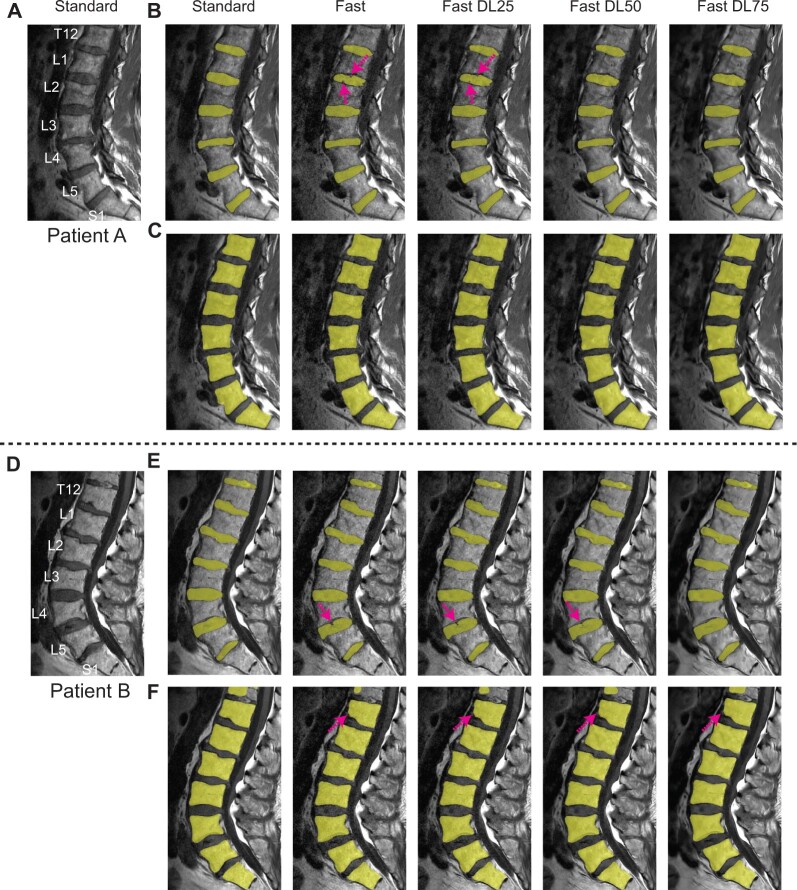
Methods: Eighteen patients were imaged with both standard protocol and fast protocol using reduced signal averages, each protocol including sagittal fat-suppressed T2-weighted, sagittal T1-weighted, and axial T2-weighted 2D fast spin-echo sequences. Fast-acquisition data was additionally reconstructed using vendor-supplied deep-learning reconstruction with three different noise reduction factors. For qualitative analysis, standard images as well as fast images with and without deep-learning reconstruction were graded by three radiologists on five different categories. For quantitative analysis, convolutional neural networks were applied to sagittal T1-weighted images to segment intervertebral discs and vertebral bodies, and disc heights and vertebral body volumes were derived.

Results: Based on noninferiority testing on qualitative scores, fast images without deep-learning reconstruction were inferior to standard images for most categories. However, deep-learning reconstruction improved the average scores, and noninferiority was observed over 24 out of 45 comparisons (all with sagittal T2-weighted images while 4/5 comparisons with sagittal T1-weighted and axial T2-weighted images). Interobserver variability increased with 50 and 75% noise reduction factors. Deep-learning reconstructed fast images with 50% and 75% noise reduction factors had comparable disc heights and vertebral body volumes to standard images (r2≥ 0.86 for disc heights and r2≥ 0.98 for vertebral body volumes).

Conclusions: This study demonstrated that deep-learning-reconstructed fast-acquisition images have the potential to provide noninferior image quality and comparable quantitative assessment to standard clinical images.

Keywords: clinical MRI; deep learning reconstruction; fast acquisition; lower back pain; lumbar spine MRI; segmentation.


Figures

Figure 1.
Figure 1.
Comparison of images from a 63-year old male patient acquired with the MSK radiology lumbar spine protocol. Standard, Fast, Fast DL25, Fast DL50, Fast DL75 images from SAG T2 FS (A), SAG T1 (B), and AX T2 (C) are all shown with the axial and sagittal locations denoted by dashed lines in the left column. In (A, B) the Modic type I changes and mild central canal stenosis are denoted (by solid arrows and dashed arrows, respectively). In (C), mild facet arthropathy and moderate right foraminal narrowing are denoted by dashed arrows and a solid arrow, respectively.
Figure 2.
Figure 2.
Comparison of images from a 73-year old male patient acquired with the neuroradiology lumbar spine protocol. Standard, Fast, Fast DL25, Fast DL50, Fast DL75 images from SAG T2 FS (A), SAG T1 (B), and AX T2 (C) are again shown. Mild central canal stenosis and small Schmorl’s nodes are denoted by dashed arrows (A) and solid arrows (B). In the axial images (C), mild facet arthropathy and mild left foraminal narrowing related to a lateral disc protrusion are identified (denoted by arrows).
Figure 3.
Figure 3.
Comparison of other anatomical structures/pathologies. (A) Images from a patient having transitional lumbosacral anatomy, moderate neuroforaminal stenosis at the L5–S1 disc level (solid arrow), and severe L4 and L5 facet arthropathy (dashed arrows). (B) Images from a patient having central annular fissure (solid arrow). These abnormal structures were well delineated on Fast DL50 images.
Figure 4.
Figure 4.
Box plots summarizing qualitative assessment. Radiologists’ scores over the 18 patients for the Standard, Fast, Fast DL25, Fast DL50, and Fast DL75 groups are compared as box plots on “Apparent SNR,” “Ability to Discern Anatomical Structures,” “Diagnostic confidence,” and “Overall Image Quality,” separately for SAG T2 FS (A–D), SAG T1 (E–H), and AX T2 (I–L) sequences. The minimum, first quartile, median, third quartile, and maximum values are shown, and outliers are denoted as red crosses.
Figure 5.
Figure 5.
Disc and vertebral body segmentation masks. (A, D) Standard SAG T1 images, and segmentation masks of discs (B, E) and vertebral bodies (C, F) attained from Standard, Fast, and Fast-DL images of two patients. Some pixels within the disc regions were missed on Fast images, as denoted by arrows in (B, E), but disc segmentation was improved on Fast-DL images. For vertebral bodies, segmentation worked well over all the vertebral bodies except the T12 vertebral body on Fast and Fast-DL images in Patient B (F).
Figure 6.
Figure 6.
Correlation plots over extracted biomarkers. Disc heights (A–D) and vertebral body volumes (E–H) measured from Fast and Fast-DL images are compared to those from Standard images. Linear equations and the correlation of determination (r2) are denoted. The highest correlation was achieved when the noise reduction factor was 50% for disc heights (r2= 0.87) and when the noise reduction factor was 75% for vertebral body volumes (r2 = 0.99).

References

    1. Hoy D, March L, Brooks P, et al.Measuring the global burden of low back pain. Best Pract Res Clin Rheumatol. 2010;24(2):155–165. - PubMed
    1. Dagenais S, Caro J, Haldeman S.. A systematic review of low back pain cost of illness studies in the United States and internationally. Spine J. 2008;8(1):8–20. - PubMed
    1. Luo X, Pietrobon R, Sun SX, Liu GG, Hey L.. Estimates and patterns of direct health care expenditures among individuals with back pain in the United States. Spine (Phila Pa 1976). 2004;29(1):79–86. - PubMed
    1. Brinjikji W, Luetmer PH, Comstock B, et al.Systematic literature review of imaging features of spinal degeneration in asymptomatic populations. AJNR Am J Neuroradiol. 2015;36(4):811–816. - PMC - PubMed
    1. Koes BW, van Tulder MW, Thomas S.. Diagnosis and treatment of low back pain. BMJ. 2006;332(7555):1430–1434. - PMC - PubMed

Publication types

MeSH terms