. 2023 May 8;8(9):e151933.
doi: 10.1172/jci.insight.151933.

Inactivating the lipid kinase activity of PI3KC2β is sufficient to rescue myotubular myopathy in mice

Affiliations

Inactivating the lipid kinase activity of PI3KC2β is sufficient to rescue myotubular myopathy in mice

Xènia Massana-Muñoz et al. JCI Insight. .

Abstract

Phosphoinositides (PIs) are membrane lipids that regulate signal transduction and vesicular trafficking. X-linked centronuclear myopathy (XLCNM), also called myotubular myopathy, results from loss-of-function mutations in the MTM1 gene, which encodes the myotubularin phosphatidylinositol 3-phosphate (PtdIns3P) lipid phosphatase. No therapy for this disease is currently available. Previous studies showed that loss of expression of the class II phosphoinositide 3-kinase (PI3K) PI3KC2β (PI3KC2B) protein improved the phenotypes of an XLCNM mouse model. PI3Ks are well known to have extensive scaffolding functions and the importance of the catalytic activity of this PI3K for rescue remains unclear. Here, using PI3KC2β kinase-dead mice, we show that the selective inactivation of PI3KC2β kinase activity is sufficient to fully prevent muscle atrophy and weakness, histopathology, and sarcomere and triad disorganization in Mtm1-knockout mice. This rescue correlates with normalization of PtdIns3P level and mTORC1 activity, a key regulator of protein synthesis and autophagy. Conversely, lack of PI3KC2β kinase activity did not rescue the histopathology of the BIN1 autosomal CNM mouse model. Overall, these findings support the development of specific PI3KC2β kinase inhibitors to cure myotubular myopathy.

Keywords: Inositol phosphates; Muscle Biology; Protein kinases; Skeletal muscle.


Figures
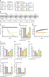
Figure 1
Figure 1. PI3KC2β kinase inhibition rescues Mtm1–/Y survival and muscle force defects.
(A) Genotypes analyzed and nomenclature used in this study. (B) Percentage of genotypes obtained on postnatal day 10. (C) Kaplan-Meier survival curve from all genotypes analyzed (n = 7). (D) Hanging test performance at 4 weeks of age (6 ≤ n ≤ 7). Maximum hanging time is 60 seconds. (E) Growth curve representing the average body weight of each group from 4 to 12 weeks (6 ≤ n ≤ 7). (FH) Muscle weight of (F) tibialis anterior (4 ≤ n ≤ 10), (G) gastrocnemius (4 ≤ n ≤ 10), and (H) soleus muscle (4 ≤ n ≤ 10) at 5 weeks. (I) Absolute muscle force (3 ≤ n ≤ 7) and (J) specific muscle force at 5 weeks (3 ≤ n ≤ 7). *P < 0.05 vs. WT/WT; P < 0.05 vs. KO/WT by Kruskal-Wallis test (D and I) or 1-way ANOVA test (F, G, H, and J).
Figure 2
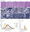
Figure 2. Inhibiting PI3KC2β activity rescues Mtm1–/Y muscle histological defects.
Muscle sections stained with (A) H&E or for (B) SDH or (C) NADH. Scale bars: 50 μm. (D) Quantification of fiber diameter (2 ≤ n ≤ 4). (E) Percentage of fibers with central accumulation of SDH staining (2 ≤ n ≤ 5). All mice analyzed at 5 weeks of age. Note the central accumulation indicated by red arrowheads in B. *P < 0.05 vs. WT/WT by Kruskal-Wallis test (E).
Figure 3
Figure 3. Inhibiting PI3KC2β activity rescues Mtm1–/Y muscle ultrastructural defects.
(A) Ultrastructural analysis by electron microscopy of TA muscles from all the analyzed genotypes at 5 weeks of age. Scale bars: 0.5 μm. (B) Violin plot showing the number of identifiable triads per sarcomere; each dot represents a sarcomere with 0, 1, or 2 recognizable triads (1 ≤ n ≤ 3; n sarcomere >39). (C) Quantification of T-tubule circularity (1 ≤ n ≤ 3; n T-tubules >33). (D) Representative images of triads from each genotype analyzed. *P < 0.05 vs. WT/WT; P < 0.05 vs. KO/WT by Kruskal-Wallis test (B and C).
Figure 4
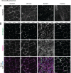
Figure 4. Mislocalization of β1 integrin but not dysferlin is improved in Mtm1–/Y muscle upon PI3KC2β inactivation.
(A) Immunolabeling of dysferlin and (B) coimmunostaining of β1 integrin and desmin in transverse TA muscle sections at 5 weeks of age. Scale bars: 10 μm.
Figure 5
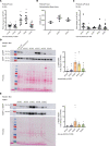
Figure 5. Increased PtdIns3P level and mTORC1 activity in Mtm1–/Y is restored by inactivating PI3KC2β.
Quantification of PtdIns3P levels by (A) ELISA (7 ≤ n ≤ 8) and (B) radiolabeling mass assay (2 ≤ n ≤ 4). (C) Quantification of PtdIns(3,4)P2 levels by ELISA (6 ≤ n ≤ 9). (D) Western blotting of TA muscle extracts (and brain when specified) probed with anti–p-S6RP and anti-S6RP antibodies, each normalized to its ponceau staining, and quantified as a ratio (n = 6). (E) Western blotting of TA muscle extracts probed with anti–p-4EBP1 and anti-4EBP1 antibodies, each normalized to its ponceau staining, and quantified as a ratio (5 ≤ n ≤ 6). *P < 0.05 vs. WT/WT; P < 0.05 vs. KO/WT by 1-way ANOVA test (A, C, and E) or Kruskal-Wallis test (D).
Figure 6
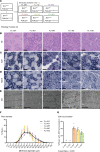
Figure 6. Inactivation of PI3KC2β is not a common therapy for different CNM forms.
(A) Genotypes analyzed after intercrossing Bin1mck–/– and Pik3c2bD1212A mice. (BD) Histological sections from 8-week-old animals stained with (B) H&E and for (C) SDH and (D) NADH. Scale bars: 50 μm. (E) Muscle ultrastructure determined by electron microscopy. Note the mitochondrial defects still present in FL+/HO animals. Scale bars: 2 μm. (F) Quantification of fiber diameter (n = 4). (G) Percentage of fibers with internalized SDH staining (4 ≤ n ≤ 7). *P < 0.05 vs. FL+/WT by Kruskal-Wallis test (G).

References

    1. Tasfaout H, et al. Centronuclear myopathies under attack: a plethora of therapeutic targets. J Neuromuscul Dis. 2018;5(4):387–406. doi: 10.3233/JND-180309. - DOI - PMC - PubMed
    1. Bitoun M, et al. Mutations in dynamin 2 cause dominant centronuclear myopathy. Nat Genet. 2005;37(11):1207–1209. doi: 10.1038/ng1657. - DOI - PubMed
    1. Nicot AS, et al. Mutations in amphiphysin 2 (BIN1) disrupt interaction with dynamin 2 and cause autosomal recessive centronuclear myopathy. Nat Genet. 2007;39(9):1134–1139. doi: 10.1038/ng2086. - DOI - PubMed
    1. Laporte J, et al. A gene mutated in X-linked myotubular myopathy defines a new putative tyrosine phosphatase family conserved in yeast. Nat Genet. 1996;13(2):175–182. doi: 10.1038/ng0696-175. - DOI - PubMed
    1. Agrawal PB, et al. SPEG interacts with myotubularin, and its deficiency causes centronuclear myopathy with dilated cardiomyopathy. Am J Hum Genet. 2014;95(2):218–226. doi: 10.1016/j.ajhg.2014.07.004. - DOI - PMC - PubMed

Publication types

Substances