. 2023 Mar 28;120(13):e2217084120.
doi: 10.1073/pnas.2217084120. Epub 2023 Mar 21.

NHA1 is a cation/proton antiporter essential for the water-conserving functions of the rectal complex in Tribolium castaneum

Affiliations

NHA1 is a cation/proton antiporter essential for the water-conserving functions of the rectal complex in Tribolium castaneum

Muhammad T Naseem et al. Proc Natl Acad Sci U S A. .

Abstract

More than half of all extant metazoan species on earth are insects. The evolutionary success of insects is linked with their ability to osmoregulate, suggesting that they have evolved unique physiological mechanisms to maintain water balance. In beetles (Coleoptera)-the largest group of insects-a specialized rectal ("cryptonephridial") complex has evolved that recovers water from the rectum destined for excretion and recycles it back to the body. However, the molecular mechanisms underpinning the remarkable water-conserving functions of this system are unknown. Here, we introduce a transcriptomic resource, BeetleAtlas.org, for the exceptionally desiccation-tolerant red flour beetle Tribolium castaneum, and demonstrate its utility by identifying a cation/H+ antiporter (NHA1) that is enriched and functionally significant in the Tribolium rectal complex. NHA1 localizes exclusively to a specialized cell type, the leptophragmata, in the distal region of the Malpighian tubules associated with the rectal complex. Computational modeling and electrophysiological characterization in Xenopus oocytes show that NHA1 acts as an electroneutral K+/H+ antiporter. Furthermore, genetic silencing of Nha1 dramatically increases excretory water loss and reduces organismal survival during desiccation stress, implying that NHA1 activity is essential for maintaining systemic water balance. Finally, we show that Tiptop, a conserved transcription factor, regulates NHA1 expression in leptophragmata and controls leptophragmata maturation, illuminating the developmental mechanism that establishes the functions of this cell. Together, our work provides insights into the molecular architecture underpinning the function of one of the most powerful water-conserving mechanisms in nature, the beetle rectal complex.

Keywords: Tribolium castaneum; cation/H+ antiporter; genetics; rectal complex; secondary cell.


Conflict of interest statement

The authors declare no competing interest.

Figures

Fig. 1.
Fig. 1.
BeetleAtlas is a comprehensive transcriptomic atlas of gene expression. (A) Adult Tribolium anatomy highlighting the tissues selected for microdissection and bulk RNA sequencing. (B) Clustered heat maps of gene expression (log2 transformed values of fragments per kilobase of transcript per million, FPKM) for select genes that are enriched in a certain tissue or life stage in Tribolium. The same genes are depicted across all samples demonstrating distinct transcriptional signatures for each tissue or ontogenetic stage.
Fig. 2.
Fig. 2.
Mining BeetleAtlas to identify molecules involved in rectal complex function. (A) Fold increase (enrichment) in gene expression of membrane channels and transporters in the rectal complex relative to the whole animal (dashed line) according to BeetleAtlas. A gene encoding a putative Nha1-like protein is most highly enriched in the rectal complex. (B) Spatial expression analysis of Nha1 shows that it is almost exclusively expressed in the hindgut and rectal complex of both larval and adult Tribolium. (C) Heat map of Nha1 expression superimposed on larval and adult anatomy. (D) Predicted exon map of Nha1 marking the epitope targeted for antibody generation and regions selected for dsRNA synthesis. Of the two fragments tested, fragment #2 produced the strongest knockdown and was therefore used in the remaining part of the study.
Fig. 3.
Fig. 3.
NHA1 localizes to specialized leptophragmata cells in the PTs of the rectal complex. (AC) Scanning electron microscopy (SEM) images showing the gross morphology of the rectal complex. A subpopulation of cells is arranged along the perirectal tubules (PTs) as a series of dilations that extend radially toward the perinephric membrane (PM), suggesting that they are leptophragmata (LP). (DF) Back-scattered electron (BSE) detection reveal that these dilations contain elements of high atomic number (Z) following AgNO3 application, confirming that these dilations are LP. (G) Maximum projection confocal microscopy images of paraffin sections of the rectal complex demonstrate that the V-ATPase localizes predominantly to the apical brush border of principal cells in the PTs (scale bar, 40 µm.) Note the cuticle of the rectal epithelium shows strong autofluorescence (arrows). (H) Maximum projection confocal microscopy images demonstrate that NHA1 is expressed in this subpopulation of LP cells along the PTs (small arrows). Zoom: note the small nucleus of the LP and the characteristic central band of F-actin spanning the apical surface of the cell (small arrows). Cross-section of PT: Subcellular localization of NHA1 demonstrates exclusive expression in the small-nucleated LP cells. Rectal complex dissected from animals injected with dsRNA targeting the Nha1 gene (Nha1-RNAi) shows a dramatic reduction in immunoreactivity confirming the specificity of the antibody. (scale bar, 100 µm.) (I) LP identity is further confirmed by coexpression of the Tiptop (Tio) transcription factor, which is a marker of the LPs [as well as secondary cells in the “free” part of the tubule (34)].
Fig. 4.
Fig. 4.
NHA1 acts as a K+(Na+)/H+ exchanger. (A) Predicted three-dimensional ribbon diagram of the NHA1 transporter embedded in the plasma membrane (B) Top view of the NHA1 protein highlighting K+ (pink) and Na+ (purple) binding; K+ is predicted to bind closer to negatively charged transport funnel compared to Na+ (Insets). (C) Root mean square deviation (RMSD) plot of NHA1 backbone (C-α) atoms in the presence of K+ (pink) and Na+ (purple) as a function of time. (D) Radius of gyration (RoG) plot of NHA1 compactness in the presence of K+ (pink) and Na+ (purple) as a function of time. (E) Representative traces of pHi and Vm of control (dark gray) or Nha1 (blue)-injected X. laevis oocytes in response to changes in bath pH, [Na+] and [K+]. The gray boxes mark replacement of the control Kulori’s solution (90 mM NaCl, 4 mM KCl, 1 mM MgCl2, 1 mM CaCl2, 5 mM HEPES, pH 7.4) with either (0Na+) Kulori’s solution where NaCl was substituted by choline-Cl (pH 8.5); where pH was adjusted to 8.5 (90K+); where the potassium concentration was increased or (0K+); where KCl was substituted by choline-Cl. Significant changes in pHi relative to the previous solution are indicated by arrows. (F) Quantitative comparison of changes in pHi of oocytes in response to different challenges (Student’s t test; n = 5 to 10, *< 0.05, **< 0.01). (G) Resting levels of pHi of oocytes from NHA1 (blue) and water-injected controls (dark gray) after incubation in Kulori’s solution pH 7.4 (Student’s t test; n = 5 to 10, **< 0.050.01). (H) Schematic representation of ion transport mediated by NHA1.
Fig. 5.
Fig. 5.
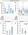
Environmental cues modulating rectal complex Nha1 expression. (A) Nha1 transcript levels in the rectal complex (n = 4 to 8) and (B) Raincloud plot of anti-NHA1 immunofluorescence (n = 23 to 45) in leptophragmata from animals exposed to different environmental conditions. Representative images of NHA1 immunofluorescence levels following exposure to each condition are shown below. Significant differences indicate pair-wise comparisons between control (food, RH 50%) and a given experimental group (one-way analysis of variance (ANOVA), *< 0.05, ****< 0.0001). (C) Nha1 transcript levels in the rectal complex (n = 5) from animals injected with DH37 (one-way ANOVA, **< 0.01) or (D) from beetles injected with dsRNA targeting the Urn8 gene (unpaired Student’s t test, *< 0.05).
Fig. 6.
Fig. 6.
Systemic water balance depends on NHA1 activity. (A) Kaplan–Meyer survival function of adult control (dsRNA targeting beta-lactamase, ampR) and Nha1-silenced animals. Adult-specific knockdown of Nha1 reduces organismal survival when exposed to low humidity conditions compared to control (RH 5%, log-rank test, n = 41 to 43). (B) Gravimetric analysis of control and Nha1-silenced animals. Desiccation-induced water loss is significantly increased in Nha1 knockdown animals relative to control (unpaired Student’s t test, n = 57, ***< 0.001). (C) Hemolymph osmotic pressure of control and Nha1-depleted beetles. Hemolymph osmolality is significantly increased in Nha1 knockdown animals relative to controls (unpaired Student’s t test, n = 3, ***< 0.05). (D) Defecation rate of Nha1-depleted animals is significantly increased relative to controls (unpaired Student’s t test, ***n = 3 groups with 10 animals per group, **< 0.05) and (E) the deposits are more circular (*> 0.05), (F) larger (**< 0.01), and (G) with a reduced dye intensity (****< 0.001). (D–G) Statistical differences were tested using unpaired Student’s t test. (H) The size of the deposits is inversely correlated with dye intensity in excreta from both Nha1-silenced (blue trend line) and control animals (gray trend line) yet the slopes of the trend lines are significantly different (*< 0.05). Note the subpopulation of large, less intense deposits (blue shaded box), which is produced almost exclusively by Nha1 knockdown beetles. (I) Representative images of excreta produced by control and Nha1 knockdown animals. Deposits produced by Nha1 knockdown animals were often associated with excess fluid, as evidenced by the dye-labeled fluid surrounding the deposit, which was never observed in control animals (scale bar, 1 mm.) (J) Ex vivo preparations of Nha1 knockdown beetles show a significantly lower rectal complex-mediated fluid reabsorption rate relative to control (unpaired Student’s t test, n = 17 to 33, ***< 0.001).
Fig. 7.
Fig. 7.
Tiptop controls NHA1 expression and leptophragmata differentiation. (A) Changes in body water in control and Nha1-silenced animals exposed to RH 90% as a function of time. Nha1-depleted animals consistently lose water over a 6-d period, while controls retain, or even increase, organismal water levels relative to their initial water levels (paired Student’s t test, n = 40 to 49, *< 0.05). (B) Before–after plot of individual measurement graphed in A at day 0 (pre) and day 6 (post). (C) Immunolocalization of NHA1 and Tio in control and Tio-silenced animals. Tio knockdown results in almost complete loss of NHA1 expression as well as in morphological defects in the leptophragmata. (D) Tio depletion similarly abolishes NHA1 expression and causes defects in leptophragmata cytoarchitecture, such as a complete loss of the central band of F-acting spanning the LP (red arrows in Bottom panels), which (E) significantly impairs water vapor absorption compared to controls in larvae of Tenebrio (unpaired Student’s t test, n = 3 to 7, *< 0.05; **< 0.01). (F) Model for the systemic control of water balance in tenebrionid beetles.

References

    1. Eggleton P., The state of the world’s insects. Annu. Rev. Environ. Res. 45, 61–82 (2020).
    1. Dow J. A. T., Halberg K. A., Terhzaz S., Davies S. A., “Drosophila as a model for neuroendocrine control of renal homeostasis“ in Model Animals in Neuroendocrinology: From Worms to Mouse to Man, M. Ludwig & G. Levkowitz, Eds. (Wiley-Blackwell, 2018), Vol. 1, pp. 81–100.
    1. Maddrell S. H. P., The functional design of the insect excretory system. J. Exp. Biol. 90, 1–15 (1981).
    1. Phillips J. E., Hanrahan J., Chamberlin M., Thomson B., “Mechanisms and control of reabsorption in insect hindgut“ in Advances in Insect Physiology, Evans P. D., Wigglesworth V. B., Eds. (Academic Press, 1987), vol. 19, pp. 329–422.
    1. J. Machin, Water balance inTenebrio molitor, L, Larvae; the effect of atmospheric water absorption. J. Comparat. Physiol. 101, 121–132 (1975).

Publication types