. 2023 Mar 21;16(777):eabo3406.
doi: 10.1126/scisignal.abo3406. Epub 2023 Mar 21.

Hedgehog-induced ZFYVE21 promotes chronic vascular inflammation by activating NLRP3 inflammasomes in T cells

Affiliations

Hedgehog-induced ZFYVE21 promotes chronic vascular inflammation by activating NLRP3 inflammasomes in T cells

Bo Jiang et al. Sci Signal. .

Abstract

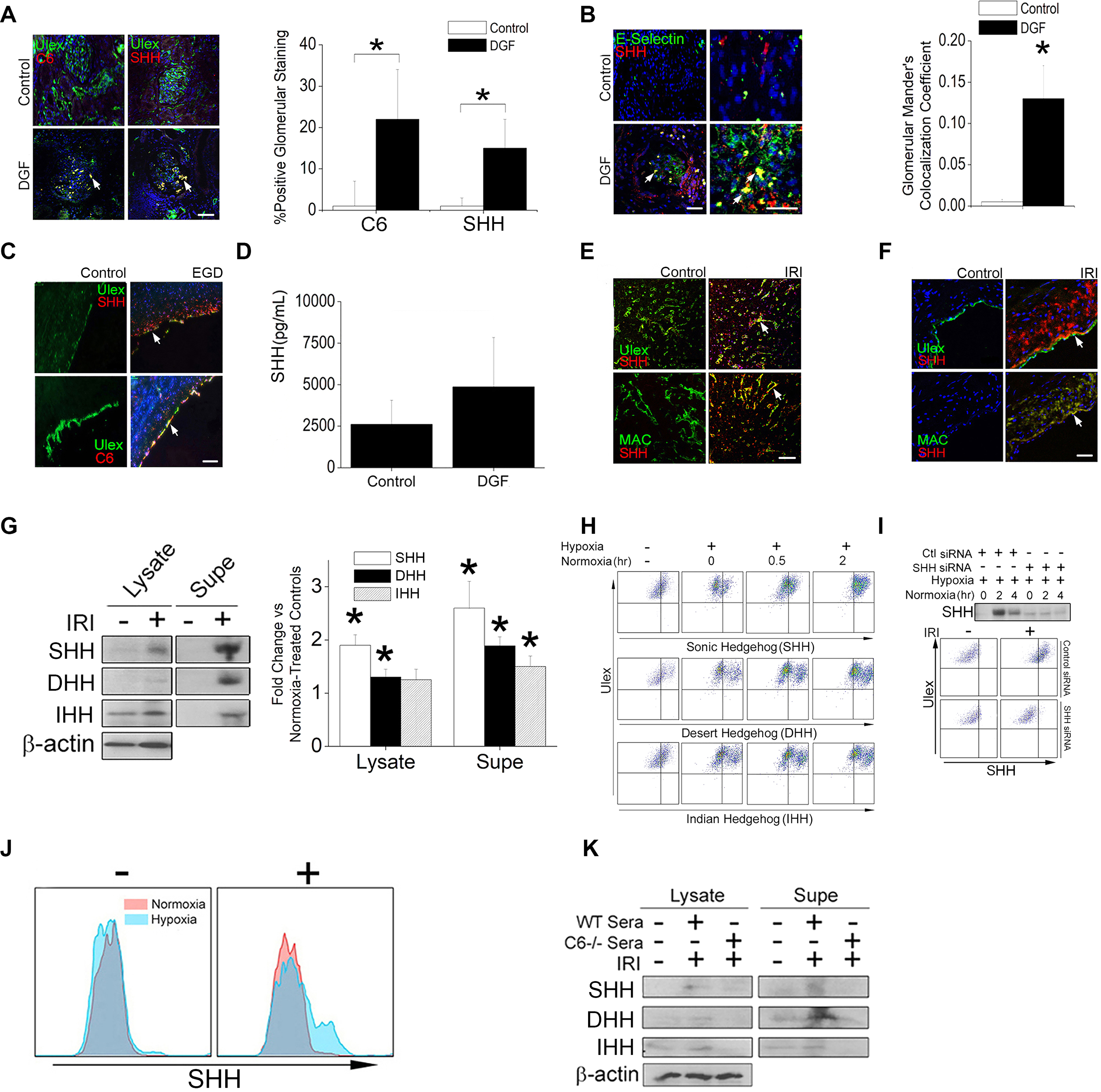
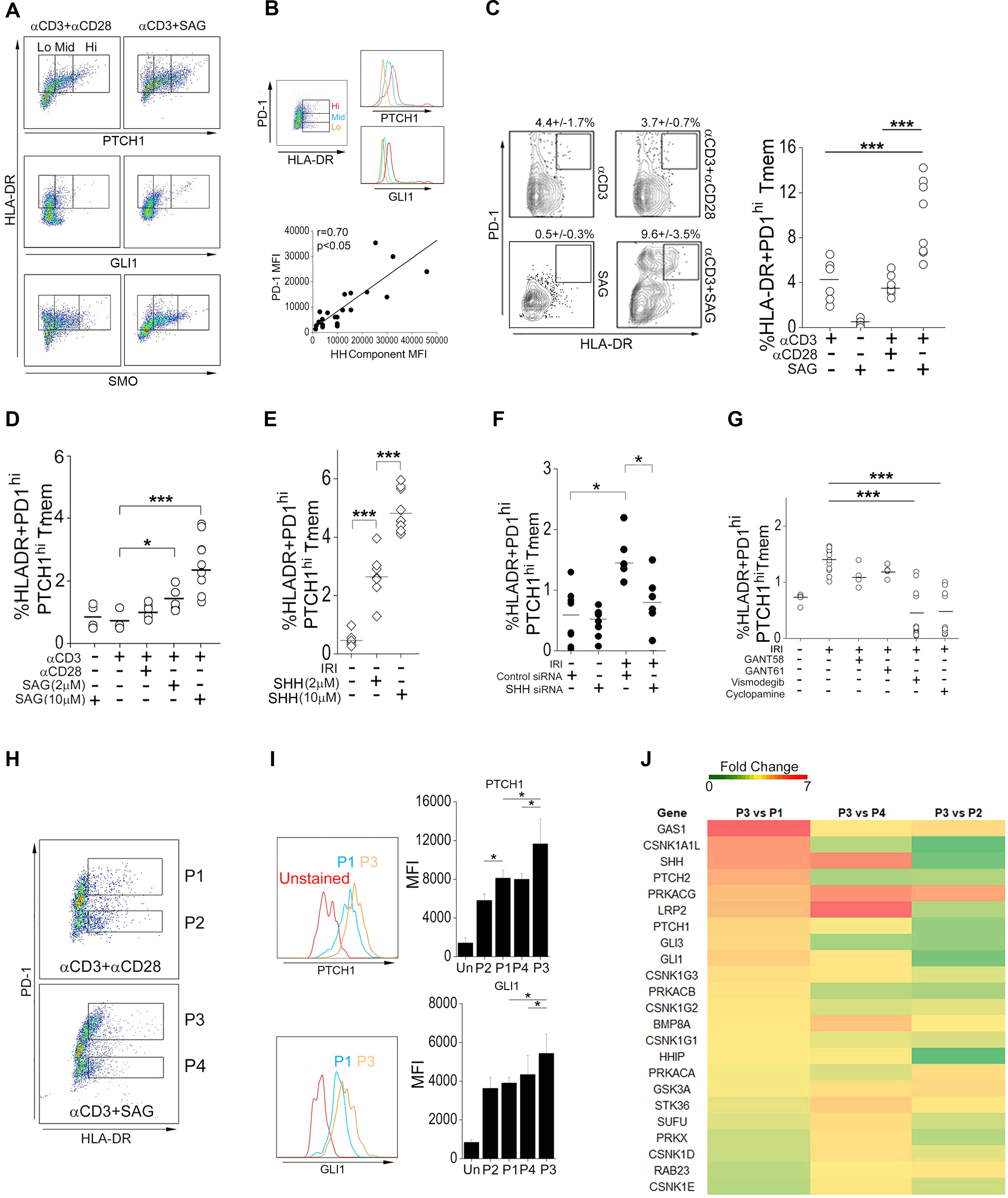
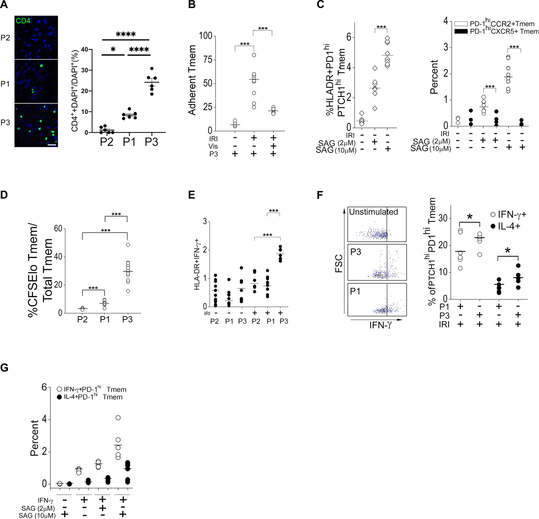
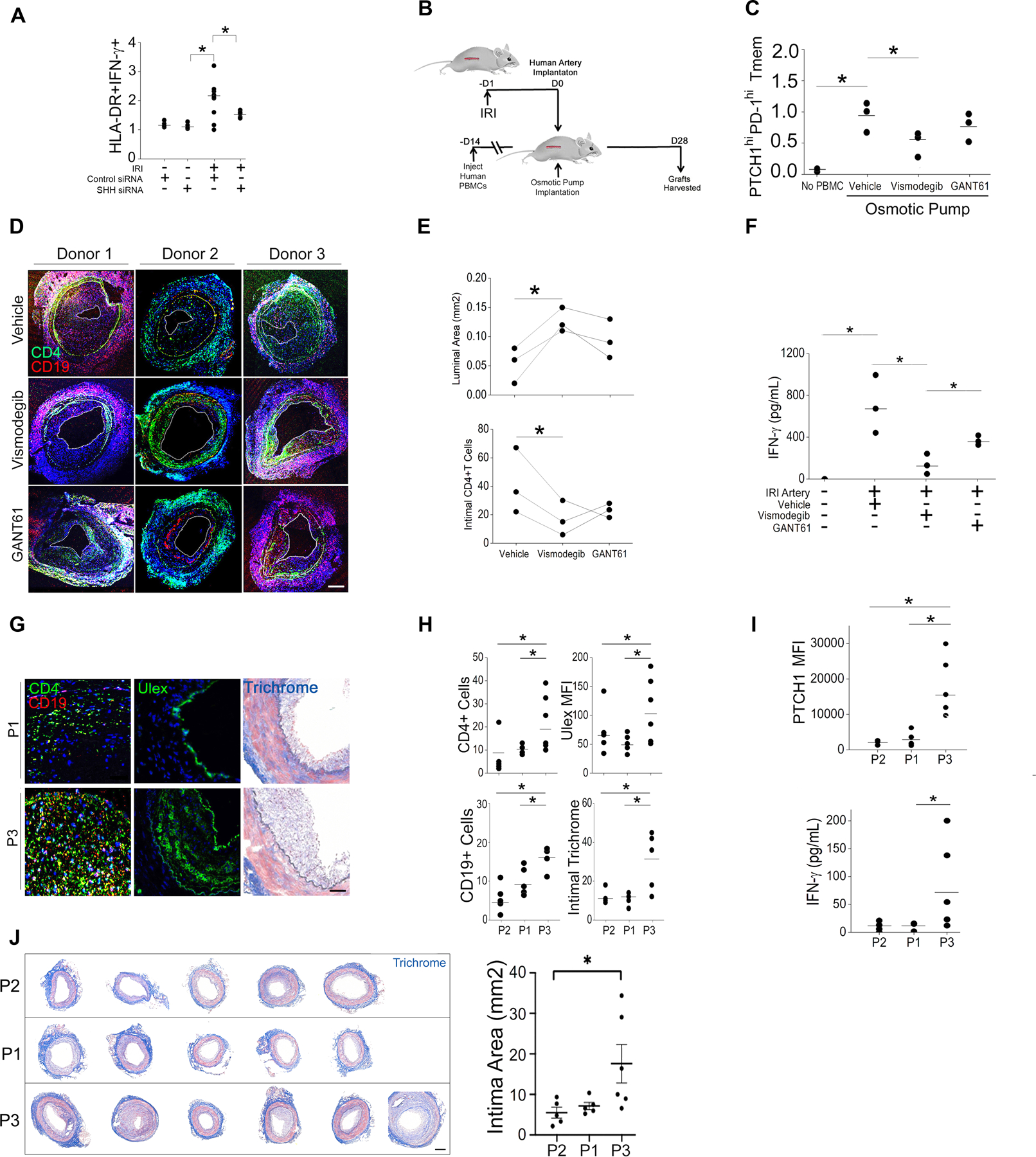
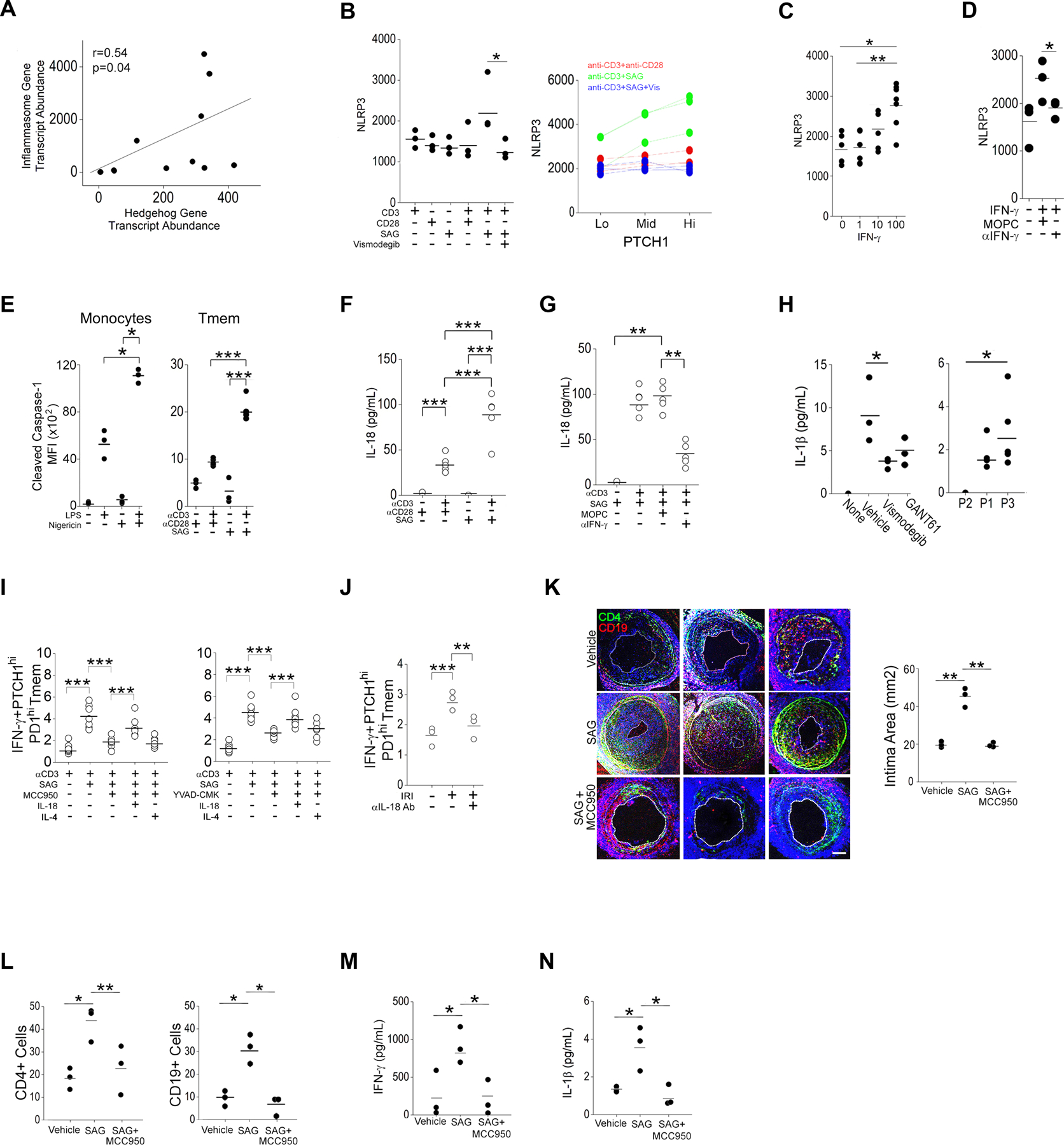
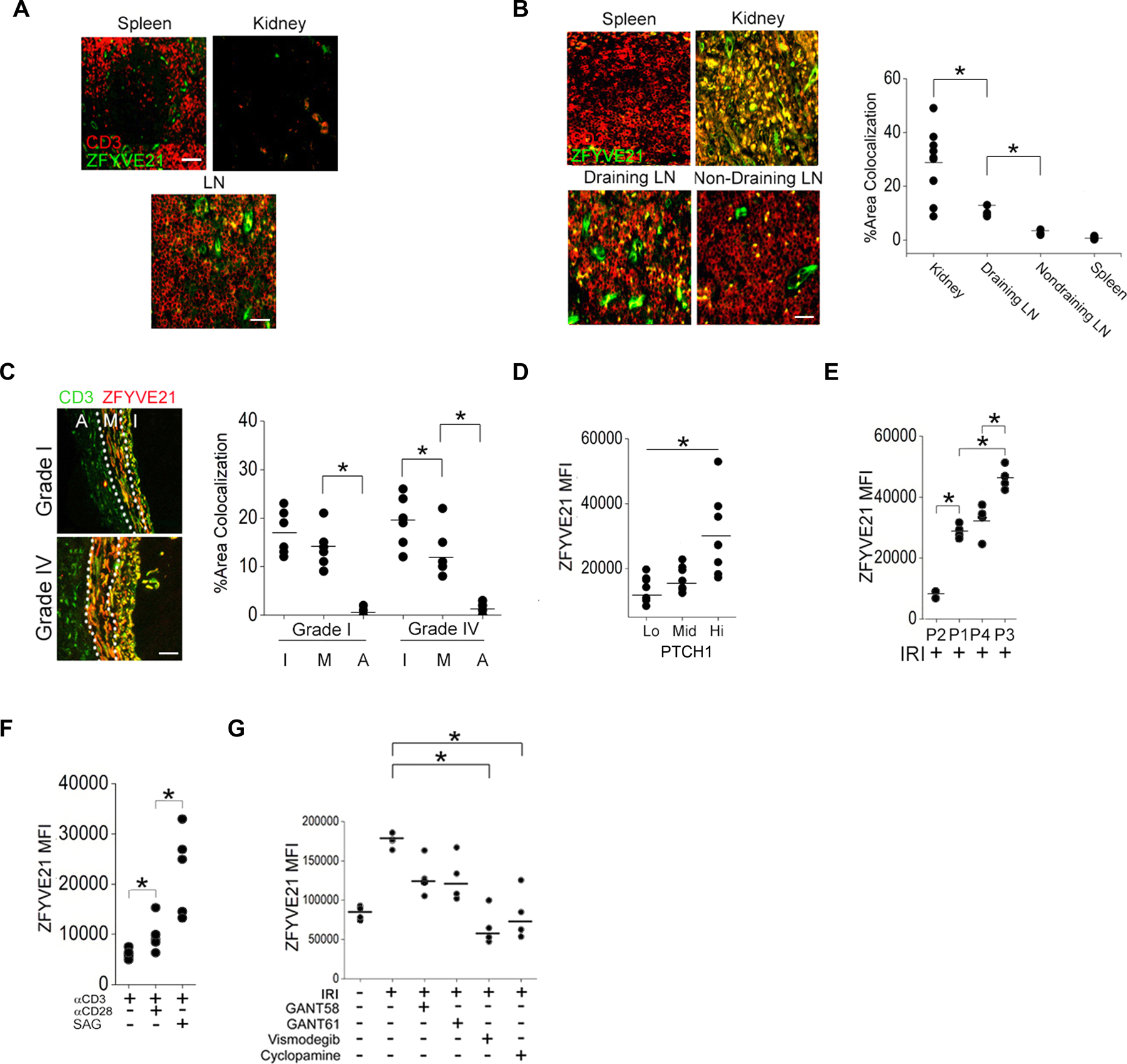
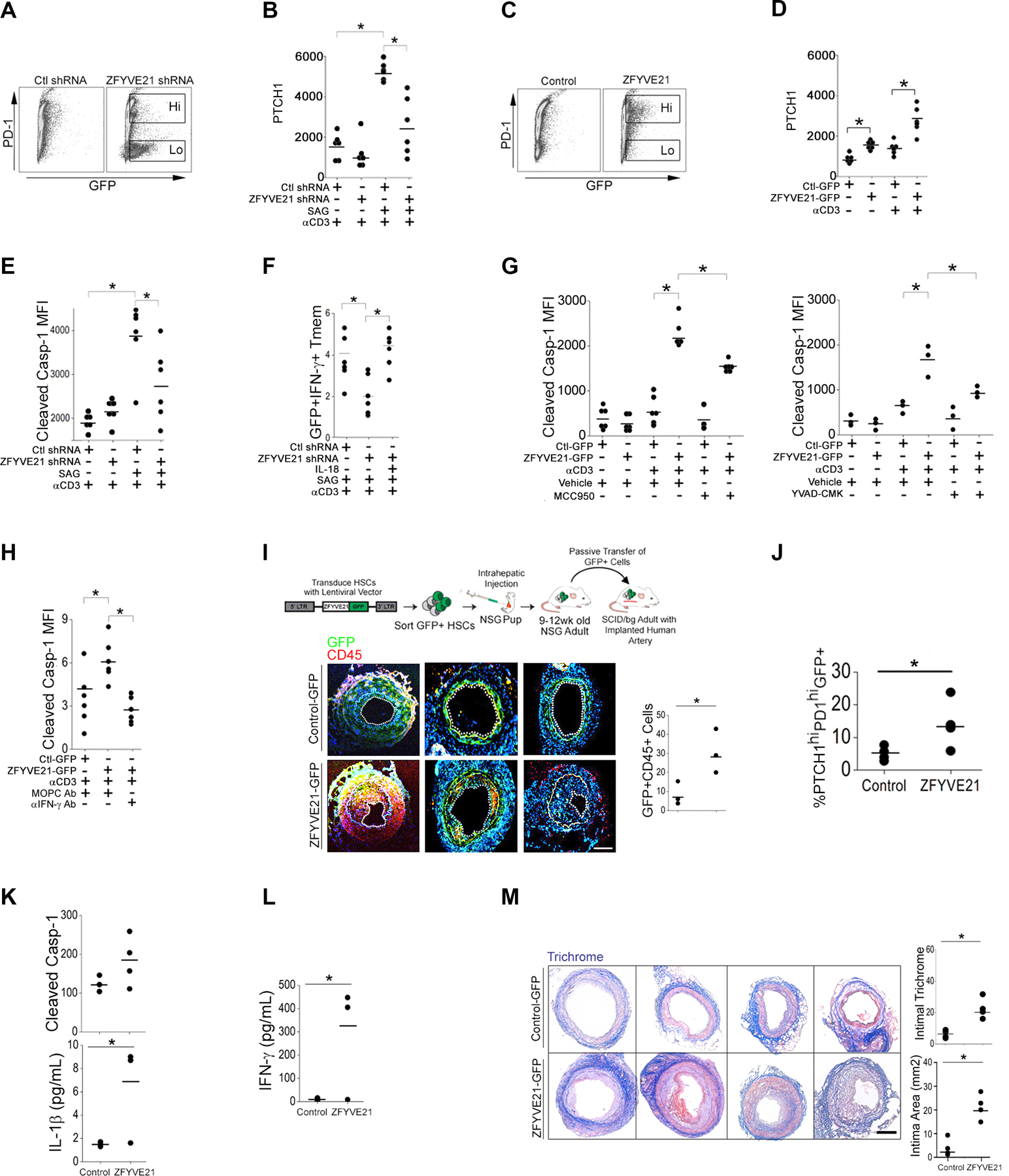
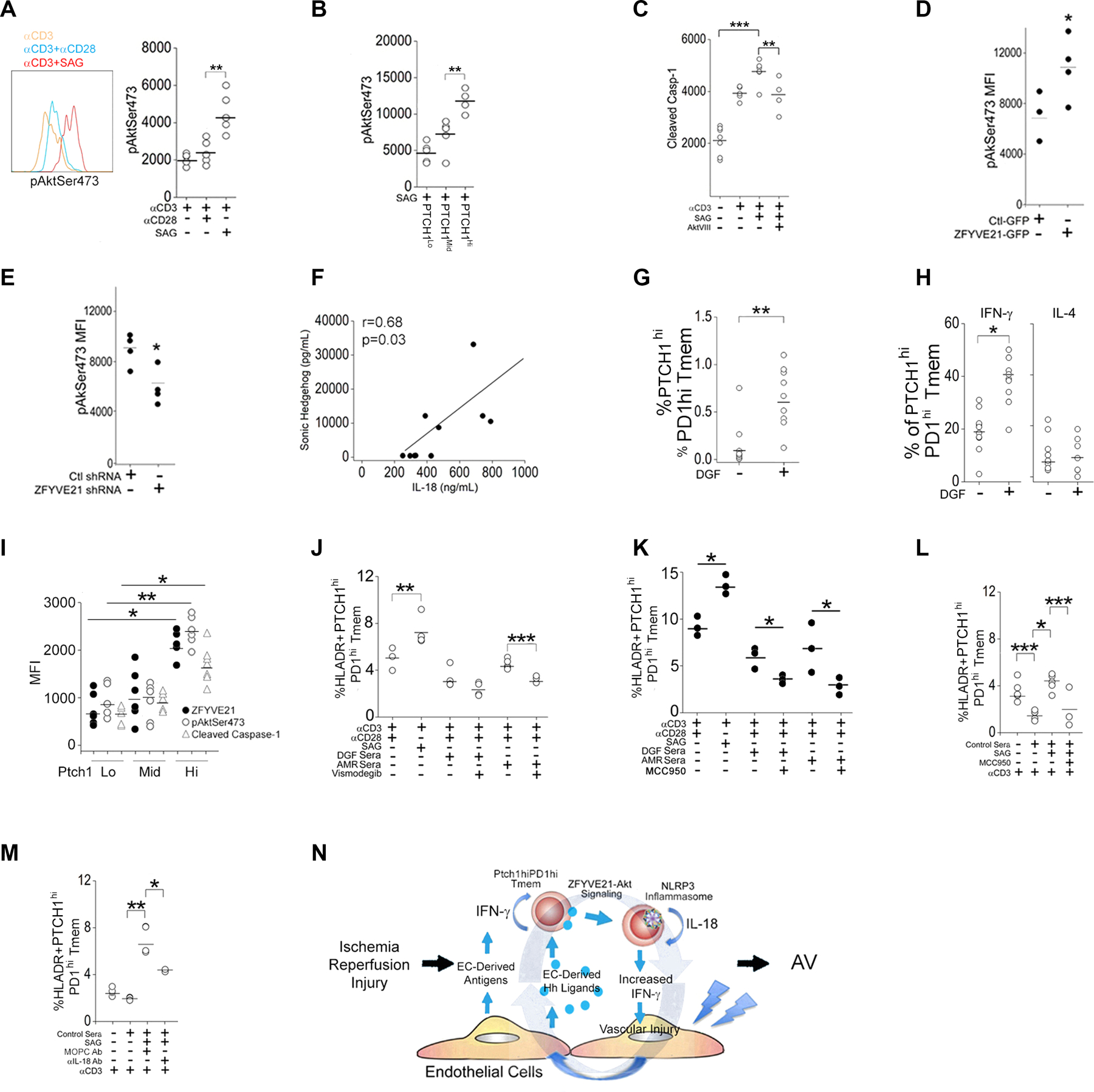
The zinc finger protein ZFYVE21 is involved in immune signaling. Using humanized mouse models, primary human cells, and patient samples, we identified a T cell-autonomous role for ZFYVE21 in promoting chronic vascular inflammation associated with allograft vasculopathy. Ischemia-reperfusion injury (IRI) stimulated endothelial cells to produce Hedgehog (Hh) ligands, which in turn induced the production of ZFYVE21 in a population of T memory cells with high amounts of the Hh receptor PTCH1 (PTCHhi cells, CD3+CD4+CD45RO+PTCH1hiPD-1hi), vigorous recruitment to injured endothelia, and increased effector responses in vivo. After priming by interferon-γ (IFN-γ), Hh-induced ZFYVE21 activated NLRP3 inflammasome activity in T cells, which potentiated IFN-γ responses. Hh-induced NLRP3 inflammasomes and T cell-specific ZFYVE21 augmented the vascular sequelae of chronic inflammation in mice engrafted with human endothelial cells or coronary arteries that had been subjected to IRI before engraftment. Moreover, the population of PTCHhi T cells producing high amounts of ZFYVE21 was expanded in patients with renal transplant-associated IRI, and sera from these patients expanded this population in control T cells in a manner that depended on Hh signaling. We conclude that Hh-induced ZFYVE21 activates NLRP3 inflammasomes in T cells, thereby promoting chronic inflammation.


Conflict of interest statement

COMPETING INTERESTS: The authors declare that they have no competing interests.

Figures

Fig. 1.
Fig. 1.. Endothelial cells produce Hh ligands following IRI.
(A to C) Immunofluorescence (IF) staining for C6, SHH, the endothelial label Ulex, and the endothelial inflammation marker E-selectin in renal (A and B, n=3) biopsies from DGF patients and in coronary artery (C, n=4) biopsies from EGD patients. Cells showing C6 and SHH were quantified in (A), and colocalization of SHH and E-selectin was quantified in (B). (D) Quantification of SHH in sera from control (n=10), DGF (n=9), and AMR (n=2) patients by enzyme immunoassay. (E) IF staining for Ulex, MAC, and SHH in HUVECs embedded in collagen-fibronectin gels, implanted adjacent to proximal femoral arteries in SCID/bg mice, and subjected to IRI injury. Controls are implants from the sham-operated contralateral artery. n=3 mice. (F) IF staining for Ulex, MAC, and SHH in human coronary artery explants that were subjected to normoxia or IRI ex vivo prior to surgical implantation into the infrarenal aortae of SCID/bg mice. N=3 mice per treatment group. (G and H) Immunoblotting (G) and FACS analysis (H) for SHH, DHH, and IHH in cell lysates and supernatants of HUVECs subjected to normoxia or IRI-simulating conditions (11). β-actin is a loading control. (I) Immunoblotting for SHH in supernatants and FACS analysis for SHH in HUVECs treated with control or SHH-targeting siRNA and subjected to normoxia or IRI-simulating conditions. (J) FACS analysis of SHH in HUVECs cultured in WT serum (+) or C6-defiecient serum (–). (K) Immunoblotting for SHH, DHH, and IHH in HUVECs cultured with WT human serum or C6-deficient serum and subjected to IRI conditions as indicated. Data in (G to K) are representative of experiments using cells from 3 different human donors. All scale bars, 200μm. For quantifications in (A), 3–6 hpf fields per patient were analyzed in a blinded fashion by 3 independent reviewers. Data are presented as mean ± SD. For quantifications in A, B, and D Student’s t-test was used for statistical comparisons.
Fig. 2.
Fig. 2.. Identification of a PTCHhi T cell subset that responds to Hh agonism.
(A) FACS analysis of PTCH1, GLI1, and SMO in HLA-DR+CD4+CD45RO+ memory T cells (Tmem) pretreated with Smoothened agonist (SAG) or soluble antibody recognizing CD28 as indicated and stimulated with immobilized antibody specific for CD3. (B) Correlation of PD-1 abundance with that of Hh signaling components PTCH1 and GLI1 in Tmem activated with CD3-specfic antibody in the presence of SAG. MFI, mean fluorescence intensity. (C and D) FACS analysis (C) and HLA-DR+PD-1hi Tmem frequency (C and D) in Tmem treated with CD3-specific antibody, SAG, and CD28-sepecific antibody as indicated. (E) HLA-DR+PD1hiPTCHhi Tmem frequency in endothelial cell (EC):T cell cocultures with or without the indicated concentrations of SHH under normoxic conditions (no IRI). (F) HLA-DR+PD1hiPTCHhiTmem frequency in EC:T cell cocultures treated with control or SHH siRNA and subjected to IRI conditions as indicated. (G) HLA-DR+PD1hiPTCHhi Tmem frequency in EC:T cell cocultures treated with or without GANT58, GANT61, vismodegib, or cyclopamine and subjected to IRI conditions as indicated. (H to J) Tmem were pretreated with CD28-specific antibody or SAG before stimulation with immobilized CD3 antibody for 48 hrs. The P1–P4 populations defined according to PD-1 abundance in HLA-DR+ Tmem (H) were further classified as PTCHlo (P2), PTCHmid (P1,P4), and PTCHhi (P3) based on their abundance of PTCH1 and GLI1 (I). Heatmap of bulk RNA-seq analysis of the P1–P4 Tmem populations (J). Experimental points reflect technical replicates. Data shown are representative of 2–4 independent experiments using cells from N=3–6 separate leukopack donors. Data are presented as mean ± SD. * indicates p<0.05. Two-way ANOVA followed by Tukey’s pairwise comparison was used for statistical comparisons.
Fig. 3.
Fig. 3.. Phenotypic analysis of PTCH hiCD4+ Tmem.
(A) Quantification of PTCHlo (P2), PTCHmid (P1), and PTCHhi (P3) Tmem adhesion to endothelial cells (ECs) in EC:T cell cocultures. Experiments were repeated 3 times. A total of 6 images per Tmem subset were quantified by 3 independent researchers. Scale bar, 200μm. (B) Quantification of the adhesion of PTCHhi (P3) Tmem in IRI-treated EC:T cell cocultures treated with vehicle or Vismodegib (Vis) as indicated. (C) Frequency of HLA-DR+PD1hiPTCHhi, PD1hiCCR2+, and PD1hiCXCR5+ Tmem frequency in IRI-treated EC:T cell cocultures treated with different concentrations of Smoothened agonist (SAG). (D) Frequency of CFSElo Tmem in PTCHlo (P2), PTCHmid (P1), or PTCHhi (P3) Tmem cocultured with IRI-treated ECs. (E) Frequency of HLA-DR+IFN-γ+ Tmem in PTCHlo (P2), PTCHmid (P1), or PTCHhi (P3) Tmem cocultured with IRI-treated ECs. (F) FACS analysis of IFN-γ production in unstimulated control, stimulated PTCHmid (P1), and PTCHhi (P3) Tmem and frequency of IL4+PTCHhiPD-1hiTmem and IFN-γ+PTCHhiPD-1hiTmem in IRI-treated EC:T cell cocultures. FSC, forward scatter. (G) Frequency of IL4+PD-1hi and IFN-γ+PD-1hi Tmem in IRI-treated EC:T cell cocultures treated with IFN-γ and different concentrations of SAG. Data in (A and B) are representative of 2–4 independent experiments using N=3 separate leukopack donors. Data in (C to G) are representative of 3 independent experiments using N=3 separate leukopack donors. Data are presented as mean ± SD. Two-way ANOVA followed by Tukey’s pairwise comparison was used for statistical comparisons.
Fig. 4.
Fig. 4.. Hh signaling promotes AV pathology.
(A) Frequency of HLA-DR+IFN-γ+ Tmem in IRI-treated EC:T cell cocultures following EC transfection with control or SHH siRNA. 2–4 independent experiments were performed using N=3 separate leukopack donors (B) Schematic of artery xenograft experimental design. SCID/bg mice were engrafted with human arteries subjected ex vivo to IRI conditions or adoptively transferred with human PBMCs then implanted with osmotic pumps delivering the pharmacological inhibitors of SMO (vismodegib) or GLI (GANT61). (C) Circulating PTCH1hiPD-1hi Tmem frequency in xenografted mice. (D and E) Immunofluorescence staining for CD4+ and CD19+ cells (D) and quantitation of luminal area and intimal CD4+ T cells coverage area (E) in human artery xenografts harvested from mice. (F) IFN-γ concertation in the sera of the xenografted mice. (G and H) SCID/bg mice bearing human artery xenografts were adoptively transferred with FACS-sorted PTCHmid (P1), PTCHlo (P2), or PTCHhi (P3) Tmem. Representative images show immunofluorescence staining for CD4, CD19, and Ulex and Masson’s trichrome staining (G). CD4+ and CD19+ cell coverage, MFI of Ulex, and intimal Masson’s trichrome staining area were quantified (H). (I) FACS analysis of PTCH1 in circulating Tmem and serum IFN-γ concentration in SCID/bg mice that were treated with FACS-sorted PTCHmid (P1), PTCHlo (P2), or PTCHhi (P3) Tmem. (J) Masson’s trichrome staining and quantification of intimal area of human artery xenografts from SCID/bg mice that were treated with FACS-sorted PTCHmid (P1), PTCHlo (P2), or PTCHhi (P3) Tmem. Data in (C–F) represent N=3 mice receiving human arteries from 3 donors. Data in (G–J) represent N=5 mice receiving P2 and P1 Tmem, N=6 mice receiving P3 Tmem. Scale bars, 200μm. *indicates p<0.05, ** indicates p<0.005. Two-way ANOVA followed by Tukey’s pairwise comparison was used for statistical comparisons.
Fig. 5.
Fig. 5.. NLRP3 inflammasome activity in PTCHhi Tmem promotes AV.
(A) Correlation coefficients (Pearson’s r) between hedgehog (Hh)- and inflammasome-associated transcripts in publicly available RNA-seq data. (B) MFI of NLRP3 in bulk Tmem and in PTCHlo, PTCHmid, and PTCHhi Tmem that were pretreated with CD28-specific antibody, Smoothened agonist (SAG), and vismodegib and stimulated with immobilized CD3 antibody as indicated. (C) MFI of NLRP3 in Tmem treated with varying doses of IFN-γ. (D) MFI of NLRP3 in Tmem treated with IFN-γ in the presence of isotype control MOPC-21 (MOPC) or IFN-γ–neutralizing antibody. (E) MFI of cleaved caspase-1in monocytes treated with LPS or nigericin and Tmem treated with CD28-specific antibody and SAG before stimualtion of CD3. (F and G) IL-18 concertation in culture supernatants from Tmem that were pretreated with CD28-specific antibody, SAG, MOPC, and IFN-γ neutralization antibody before stimulation of CD3 antibody as indicated. (H) IL-1β concentration in the sera from mice bearing artery xenografts and treated with the indicated drugs (Fig. 4D) or adoptively transferred with FACS-sorted PTCHmid (P1), PTCHlo (P2), and PTCHhi (P3) Tmem (Fig. 4G). (I and J) Frequency of IFN-γ+PTCHhiPD-1hi Tmem in T cells pretreated with MCC950 or Ac-YVAD-CMK, SAG, IL-18, and IL-4 as indicated before CD3 stimulation (I), or pretreated with IL-18 neutralization antibody prior to CD3 stimulation in EC:T cell cocultures under IRI conditions. (K to N) SCID/bg mice were engrafted with human arteries, adoptively transferred with PBMCs, and implanted with osmotic pumps eluting SAG or SAG + MCC950. Immunofluorescence staining for CD4+ and CD19+ cells (K) was used to quantify the intima area (K) and intima CD4+ and CD19+ cell coverage (L) in the human artery xenografts. IFN-γ+ (M) and IL-1β (N) serum concentrations were quantified. Data in (A to J) are representative of 2–4 experiments using N=3 separate leukopack donors. Data in (K to N) represent N=3 mice implanted with human artery from 3 donors per group. Scale bars, 200μm. * indicates p<0.05, ** indicates p<0.01. Two-way ANOVA followed by Tukey’s pairwise comparison was used for statistical comparisons.
Fig. 6.
Fig. 6.. Hh agonism induces ZFYVE21 in PTCHhi Tmem.
(A to C) CD3 and ZFYVE21 staining in tissues from control renal transplant patients (A, N=3), renal transplant patients with CAMR (B, N=3), or heart transplant patients with AV (C, N=4). ZFYVE21 and CD3 colocalization was quantified in (B) and (C). (D) Abundance of ZFYVE21 in PTCHlo, PTCHmid, and PTCHhi Tmem treated with SAG and stimulated with CD3-specific antibody. (E) Abundance of of ZFYVE21 in PTCHlo (P2), PTCHmid (P1,P4), and PTCHhi (P3) Tmem in IRI-treated EC:T cell cocultures. (F) Abundance of ZFYVE21 in PTCH1hi Tmem following stimulation with antibodies against CD3 and CD28 in the absence or presence of SAG as indicated. (G) Abundance of ZFYVE21 in PTCH1hi Tmem in IRI-treated EC:T cell cocultures in the presence of GANT58, GANT61, vismodegib, or cyclopamine as indicated. Experimental points reflect technical replicates. Data in (D–G) are representative of 4–5 experiments using N=3 separate leukopack donors. Scale bars, 200μm. * indicates p<0.05, ** indicates p<0.01. Two-way ANOVA followed by Tukey’s pairwise comparison was used for statistical comparisons.
Fig. 7.
Fig. 7.. Hh-induced ZFYVE21 activates NLRP3 inflammasomes in T cells.
(A and B) Abundance of PD-1 (A) in Tmem expressing control (Ctl) or ZFYVE21 shRNA GFP-marked constructs and abundance of PTCH1 (B) when these cells were treated with SAG and stimulated with immobilized CD3-specific antibody. (C and D) Abundance of PD-1 (C) in Tmem overexpressing GFP-tagged ZFYVE21 or empty vector (Ctl-GFP) and abundance of PTCH1 (D) when these cells were stimulated with CD3-specific antibody. (E and F) Quantification of cleaved casp-1 (E) and frequency of GFP+IFNγ+ Tmem (F) in Tmem expressing control or ZFYVE21 shRNA, treated with SAG and IL-18 as indicated, and stimulated with immobilized CD3-specific antibody. (G and H) Quantification of cleaved casp-1 in Tmem overexpressing ZFYVE21-GFP or GFP alone following stimulation with immobilized CD3-specific antibody. Cells were treated with MCC950 or YVAD-CMK (G) or with IFN-γ neutralization antibody (H) as indicated. MOPC is an isotype-specific control for IFN-γ antibody. Data in (A to H) represent experiments from N=6 separate leukopack donors. (I to M) Human CD34 stem cells (HSCs) transduced with control GFP (N=4) or ZFYVE21-GFP expression (N=4) constructs were intrahepatically injected into NSG pups. PBMCs and splenocytes from these adult animals were passively transferred into SCID/bg hosts bearing human artery xenografts. Immunofluorescence images show CD45 and GFP in tissue grafts and were used to quantify GFP+CD45+ cells (I). PTCHhiPDhiGFP+ PBMC frequency (J), cleaved casp-1 and IL-1β concentration in PBMCs (K), and IFN-γ concentration in sera (L) were quantified in the mice receiving xenografts and donor PBMCs. Xenografts were harvested and stained with Masson’s trichrome (M). N=3–4 mice implanted with human arteries from 3 donors were used in each group. Scale bars, 200μm. *indicates p<0.05. Two-way ANOVA followed by Tukey’s pairwise comparison (B, D, E, F, G, H) and Student’s t-test (I-M) were used for statistical comparisons.
Fig. 8.
Fig. 8.. Hh-induced ZFYVE21-Akt-Casp1 signaling in Tmem from DGF patients.
(A) Quantification of pAktSer473 in Tmem pretreated with antibody against CD28 or Smoothened agonist (SAG) prior to stimulation with antibody against CD3. (B) Quantification of pAktSer473 in FACS-sorted PTCHlo, PTCHmid, and PTCHhi Tmem. (C) Quantification of cleaved Casp-1 in Tmem pretreated with SAG or AktVIII prior to CD3 stimulation. (D and E) Quantification of pAktSer473 in Tmem overexpressing ZFYVE21-GFP (D) or expressing ZFYVE21 shRNA (E) and stimulated with antibody against CD3. (F) Pearson correlation between SHH and IL-18 in DGF patient sera (N=7). (G) PTCHhiPD-1hi Tmem frequency in control (–) and DGF (+) PBMCs. (H) IFN-γ and IL-4+ PTCHhiPD-1hi Tmem frequency in control and DGF PBMCs. (I) Quantification of ZFYVE21, pAktSer473, and cleaved casp-1 in PTCHlo, PTCHmid, and PTCHhi Tmem. (J to L) HLA-DR+PTCHhiPD-1hi Tmem frequency in non-autologous Tmem from a healthy donor stimulated with an antibody against CD3 in the presence of CD28-specific antibody, SAG, MCC950, or vismodegib as indicated. DGF or AMR sera (J and K) or control sera (L) were used as indicated. (M) HLA-DR+PTCHhiPD-1hi Tmem frequency in non-autologous Tmem from a healthy donor stimulated with an antibody specific for CD3 in control serum in the presence of SAG or antibodies specific for MOPC or IL-18 as indicated. (N) Working model for an Hh-induced ZFYVE21-Akt-Casp-1 signaling axis in Tmem. Experimental points in (A–E) and (G–M) reflect technical replicates and were repeated 2–3 times using N=6 separate leukopack donors. * indicates p<0.05, ** indicates p<0.01. Two-way ANOVA followed by Tukey’s pairwise comparison (A-C, I-L) and Student’s t-test (D, E, G, H) were used for statistical comparisons.

Comment in

Similar articles

Cited by

References

    1. Uekita T, Gotoh I, Kinoshita T, Itoh Y, Sato H, Shiomi T, Okada Y, Seiki M. Membrane-type 1 matrix metalloproteinase cytoplasmic tail-binding protein-1 is a new member of the Cupin superfamily. A possible multifunctional protein acting as an invasion suppressor down-regulated in tumors. J Biol Chem. 2004. 279:12734–43. - PubMed
    1. Hoshino D, Nagano M, Saitoh A, Koshikawa N, Suzuki T, Seiki M. The phosphoinositide-binding protein ZF21 regulates ECM degradation by invadopodia. PLoS One. 2013. 8:e50825. - PMC - PubMed
    1. Nagano M, Hoshino D, Sakamoto T, Akizawa T, Koshikawa N, Seiki M. ZF21 is a new regulator of focal adhesion disassembly and a potential member of the spreading initiation center. Cell Adh Migr. 2011. 5:23–28. - PMC - PubMed
    1. Nagano M, Hoshino D, Toshima J, Seiki M, Koshikawa N. NH2 -terminal fragment of ZF21 protein suppresses tumor invasion via inhibiting the interaction of ZF21 with FAK. Cancer Sci. 2020. 111:4393–4404. - PMC - PubMed
    1. Nagano M, Hoshino D, Koshiba S, Shuo T, Koshikawa N, Tomizawa T, Hayashi F, Tochio N, Harada T, Akizawa T, Watanabe S, Handa N, Shirouzu M, Kigawa T, Yokoyama S, Seiki M. ZF21 protein, a regulator of the disassembly of focal adhesions and cancer metastasis, contains a novel noncanonical pleckstrin homology domain. J Biol Chem. 2011. 286:31598–609. - PMC - PubMed

Publication types

MeSH terms

Substances