. 2023 Mar;13(3):220373.
doi: 10.1098/rsob.220373. Epub 2023 Mar 22.

The lipid linked oligosaccharide polymerase Wzy and its regulating co-polymerase, Wzz, from enterobacterial common antigen biosynthesis form a complex

Affiliations

The lipid linked oligosaccharide polymerase Wzy and its regulating co-polymerase, Wzz, from enterobacterial common antigen biosynthesis form a complex

Miriam Weckener et al. Open Biol. 2023 Mar.

Abstract

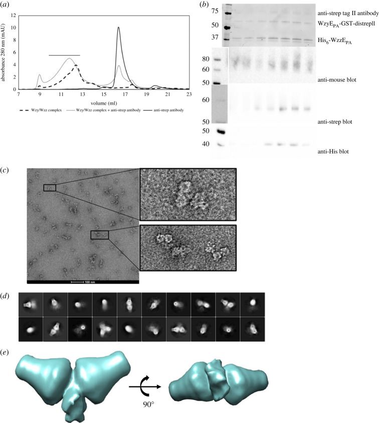
The enterobacterial common antigen (ECA) is a carbohydrate polymer that is associated with the cell envelope in the Enterobacteriaceae. ECA contains a repeating trisaccharide which is polymerized by WzyE, a member of the Wzy membrane protein polymerase superfamily. WzyE activity is regulated by a membrane protein polysaccharide co-polymerase, WzzE. Förster resonance energy transfer experiments demonstrate that WzyE and WzzE from Pectobacterium atrosepticum form a complex in vivo, and immunoblotting and cryo-electron microscopy (cryo-EM) analysis confirm a defined stoichiometry of approximately eight WzzE to one WzyE. Low-resolution cryo-EM reconstructions of the complex, aided by an antibody recognizing the C-terminus of WzyE, reveals WzyE sits in the central membrane lumen formed by the octameric arrangement of the transmembrane helices of WzzE. The pairing of Wzy and Wzz is found in polymerization systems for other bacterial polymers, including lipopolysaccharide O-antigens and capsular polysaccharides. The data provide new structural insight into a conserved mechanism for regulating polysaccharide chain length in bacteria.

Keywords: Wzy; lipid; oligosaccharides; polymerase; regulating.


Conflict of interest statement

We declare we have no competing interests.

Figures

Figure 1.
Figure 1.
Isolation of Wzy : Wzz complexes. (a) SDS–PAGE of purifications of different combinations of affinity-tagged WzyEPA/WzzEPA complexes. The left panel shows GST-WzzEPA in complex with WzyEPA-His10 and was purified by first applying the protein to glutathione 4B resin and then applying the eluate to cobalt resin. Note that the GST fusion substantially changes the size of the corresponding Wzz fusion. The predicted molecular weights of the fusion proteins are 66 kDa for GST-WzzEPA, 55.5 kDa for WzyEPA-His10, 57 kDa for WzyEPA-distrepII and 42 kDa for His6-WzzEPA. M, marker; FT, flow-through; W, wash and E, eluate. E2 denotes the eluate from the second affinity column. The right panel shows the SDS–PAGE gel of the purification of the His6-WzzEPA/WzyEPA-distrepII complex by streptactin resin followed by cobalt resin. (b) Western blot images of samples taken during tandem affinity purification of His6-WzzEPA and WzyEPA-distrepII by streptactin pulldown followed by TALON affinity chromatography. The western blots on the left are probing the samples taken after streptactin pulldown with the anti-His antibody recognizing His6-WzzEPA and the anti-strepII antibody recognizing WzyEPA-distrepII. The western blots on the right show the samples taken during the cobalt affinity column, probed with the same antibodies as before. The predicted molecular weights of the fusion proteins are 57 kDa for WzyEPA-distrepII and 42 kDa for His6-WzzEPA. E1–6 denote eluate fractions 1–6, collected during elution. (c) The SEC profiles of individually expressed and purified WzyEPA-distrepII and His6-WzzEPA and the co-expressed and co-purified WzyEPA-distrepII: His6-WzzEPA complex.
Figure 2.
Figure 2.
Identification of the in vivo WzyEPA : WzzEPA complex by FRET. (a) Overview of the domain architecture of the different fluorophore-labelled proteins. (b) Various combinations of fluorophore-labelled WzyEPA, WzzEPA and control proteins in E. coli lysates probed by western immunoblot analysis. (c). FRET was measured for the combinations of PglL, WzyEPA and WzzEPA and corrected for background fluorescence and fluorophore bleed-through. n = 18; the data represent triplicates and error bars are standard deviation.
Figure 3.
Figure 3.
Structure of WzzEPA. (a) WzzEPA (residue I39–V337) viewed from the side, the protein is represented in cartoon form (left) and in space fill (right). The periplasmic regions enclose a large central lumen. The transmembrane helices create a cavity in the membrane with channels. Residues 1–39 and 338–348 were not visualized. (b) WzzEPA viewed from the periplasm looking into the cytoplasm through the protein. The protein is represented in cartoon form (left) and in space fill (right). Space fill shows that the entrance to the lumen from the periplasm is almost occluded. (c) The monomers (coloured blue and orange) of WzzEPA are found in two conformations, distinguished by their structures at the periplasmic tip (A222-P280), highlighted in box. The monomers show an RMSD of 1.3 Å over 276 Cα atoms. (d) Motif 1 (W58-A204 and R310-E317), the two transmembrane helices (TM1 38–53; TM2 326–337) and the connecting loops are structurally conserved in WzzEPA (blue) and in WzcEC (pink). (e) The side and top view of motif 1 (defined as (d)). Motif 1 forms a ring at the periplasmic face of the membrane. Two regions of structure the L-loop (T153-D163) and a loop–helix–loop (Q78-I100) that reach into lumen are highlighted. The L-loop (T153-D163) lies closest to the membrane surface.
Figure 4.
Figure 4.
WzyEPA is located within WzzEPA. (a) Addition of the anti-strep tag II antibody leads to a shift in elution volume of the complex in SEC, as well as to a broadening of the elution peak. Control samples are shown. (b) The fractions under the broad peak (indicated by a line above the peak) were analysed by SDS–PAGE and western immunoblot. A polyclonal anti-mouse secondary antibody recognizes the mouse-derived anti-strep tag II antibody bound to WzyEPA-GST-distrepII; the HRP-coupled anti-strep tag II antibody was used to detect WzyEPA-GST-distrepII and the anti-His antibody was used to probe His6-WzzEPA. For the western immunoblots, the lanes containing the molecular weight markers have been spliced from the same blot showing the protein lanes in order to align the lanes on the immunoblot containing sample from the same fractions. For the anti-strep immunoblot, a photograph had to be taken of the coloured marker as the antibody did not recognize it. The full-size images of the immunoblots can be found in the electronic supplementary material. (c) The concentrated peak fractions were applied to cryo-EM grids and negatively stained with uranyl formate. The inset shows dimeric WzzEPA particles, which were attributed to the antibody cross-linking of WzyEPA. (d) The reference-free 2D class averages from cryo-EM of the concentrated peak fractions. (e) The initial model was generated from 2D class averages obtained from cryo-EM experiments (d) showing two WzzEPA molecules adjacent to each other.
Figure 5.
Figure 5.
Model for the process of chain length regulation in the Wzx/Wzy pathway. (a) AlphaFold model of WzyEPA docked into EM structure of WzzEPA. The model of WzyEPA was obtained from AlphaFold (ID: Q6CZF1) and placed by hand into the transmembrane cavity of WzzE. (b) The molecular measuring cylinder model of chain regulation of Wzy by Wzz (PCP-1) in ECA and OPS biosynthesis. (c) The molecular timer mechanism of chain regulation of Wzy by Wzc (PCP-2a) in CPS biosynthesis.

References

    1. Sun J, Rutherford ST, Silhavy TJ, Huang KC. 2022. Physical properties of the bacterial outer membrane. Nat. Rev. Microbiol. 20, 236-248. ( 10.1038/s41579-021-00638-0) - DOI - PMC - PubMed
    1. Rojas ER, et al. 2018. The outer membrane is an essential load-bearing element in Gram-negative bacteria. Nature 559, 617-621. ( 10.1038/s41586-018-0344-3) - DOI - PMC - PubMed
    1. Whitfield C, Williams DM, Kelly SD. 2020. Lipopolysaccharide O-antigens-bacterial glycans made to measure. J. Biol. Chem. 295, 10 593-10 609. ( 10.1074/jbc.REV120.009402) - DOI - PMC - PubMed
    1. di Lorenzo F, Duda KA, Lanzetta R, Silipo A, de Castro C, Molinaro A. 2022. A journey from structure to function of bacterial lipopolysaccharides. Chem. Rev. 122, 15 767-15 821. ( 10.1021/acs.chemrev.0c01321) - DOI - PubMed
    1. Whitfield C, Trent MS. 2014. Biosynthesis and export of bacterial lipopolysaccharides. Annu. Rev. Biochem. 83, 99-128. ( 10.1146/annurev-biochem-060713-035600) - DOI - PubMed

Publication types