. 2023 Mar 21;14(1):1557.
doi: 10.1038/s41467-023-36942-x.

A symbiotic physical niche in Drosophila melanogaster regulates stable association of a multi-species gut microbiota

Affiliations

A symbiotic physical niche in Drosophila melanogaster regulates stable association of a multi-species gut microbiota

Ren Dodge et al. Nat Commun. .

Abstract

The gut is continuously invaded by diverse bacteria from the diet and the environment, yet microbiome composition is relatively stable over time for host species ranging from mammals to insects, suggesting host-specific factors may selectively maintain key species of bacteria. To investigate host specificity, we used gnotobiotic Drosophila, microbial pulse-chase protocols, and microscopy to investigate the stability of different strains of bacteria in the fly gut. We show that a host-constructed physical niche in the foregut selectively binds bacteria with strain-level specificity, stabilizing their colonization. Primary colonizers saturate the niche and exclude secondary colonizers of the same strain, but initial colonization by Lactobacillus species physically remodels the niche through production of a glycan-rich secretion to favor secondary colonization by unrelated commensals in the Acetobacter genus. Our results provide a mechanistic framework for understanding the establishment and stability of a multi-species intestinal microbiome.


Conflict of interest statement

The authors declare no competing interests.

Figures

Fig. 1
Fig. 1. LpWF stably colonizes the fly gut with spatial specificity.
A Colonization assay schematic with initial dosing on day 0 and serial transfers to sterile food daily for 3 days before analysis. B Gut diagram. C Microscopy of LpWF-mCherry colonization in a whole gut after clearing transient cells shows a specific colonization zone in the foregut. Shown is a maximum-intensity z-projection. D The proventriculus is a major site of LpWF colonization. E Ai colonization is also specific to the proventriculus lumen and crop duct (see also Fig. S2). F CFU densities from regions dissected in B. n = 23 individual guts/region from three biological replicates. Columns represent means. Error bars are S.D. G Microsurgery was carried out to remove the crop. H LpWF colonizes the foregut of flies with the crop removed (n = 15/15). I TEM cross-section of proventriculus inner lumen. Representative image of n = 3 biological replicates. J Detail of (I). Scale bars are defined in the figure panels. Source data are provided as a Source Data file.
Fig. 2
Fig. 2. Kinetic properties of bacterial association suggest the existence of a niche in the proventriculus.
A Saturation occurs over a colonization time course of germ-free flies by LpWF. Data points are mean of log10(CFUs) in n ≥ 48 flies/data point. Error bars represent 1 s.e.m. Inset: 20-day time course after inoculation with 106 CFUs (data from). B Bacterial pulse-chase experimental design: flies were first pre-colonized with LpWF-mCherry, then fed an excess of unlabeled LpWF (blue) daily on fresh food. C Acterial cell turnover quantified by pulse-chase time course of Lp-mCherry-pre-colonized flies continuously fed unlabeled LpWF or Ai-GFP-pre-colonized flies continuously fed unlabeled Ai. Data points are mean of log10(CFUs) in n ≥ 34 flies/data point. Error bars represent 1 s.e.m. D Colonization efficiency quantified by dose response to colonization of individual flies. CFUs were measured at 3 dpi of the second colonizer. n = 24 flies/dose, error bars represent 1 standard error of the proportion. Limit of detection: 50 CFUs. E Spatial structure of colonization dynamics in the proventriculus for a fly pre-colonized with LpWF-mCherry (red) invaded by LpWF-GFP and imaged 1 h post inoculation (hpi). F Optical x,z-slice. Source data are provided as a Source Data file.
Fig. 3
Fig. 3. Ai and LpWF occupy separate niches within the proventriculus.
A Strain interactions influence colonization efficiency, as seen by dose-response curves for Ai fed to germ-free flies (open green circles), Ai-pre-colonized flies (filled yellow circles), or Lp-pre-colonized flies (black-filled green squares). Z-test of differences in proportion versus Ai into germ-free flies: dose 102.3 CFUs/fly, p = 8.1×10⁻⁴; dose 103.7 CFUs/fly: p = 4.8 × 10−9; dose 105 CFUs/fly: p = 8.7 × 10−6. n ≥ 12 flies/data point. Error bars represent 1 standard error of the proportion. B Ai abundance at 5 dpi does not differ between flies mono-colonized with Ai versus pre-colonized with LpWF then fed Ai. n ≥ 65 flies/treatment; two-tailed unpaired t-test, p = 0.38; ns indicates not significant. C LpWF abundance 5 dpi does not differ between flies mono-colonized with LpWF versus pre-colonized with Ai then fed LpWF. n ≥ 53 flies/treatment; two-tailed unpaired t-test; p = 0.06; ns indicates not significant. B, C: Center of box is median; box encloses 25th to 75th percentiles; whiskers indicate minimum and maximum. D Confocal microscopy of Lp and Ai co-colonization. Ai (green) and LpWF (red) occupied the same regions of the foregut 1 dpi. Scale bar: 100 µm. E, F x,z-section of Ai and LpWF sectors. G TEM cross-section of Ai and LpWF co-colonizing the anterior proventriculus. Scale bar: 5 µm. H Detail of (G) with LpWF and Ai cells pseudocolored. Scale bar: 2 µm. Source data are provided as a Source Data file.
Fig. 4
Fig. 4. Colonization of the niche induces morphological alteration of the proventriculus.
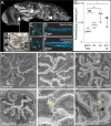
A Xray µCT model of a whole fly. Cutaway shows (1) exposed proventriculus (also inset of (B)), (2) anterior midgut, and (3) posterior midgut. B Detail of proventriculus. C Cross-section of a germ-free proventriculus inner lumen. Scale bar: 5 µm. D Germ-free proventriculus inner lumen volume rendering. E LpWF-colonized proventriculus inner lumen cross-section. Scale bar: 5 µm. F LpWF proventriculus inner lumen volume rendering. G Cardia volume calculated from surface models (n = 3 to 4 flies per condition; p = 0.0025, one-way ANOVA relative to germ-free; Tukey’s correction for multiple comparisons; GF vs. Lp p = 0.020; GF vs. Lp+Ai p = 0.022.). HM Transmission electron microscopy transverse cross-section of anterior proventriculus in (H) germ-free fly, (I) conventionally-reared fly (only lab fly bacteria; no LpWF), ( J, K) 1 hpi with LpWF, (L, M) 3 dpi colonized with LpWF (see Fig. S7). n ≥ 3 biological replicates per treatment for TEM. Yellow arrowheads indicate lumen space. Source data are provided as a Source Data file.
Fig. 5
Fig. 5. Lectin staining reveals a glycan-rich matrix associated with the foregut niche.
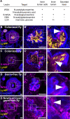
A Table of lectins that stained the proventriculus. Lectin staining of proventriculus transverse sections for B WGA in a colonized fly, C sWGA in a colonized fly, D WGA in a germ-free fly, and E WGA in a newly eclosed germ-free fly before first food ingestion. Scale bars: 20 µm. Arrowheads indicate lectin staining in the interior proventriculus lumen. n ≥ 3 biological replicates per treatment. CF: calcofluor.

References

    1. Durack J, Lynch SV. The gut microbiome: relationships with disease and opportunities for therapy. J. Exp. Med. 2019;216:20–40. doi: 10.1084/jem.20180448. - DOI - PMC - PubMed
    1. Baumgartner M, et al. Mucosal biofilms are an endoscopic feature of irritable bowel syndrome and ulcerative colitis. Gastroenterology. 2021;161:1245–1256.e20. doi: 10.1053/j.gastro.2021.06.024. - DOI - PMC - PubMed
    1. Lee J, et al. Young versus aged microbiota transplants to germ-free mice: increased short-chain fatty acids and improved cognitive performance. Gut Microbes. 2020;12:1–14. doi: 10.1080/19490976.2020.1814107. - DOI - PMC - PubMed
    1. Fisher CK, Mehta P. Identifying keystone species in the human gut microbiome from metagenomic timeseries using sparse linear regression. PLoS One. 2014;9:e102451. doi: 10.1371/journal.pone.0102451. - DOI - PMC - PubMed
    1. Subramanian S, et al. Persistent gut microbiota immaturity in malnourished Bangladeshi children. Nature. 2014;510:417–421. doi: 10.1038/nature13421. - DOI - PMC - PubMed

Publication types