

Bronchial tuberculosis with recurrent spontaneous pneumothorax: A case report
- PMID: 36944976
- PMCID: PMC10029200
- DOI: 10.1186/s12890-023-02374-y
Bronchial tuberculosis with recurrent spontaneous pneumothorax: A case report
Erratum in
-
Correction to: Bronchial tuberculosis with recurrent spontaneous pneumothorax: A case report.BMC Pulm Med. 2023 Jun 12;23(1):204. doi: 10.1186/s12890-023-02461-0. BMC Pulm Med. 2023. PMID: 37308921 Free PMC article. No abstract available.
Abstract
Background: Spontaneous pneumothorax associated with tuberculosis due to clinical manifestations, imaging findings and negative pleural biopsy is rare.
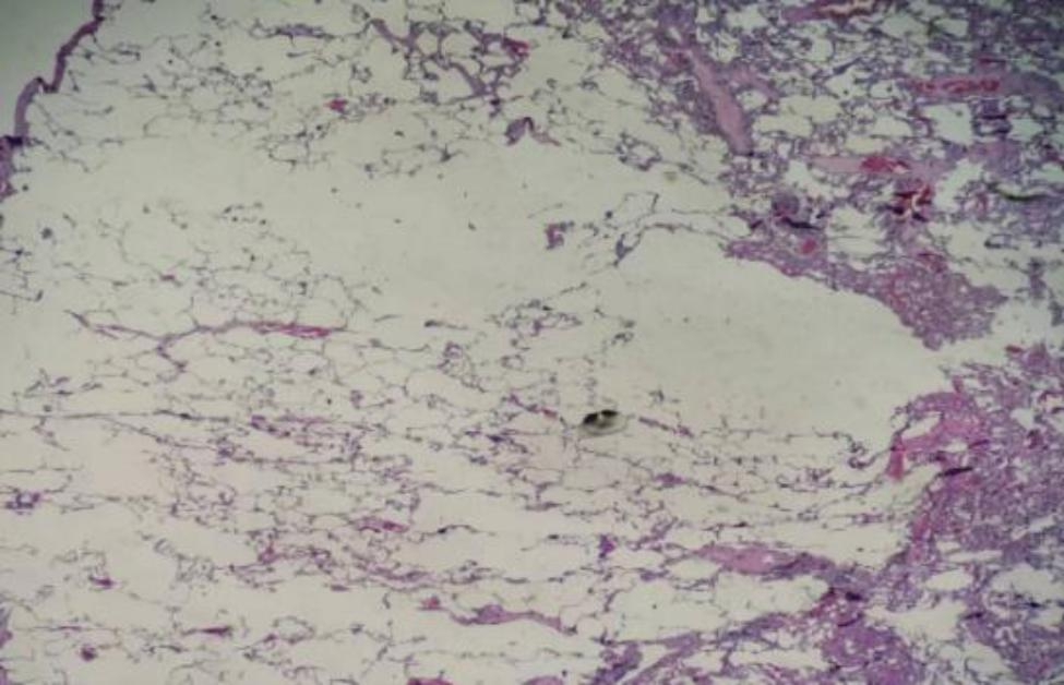
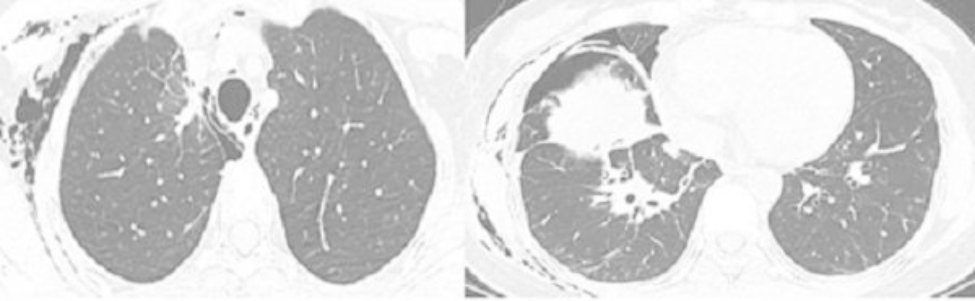
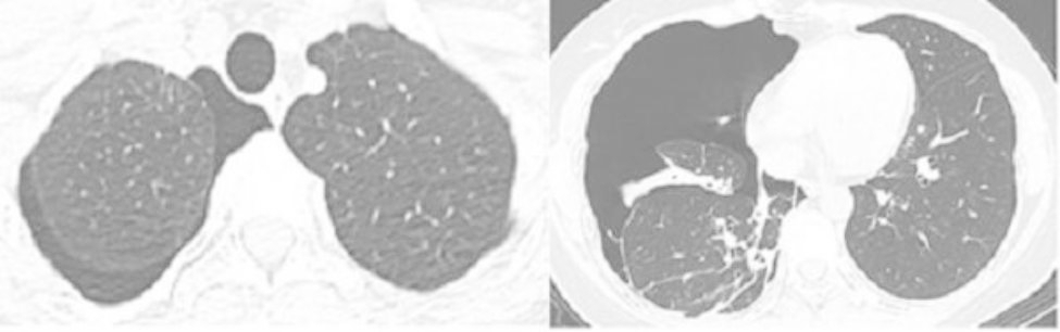
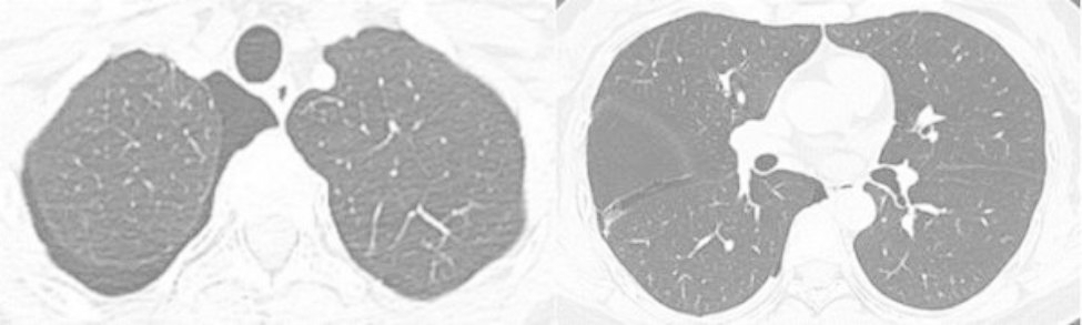
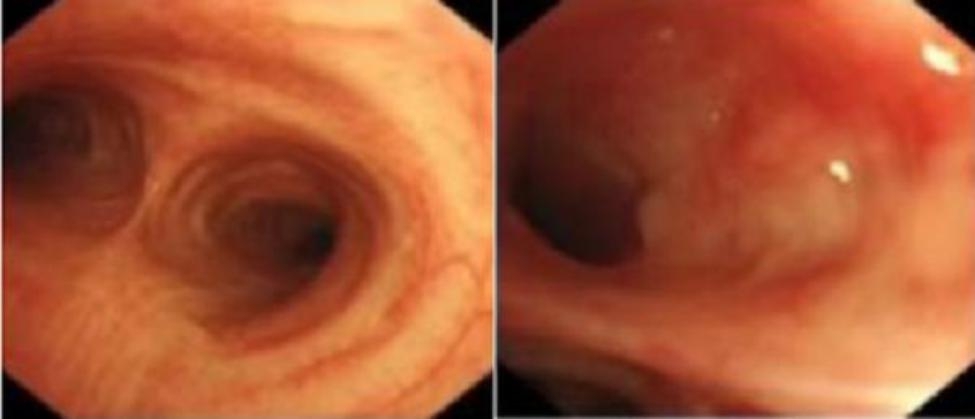
Case report: A 43-year-old young woman went to the hospital several times because of recurrent dyspnea and was diagnosed with a right spontaneous pneumothorax. She underwent multiple closed thoracic drainage procedures, but the pneumothorax was not completely resolved. Pleural biopsy pathology was chronic inflammation; there was no evidence of tuberculosis. A small amount of pneumothorax persisted, intermittent dyspnea became more severe, and pneumothorax increased. Bronchoscopy showed thickening of the left lung lingular segment mucosa, and the bronchial lavage fluid gene X-PERT/rifampicin resistance test was positive. After one month of anti-tuberculosis treatment, the symptoms of short breath were completely relieved, and chest computerized tomography (CT) showed complete resolution of the right pneumothorax.
Conclusions: When searching for the cause of spontaneous pneumothorax, people should not overlook tuberculosis-related secondary pneumothorax, which should be diagnosed and treated as soon as possible.
Keywords: Bronchial tuberculosis; Secondary pneumothorax; Spontaneous pneumothorax.
© 2023. The Author(s).
Conflict of interest statement
The authors declare that they have no competing interests.
Figures


References
-
- McKnight CL, Burns B. Pneumothorax. In: StatPearls [Internet]. Treasure Island (FL): StatPearls Publishing; 2022 [cited 2022 Dec 10]. Available from: http://www.ncbi.nlm.nih.gov/books/NBK441885/
-
- Baumann MH, Noppen M, Pneumothorax. Respirology. 2004 Jun;9(2):157–64. - PubMed
-
- Baumann MH, Strange C, Heffner JE, Light R, Kirby TJ, Klein J, et al. Management of spontaneous pneumothorax: an american college of chest Physicians Delphi consensus statement. Chest. 2001 Feb;119(2):590–602. - PubMed
-
- Bintcliffe O, Maskell N. Spontaneous pneumothorax. BMJ. 2014 May;8:348:g2928. - PubMed
Publication types
MeSH terms
LinkOut - more resources
Full Text Sources
Medical
