. 2023 Mar 21;20(1):82.
doi: 10.1186/s12974-023-02765-2.

Stellate ganglion block ameliorated central post-stroke pain with comorbid anxiety and depression through inhibiting HIF-1α/NLRP3 signaling following thalamic hemorrhagic stroke

Affiliations

Stellate ganglion block ameliorated central post-stroke pain with comorbid anxiety and depression through inhibiting HIF-1α/NLRP3 signaling following thalamic hemorrhagic stroke

Zhong-Mou Shi et al. J Neuroinflammation. .

Abstract

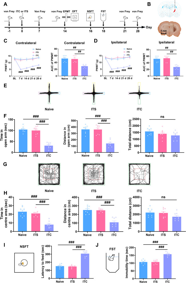
Background: Central post-stroke pain (CPSP) is an intractable and disabling central neuropathic pain that severely affects patients' lives, well-being, and socialization abilities. However, CPSP has been poorly studied mechanistically and its treatment remains challenging. Here, we used a rat model of CPSP induced by thalamic hemorrhage to investigate its underlying mechanisms and the effect of stellate ganglion block (SGB) on CPSP and emotional comorbidities.

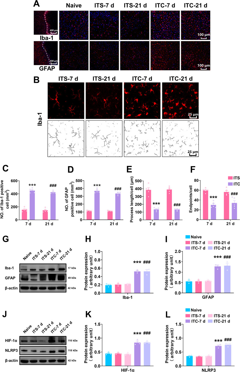
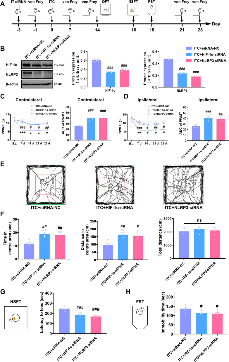
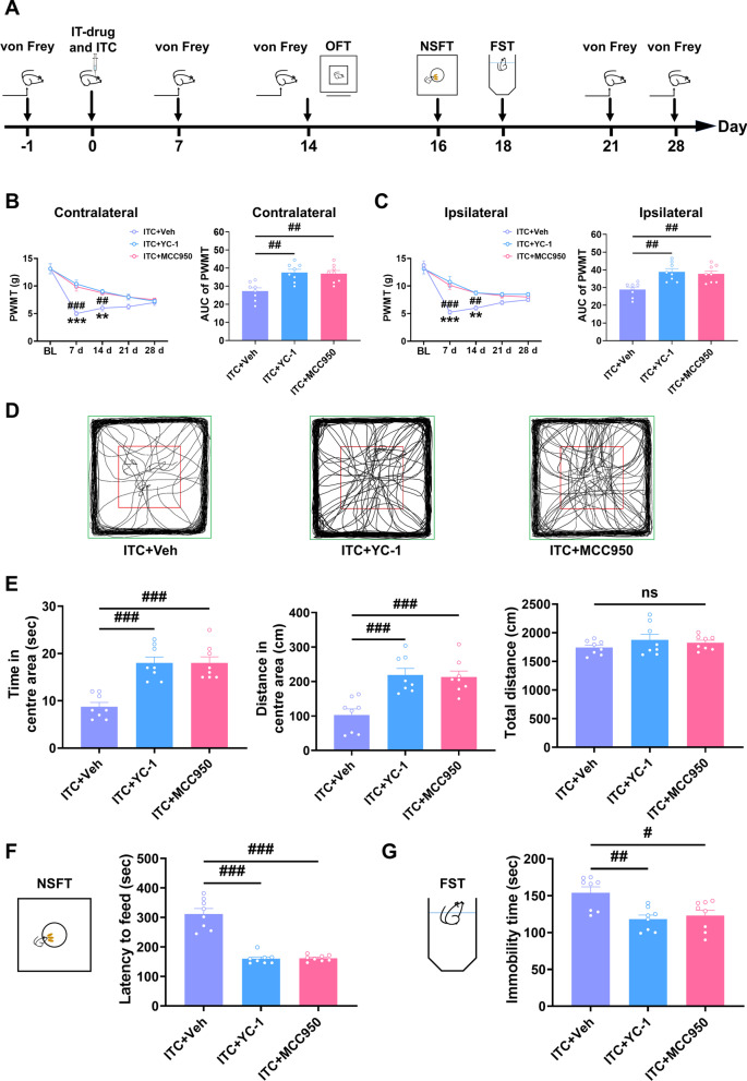
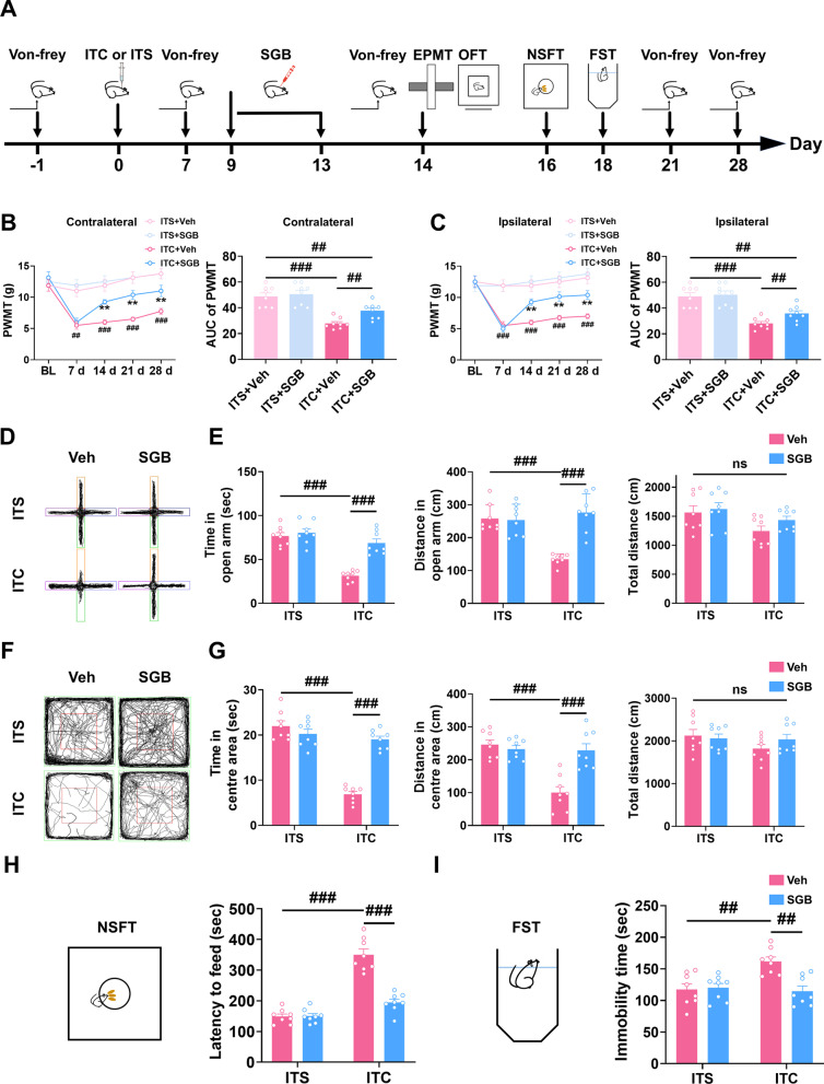
Methods: Thalamic hemorrhage was produced by injecting collagenase IV into the ventral-posterolateral nucleus (VPL) of the right thalamus. The up-and-down method with von Frey hairs was used to measure the mechanical allodynia. Behavioral tests were carried out to examine depressive and anxiety-like behaviors including the open field test (OFT), elevated plus maze test (EPMT), novelty-suppressed feeding test (NSFT), and forced swim test (FST). The peri-thalamic lesion tissues were collected for immunofluorescence, western blotting, and enzyme-linked immunosorbent assay (ELISA). Genetic knockdown of thalamic hypoxia-inducible factor-1α (HIF-1α) and NOD-like receptor thermal protein domain associated protein 3 (NLRP3) with microinjection of HIF-1α siRNA and NLRP3 siRNA into the VPL of thalamus were performed 3 days before collagenase injection into the same regions. Microinjection of lificiguat (YC-1) and MCC950 into the VPL of thalamus were administrated 30 min before the collagenase injection in order to inhibited HIF-1α and NLRP3 pharmacologically. Repetitive right SGB was performed daily for 5 days and laser speckle contrast imaging (LSCI) was conducted to examine cerebral blood flow.

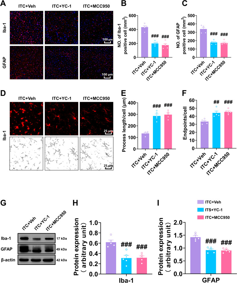
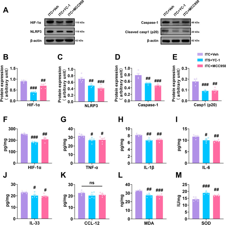
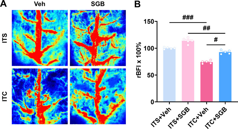
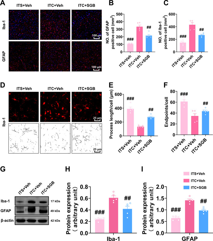
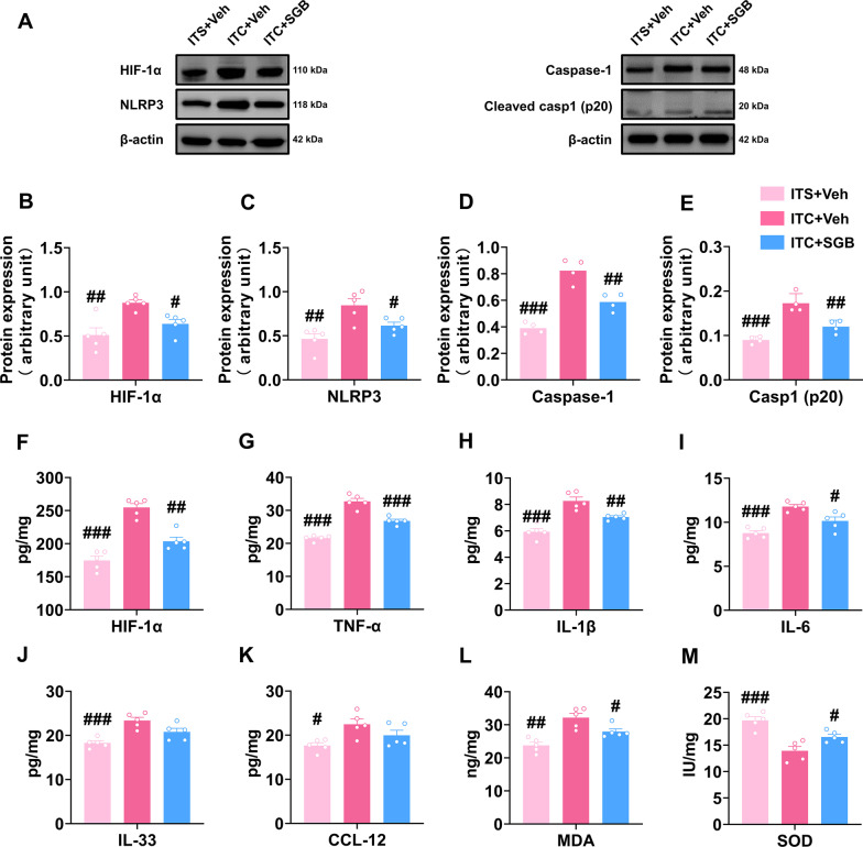
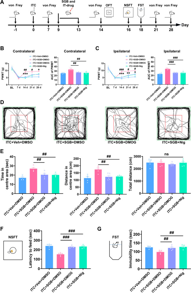
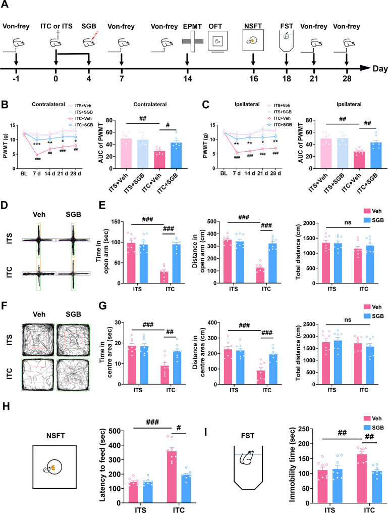
Results: Thalamic hemorrhage caused persistent mechanical allodynia and anxiety- and depression-like behaviors. Accompanying the persistent mechanical allodynia, the expression of HIF-1α and NLRP3, as well as the activities of microglia and astrocytes in the peri-thalamic lesion sites, were significantly increased. Genetic knockdown of thalamic HIF-1α and NLRP3 significantly attenuated mechanical allodynia and anxiety- and depression-like behaviors following thalamic hemorrhage. Further studies revealed that intra-thalamic injection of YC-1, or MCC950 significantly suppressed the activation of microglia and astrocytes, the release of pro-inflammatory cytokines, the upregulation of malondialdehyde (MDA), and the downregulation of superoxide dismutase (SOD), as well as mechanical allodynia and anxiety- and depression-like behaviors following thalamic hemorrhage. In addition, repetitive ipsilateral SGB significantly restored the upregulated HIF-1α/NLRP3 signaling and the hyperactivated microglia and astrocytes following thalamic hemorrhage. The enhanced expression of pro-inflammatory cytokines and the oxidative stress in the peri-thalamic lesion sites were also reversed by SGB. Moreover, LSCI showed that repetitive SGB significantly increased cerebral blood flow following thalamic hemorrhage. Most strikingly, SGB not only prevented, but also reversed the development of mechanical allodynia and anxiety- and depression-like behaviors induced by thalamic hemorrhage. However, pharmacological activation of thalamic HIF-1α and NLRP3 with specific agonists significantly eliminated the therapeutic effects of SGB on mechanical allodynia and anxiety- and depression-like behaviors following thalamic hemorrhage.

Conclusion: This study demonstrated for the first time that SGB could improve CPSP with comorbid anxiety and depression by increasing cerebral blood flow and inhibiting HIF-1α/NLRP3 inflammatory signaling.

Keywords: Anxiety; Central post-stroke pain; Depression; Hypoxia-inducible factor 1α; NLRP3; Neuroinflammation; Stellate ganglion block; Thalamus.


Conflict of interest statement

The authors have no conflict or competing interests.

Figures

Fig. 1
Fig. 1
ITC rats exhibited mechanical allodynia and anxiodepressive-like behaviors. A Experimental timeline for surgical procedure and behavior tests. B Schematic diagram showing the injection site of ITC (top) and representative photomicrograph of brain slice showing the hemorrhagic lesion location following ITC (bottom). Scale bar = 1 mm. C The PWMT of the contralateral hindpaw was significantly decreased 7 days after ITC and persisted throughout the testing period, as did the area under curve of the contralateral hindpaw PWMT (n = 8, PWMT: group, F2, 21 = 15.53, day, F4, 84 = 25.63, group × day, F8, 84 = 16.45, ###p < 0.001 vs Naive; AUC: F2, 21 = 27.71, ##p < 0.01). D The PWMT of the ipsilateral hindpaw was significantly decreased 7 days after ITC and persisted throughout the testing period, as did the area under curve of the ipsilateral hindpaw PWMT (n = 8, PWMT: group, F2, 21 = 22.95, day, F4, 84 = 29.24, group × day, F4, 84 = 19.49, ###p < 0.001 vs Naive; AUC: F2, 21 = 19.18, ##p < 0.01). E Representative track plot in the EPMT. F ITC decreased the time spent and traveled distance in the open arms, but had no effect on the total traveled distance in EPMT (n = 8, time in open arm: F2, 21 = 61.14, ###p < 0.001; distance in open arm: F2, 21 = 32.31, ###p < 0.001; total distance: ns, no significance). G Representative track plot in the OFT. H ITC decreased the time spent and traveled distance in central area, but had no effect on the total traveled distance in the OFT (n = 8, time in central area: F2, 21 = 28.00, ###p < 0.001; distance in central area: F2, 21 = 35.95, ###p < 0.001; total distance: ns, no significance). I ITC increased the latency to feed in the NSFT (n = 8, F2, 21 = 38.23, ###p < 0.001). J ITC increased the immobility time in the FST (n = 8, F2, 21 = 97.32, ###p < 0.001). Data are expressed as mean ± SEM, one-way ANOVA followed by the Tukey test
Fig. 2
Fig. 2
ITC activated microglia and astrocytes and upregulated HIF-1α and NLRP3 in the peri-thalamic lesion sites of rats. A Representative immunofluorescence images showing the time course expressions of GFAP and Iba-1, in the peri-thalamic lesion sites. Scale bar = 100 μm. B Representative magnified images of microglia (top) and the corresponding black-and-white, skeletonized images (bottom) in the peri-thalamic lesion sites. Scale bar = 25 μm. C, D Quantification of cell number showed ITC increased Iba-1 (C) and GFAP (D) positive cells in in the peri-thalamic lesion sites at 7 and 21 d n = 5, Iba-1: F3, 16 = 167.2, ***p < 0.001 vs ITS-7 d, ###p < 0.001 vs ITS-21 d; n = 5, GFAP: F3, 16 = 243.1, ***p < 0.001 vs ITS-7 d, ###p < 0.001 vs ITS-21 d). E Quantification of process length showed ITC reduced the process length of microglia at 7 and 21 d (n = 5, F3, 16 = 20.71, ***p < 0.001 vs ITS-7 d, ###p < 0.001 vs ITS-21 d). F Quantification of endpoint showed ITC decreased the endpoints in microglia at 7 and 21 d (n = 5, F3, 16 = 22.08, ***p < 0.01 vs ITS-7 d, ###p < 0.001 vs ITS-21 d). G Representative western blots of Iba-1 and GFAP expression in total proteins of the peri-thalamic lesion sites in naïve, ITS, and ITC rats. H Quantitative summary result showed ITC increased Iba-1 expression at 7 and 21 d, while ITS had no effect (n = 3, F4, 10 = 46.93, ***p < 0.001 vs ITS-7 d, ###p < 0.01 vs ITS-21 d). I Quantitative summary result showed ITC increased GFAP expression at 7 and 21d, while ITS had no effect (n = 3, F4, 10 = 33.41, ***p < 0.001 vs ITS- 7d, ###p < 0.001 vs ITS-21 d). J Representative western blots of HIF-1α and NLRP3 expression in total proteins of the peri-thalamic lesion sites in naïve, ITS, and ITC rats. K Quantitative summary result showed ITC increased HIF-1α expression at 7 and 21 d, while ITS had no effect (n = 3, F4, 10 = 91.24, ***p < 0.001 vs ITS-7 d, ###p < 0.001 vs ITS-21 d). L Quantitative summary result showed ITC increased NLRP3 expression at 7 and 21 d, while ITS had no effect (n = 3, F4, 10 = 67.22, ***p < 0.001 vs ITS-7 d, ###p < 0.001 vs ITS-21 d). Data are expressed as mean ± SEM, one-way ANOVA followed by Tukey test
Fig. 3
Fig. 3
Intra-thalamic injection of HIF-1α siRNA or NLRP3 siRNA significantly prevented ITC-induced mechanical allodynia and anxiodepressive-like behaviors in rats. A The experimental timeline of surgical procedure and behavior tests. B Both HIF-1α siRNA and NLRP3 siRNA silenced the expression of HIF-1α and NLRP3 in total proteins of the peri-thalamic lesion sites in ITC rats. (n = 5, HIF-1α: F2, 12 = 143.9, ###p < 0.001 vs ITC + siRNA-NC; NLRP3: F2, 12 = 248.2, ###p < 0.001 vs ITC + siRNA-NC). C Temporal changes of PWMT in contralateral hindpaw after thalamic hemorrhagic stroke and the area under curve of the contralateral hindpaw PWMT (n = 5, PWMT: group, F2, 12 = 13.49, day, F4, 48 = 14.54, group × day, F8, 48 = 2.182, #p < 0.05, ##p < 0.01, ###p < 0.001 ITC + siRNA-NC vs ITC + HIF-1α-siRNA, *p < 0.05, ***p < 0.001, ITC + siRNA-NC vs ITC + NLRP3-siRNA; AUC: F2, 12 = 20.89, ###p < 0.001). D Temporal changes of PWMT in ipsilateral hindpaw after thalamic hemorrhagic stroke and the area under curve of the ipsilateral hindpaw PWMT (n = 5, PWMT: group, F2, 12 = 11.54, day, F4, 48 = 21.28, group × day, F12, 48 = 2.796, #p < 0.05, ##p < 0.01, ###p < 0.001, ITC + siRNA-NC vs ITC + HIF-1α-siRNA, *p < 0.05, **p < 0.01, ITC + siRNA-NC vs ITC + NL; RP3-siRNA; AUC: F2, 12 = 16.86, ##p < 0.01, ###p < 0.001). E Representative track plot in the OFT. F HIF-1α siRNA and NLRP3 siRNA increased the time spent and traveled distance traveled in central area, and but had no effect on the total traveled distance in the OFT (n = 5, time in central area: F2, 12 = 10.51, ##p < 0.01; distance in central area: F2, 21 = 9.466, #p < 0.05; total distance: ns, no significance). G HIF-1α siRNA and NLRP3 siRNA decreased the latency to feed in the NSFT (n = 5, F2, 12 = 24.43, ###p < 0.001). H HIF-1α siRNA and NLRP3 siRNA decreased the immobility time in the FST (n = 5, F2, 12 = 5.035, #p < 0.05). Data are expressed as mean ± SEM, one-way ANOVA followed by Tukey test
Fig. 4
Fig. 4
Intra-thalamic injection of YC-1 or MCC950 significantly prevented ITC-induced mechanical allodynia and anxiodepressive-like behaviors in rats. A The experimental timeline of surgical procedure and behavior tests. B Intra-thalamic injections of YC-1 and MCC950 increased the PWMT of the contralateral hindpaw and the area under the curve of the PWMT following ITC (n = 8, PWMT: group, F2, 21 = 6.30, day, F4, 84 = 94.81, group × day, F8, 84 = 7.687, ###p < 0.001, ITC + YC-1 vs ITC + Veh, **p < 0.001, ITC + Veh vs ITC + MCC950 ; AUC: F2, 21 = 10.48, ##p < 0.01). C Intra-thalamic injections of YC-1 and MCC950 increased the PWMT of the ipsilateral hindpaw and the area under the curve of the PWMT following ITC (n = 8, PWMT: group, F2, 21 = 6.485, day, F4, 84 = 81.73, group × day, F8, 84 = 8.051, ###p < 0.001, ITC + YC-1 vs ITC + Veh, **p < 0.01, ***p < 0.001, ITC + Veh vs ITC + MCC950; AUC: F2, 21 = 10.70, ##p < 0.01). D Representative track plot in the OFT. E YC-1 and MCC950 increased the time spent and traveled distance traveled in central area, and but had no effect on the total traveled distance in the OFT (n = 8, time in central area: F2, 21 = 21.06, ###p < 0.001; distance in central area: F2, 21 = 13.42, ###p < 0.001; total distance: ns, no significance). F YC-1 and MCC950 decreased the latency to feed in the NSFT (n = 8, W2, 11.78 = 31.52, ###p < 0.001). G YC-1 and MCC950 decreased the immobility time in the FST (n = 8, F2, 21 = 7.689, #p < 0.05, ##p < 0.01). Data are expressed as mean ± SEM, one-way ANOVA followed by Tukey test; Welch ANOVA followed by Tamhane’s T2 test
Fig. 5
Fig. 5
Intra-thalamic injection of YC-1 or MCC950 suppressed ITC-induced activation of microglia and astrocytes in peri-thalamic lesion sites. A Representative immunofluorescence images showing the expressions of GFAP and Iba-1 in the peri-thalamic lesion sites. Scale bar = 100 μm. B Quantification of cell number showed YC-1 or MCC950 decreased Iba-1 positive cells in the peri-thalamic lesion sites in ITC rats (n = 5, F2,12 = 56.07, ###p < 0.001 vs ITC + Veh). C Quantification of cell number showed YC-1 or MCC950 decreased GFAP-positive cells in the peri-thalamic lesion sites in ITC rats (n = 5, F2,12 = 44.62, ###p < 0.001 vs ITC + Veh). D Representative magnified images of microglia (top) and the corresponding black-and-white, skeletonized images (bottom) in the peri-thalamic lesion sites. Scale bar = 25 μm. E Quantification of process length showed YC-1 or MCC950 increased the process length of microglia in the peri-thalamic lesion sites (n = 5, F2,12 = 44.37, ###p < 0.001 vs ITC + Veh). F Quantification of endpoint showed YC-1 or MCC950 increased the endpoints in microglia in the peri-thalamic lesion sites (n = 5, F2,12 = 22.58, ##p < 0.01, ###p < 0.001 vs ITC + Veh). G Representative western blots of Iba-1 and GFAP expression in total proteins of the peri-thalamic lesion sites in ITC rats. H Quantitative summary result showed YC-1 or MCC950 decreased Iba-1 expression in the peri-thalamic lesion sites in ITC rats (n = 5, F2,12 = 16.17, ###p < 0.01 vs ITC + Veh). I Quantitative summary result showed YC-1 or MCC950 decreased GFAP expression in the peri-thalamic lesion sites in ITC rats (n = 5, F2,12 = 37.29, ###p < 0.001 vs ITC + Veh). Data are expressed as mean ± SEM, one-way ANOVA followed by the Tukey test
Fig. 6
Fig. 6
Intra-thalamic injection of YC-1 or MCC950 reduced ITC-induced local inflammation and oxidative stress in the peri-thalamic lesion sites in rats. A Representative western blots of HIF-1α, NLRP3, and Caspase-1, and cleaved caspase-1 expression in total proteins of the peri-thalamic lesion sites in ITC rats. B–E Quantitative summary result showed intra-thalamic injection of YC-1 or MCC950 reduced HIF-1α (B), NLRP3 (C), Caspase-1 (D), and cleaved caspase-1 (E) expression in the peri-thalamic lesion sites in ITC rats (n = 5, HIF-1α: F2,12 = 48.89, ##p < 0.01, ###p < 0.001 vs ITC + Veh; NLRP3: F2,12 = 18.71, ##p < 0.01, ###p < 0.001 vs ITC + Veh; n = 4, Caspase-1: F2,9 = 20.56, ##p < 0.01, ###p < 0.001 vs ITC + Veh; cleaved caspase-1 p20: F2,9 = 19.36, ##p < 0.01, ###p < 0.001 vs ITC + Veh). F ELISA result showed intra-thalamic injection of YC-1 or MCC950 reduced HIF-1α in the peri-thalamic lesion sites in ITC rats (n = 5, F2,12 = 38.82, ##p < 0.01, ###p < 0.001 vs ITC + Veh). G–K ELISA results showed intra-thalamic injection of YC-1 or MCC950 reduced TNF-α, IL-1β, IL-6 and IL-33 expression, but had no effect on CCL-12 expression in the peri-thalamic lesion sites (n = 5, TNF-α: F2,12 = 5.292, #p < 0.05 vs ITC + Veh; IL-1β: F2,12 = 23.33, ##p < 0.01 vs ITC + Veh; IL-6: F2,12 = 9.249, #p < 0.05, ##p < 0.01 vs ITC + Veh; IL-33: F2,12 = 5.978, #p < 0.05 vs ITC + Veh). L ELISA result showed intra-thalamic injection of YC-1 or MCC950 reduced the expression of antioxidant factor MDA in the peri-thalamic lesion sites (n = 5, F2,12 = 16.69, ##p < 0.01, ###p < 0.001 vs ITC + Veh). M ELISA result showed intra-thalamic injection of YC-1 or MCC950 increased the expression of oxidative factor SOD in the peri-thalamic lesion sites (n = 5, F2,12 = 21.45, ##p < 0.01, ###p < 0.001 vs ITC + Veh). Data are expressed as mean ± SEM, one-way ANOVA followed by the Tukey test
Fig. 7
Fig. 7
Post-treatment with repetitive SGB reversed the development of mechanical allodynia and anxiodepressive-like behaviors in CPSP rats. A The experimental timeline of surgical procedure and behavior tests. B Temporal changes of PWMT in contralateral hindpaw after thalamic hemorrhagic stroke and the area under curve of the contralateral hindpaw PWMT (n = 8, PWMT: group, F3,28 = 14.78, day, F4,112 = 29.22, group × day, F12,112 = 7.669, **p < 0.01 vs ITC + Veh, ##p < 0.01, ###p < 0.001 vs ITS + Veh; AUC: F3,28 = 18.74, ##p < 0.01, ###p < 0.001). C Temporal changes of PWMT in ipsilateral hindpaw after thalamic hemorrhagic stroke and the area under curve of the ipsilateral hindpaw PWMT (n = 8, PWMT: group, F3,28 = 15.01, day, F4,122 = 26.73, group × day, F12,112 = 9.919, **p < 0.01 vs ITC + Veh, ###p < 0.001 vs ITS + Veh; AUC: F2,21 = 19.21, ##p < 0.01, ###p < 0.001). D Representative track plot in the EPMT. E Repetitive SGB increased the time spent and traveled distance in the open arm in ITC group, but had no effect on ITS group (n = 8, time in open arm; F3,28 = 32.45, ###p < 0.001; distance in open arm; W3.0,13.63 = 38.47, ###p < 0.001, total distance: ns, no significance). F Representative track plot in the OFT. G Repetitive SGB increased the time spent and traveled distance in the central area in ITC group, but had no effect on ITS group (n = 8, time in central area: F3,28 = 58.17, ###p < 0.001; distance in central F3,28 = 17.36, ###p < 0.001; total distance: ns, no significance). H Repetitive SGB decreased the latency to feed in the NSFT in ITC group, but had no effect on ITS group (n = 8, F3,28 = 60.17, ###p < 0.001). I Repetitive SGB decreased the immobility time in the FST in ITC group, but had no effect on ITS group (n = 8, F3,28 = 8.058, ##p < 0.01). Data are expressed as mean ± SEM, one-way or two-way ANOVA followed by Tukey test, Welch ANOVA followed by Tamhane’s T2 test
Fig. 8
Fig. 8
Repetitive SGB increased the cerebral blood flow of CPSP rats. A Representative images of cerebral blood flow. B Quantitative summary result showed ITC decreased the cerebral blood flow, while repetitive SGB enhanced cerebral blood flow in ITC rats (n = 5, F3, 16 = 112.2, #p < 0.05, ##p < 0.01, ###p < 0.001)
Fig. 9
Fig. 9
Post-treatment with repetitive SGB inhibited the activation of microglia and astrocytes in peri-thalamic lesion sites of CPSP rats. A Representative immunofluorescence images showing the expressions of GFAP and Iba-1 in the peri-thalamic lesion sites. Scale bar = 100 μm. B, C Quantification of cell number showed repetitive SGB decreased the total number of GFAP (B) and Iba-1 (C) positive cells in the peri-thalamic lesion sites of CPSP rats (n = 5, Iba-1 F2,12 = 40.67, ##p < 0.01, ###p < 0.001 vs ITC + Veh; GFAP F2,12 = 99.46, ##p < 0.01, ###p < 0.001 vs ITC + Veh). D Representative magnified images of microglia (top) and the corresponding black-and-white, skeletonized images (bottom) in the peri-thalamic lesion sites. Scale bar = 25 μm. E Quantification of process length showed repetitive SGB increased process length of microglia in the peri-thalamic lesion sites of CPSP rats (n = 5, F2,12 = 59.03, ##p < 0.01, ###p < 0.001 vs ITC + Veh). F Quantification of endpoint showed repetitive SGB increased endpoint of microglia in the peri-thalamic lesion sites of CPSP rats (n = 5, F2,12 = 15.62, ##p < 0.01, ###p < 0.001 vs ITC + Veh). g Representative western blots of Iba-1 and GFAP expression in total proteins of the peri-thalamic lesion sites, H, I Quantitative summary result showed repetitive SGB decreased Iba-1 (H) and GFAP (I) expression in the peri-thalamic lesion sites of CPSP rats (n = 5, Iba-1: F2,12 = 28.08, ##p < 0.01, ###p < 0.001 vs ITC + Veh; GFAP; F2,12 = 62.45, ##p < 0.01, ###p < 0.001 vs ITC + Veh). Data are expressed as mean ± SEM, one-way ANOVA followed by the Tukey test
Fig. 10
Fig. 10
Post-treatment with repetitive SGB reversed local inflammation and oxidative stress in the peri-thalamic lesion sites of CPSP rats. A Representative western blotting of HIF-1α, NLRP3, Caspase-1, and cleaved caspase-1 expression in the peri-thalamic lesion sites. B–E Quantitative summary result showed repetitive SGB decreased HIF-1α (B), NLRP3 (C), Caspase-1 (D), and cleaved caspase-1 (E) expression in the peri-thalamic lesion sites of CPSP rats (n = 5, HIF-1α: F2,12 = 9.896, ##p < 0.01, ###p < 0.001 vs ITC + Veh; NLRP3: F2,12 = 10.40, #p < 0.05, ##p < 0.01 vs ITC + Veh; n = 4, Caspase-1: F2,9 = 38.72, ##p < 0.01, ###p < 0.001 vs ITC + Veh; cleaved caspase-1 p20: F2,9 = 28.21, ##p < 0.01, ###p < 0.001 vs ITC + Veh). F ELISA result showed repetitive SGB decreased HIF-1α expression in the peri-thalamic lesion sites of CPSP rats (n = 5, F2,12 = 42.10, ##p < 0.01, ###p < 0.001 vs ITC + Veh). G–K ELISA results showed repetitive SGB decreased TNF-α, IL-1β and IL-6 in the peri-thalamic lesion sites of CPSP rats, but had no effect on IL-33 and CCL-12 (n = 5, TNF-α: F2,12 = 72.48, ###p < 0.001 vs ITC + Veh; IL-1β: F2,12 = 26.98, ##p < 0.01, ###p < 0.001 vs ITC + Veh; IL-6: F2,12 = 20.58, #p < 0.05, #p < 0.05, ###p < 0.001 vs ITC + Veh; IL-33: F2,12 = 12.82, ###p < 0.001 vs ITC + Veh; CCL-12: F2,12 = 5.570, #p < 0.05 vs ITC + Veh). L ELISA result showed repetitive SGB decreased the expression of antioxidant factor MDA in the peri-thalamic lesion sites of CPSP rats (n = 5, F2,12 = 15.28, #p < 0.05, ##p < 0.01 vs ITC + Veh). M ELISA result showed repetitive SGB increased the expression level of oxidative factor SOD in the peri-thalamic lesion sites of CPSP rats (n = 5, F2,12 = 17.52, #p < 0.05, ###p < 0.001 vs ITC + Veh). Data are expressed as mean ± SEM, one-way ANOVA followed by the Tukey test
Fig. 11
Fig. 11
Intra-thalamic injection of DMOG or Nigericin eliminated the therapeutic effects of SGB on mechanical allodynia and anxiety and depression in CPSP rats. A The experimental timeline of surgical procedure and behavior tests. B Intra-thalamic injections of DMOG or Nigericin decreased the PWMT of the contralateral hindpaw and the area under the curve of the PWMT in ITC rats given SGB (n = 5, PWMT: group, F3,16 = 9.517, day, F4,64 = 59.90, group × day, F12,64 = 3.019, #p < 0.05, ##p < 0.01, ITC + SGB + DSMO vs ITC + SGB + DMOG, *p < 0.05, p < 0.001, ITC + SGB + DMSO vs ITC + SGB + Nig; AUC: F3,16 = 14.57, ##p < 0.01). C Intra-thalamic injections of DMOG or Nigericin decreased the PWMT of the ipsilateral hindpaw and the area under the curve of the PWMT in ITC rats given SGB (n = 5, PWMT: group, F3,16 = 17.76, day, F4,64 = 60.99, group × day, F12,64 = 2.905, #p < 0.05, ###p < 0.001, ITC + SGB + DMSO vs ITC + SGB + DMOG, *p < 0.05, **p < 0.01,***p < 0.001, ITC + SGB + DMSO vs ITC + SGB + Nig; AUC: F3,16 = 29.48 ###p < 0.001). D Representative track plot in the OFT. E DMOG and Nigericin decreased the time spent and traveled distance traveled in central area, and but had no effect on the total traveled distance in ITC rats given SGB (n = 5, time in central area: F3,16 = 12.60, ##p < 0.01; distance in central area: F3,16 = 8.491, ##p < 0.01; total distance: ns, no significance). F DMOG and Nigericin increased the latency to feed in the NSFT in ITC rats given SGB (n = 5, F3,16 = 27.42, ###p < 0.001). G DMOG and Nigericin increased the immobility time in the FST in ITC rats given SGB (n = 5, F3,16 = 8.085, ##p < 0.01). Data are expressed as mean ± SEM, one-way ANOVA followed by Tukey test
Fig. 12
Fig. 12
Pretreatment with repetitive SGB prevents the development of mechanical allodynia and anxiodepressive-like behaviors in ITC rats. A The experimental timeline of surgical procedure and behavior tests. B Temporal changes of PWMT in contralateral hindpaw after thalamic hemorrhagic stroke and the area under curve of the contralateral hindpaw PWMT (n = 8, PWMT: group, F3,28 = 9.268, day, F4,112 = 10.48, group × day, F12,112 = 6.246, *p < 0.05, **p < 0.01 vs ITC + Veh, ###p < 0.001 vs ITS + Veh; AUC: F3,28 = 18.74, #p < 0.05, ##p < 0.01). C Temporal changes of PWMT in ipsilateral hindpaw after thalamic hemorrhagic stroke and the area under curve of the ipsilateral hindpaw PWMT (n = 8, PWMT: group, F3,28 = 10.48, day, F4,122 = 17.65, group × day, F12,112 = 7.005, *p < 0.05, **p < 0.01 vs ITC + Veh, ###p < 0.001 vs ITS + Veh; AUC: F2,21 = 19.21, ##p < 0.01). D Representative track plot in the EPMT. E Pretreatment with repetitive SGB increased the time spent and traveled distance in the open arm in ITC group, but had no effect on ITS group (n = 8, time in open arm: F3,28 = 35.13, ###p < 0.001; distance in open arm: F3,28 = 51.39, ###p < 0.001; total distance: ns, no significance). F Representative track plot in the OFT. G Pretreatment with repetitive SGB increased the time spent and traveled distance in central area in ITC group, but had no effect on ITS group (n = 8, time in central area: F3,28 = 11.38, ##p < 0.01, ###p < 0.001; distance in central area: F3,28 = 14.80, ###p < 0.001; total distance: ns, no significance). H Pretreatment with repetitive SGB decreased the latency to feed in the NSFT in ITC group, but had no effect on ITS group (n = 8, W3.0,15.05 = 23.77, #p < 0.05, ###p < 0.001). I Pretreatment with repetitive SGB decreased the immobility time in the FST in ITC group, but had no effect on ITS group (n = 8, F2,21 = 8.253, ##p < 0.01). Data are expressed as mean ± SEM, one-way or two-way ANOVA followed by Tukey test, Welch ANOVA followed by Tamhane’s T2 test

References

    1. Delpont B, Blanc C, Osseby GV, Hervieu-Begue M, Giroud M, Bejot Y. Pain after stroke: a review. Rev Neurol (Paris) 2018;174:671–674. doi: 10.1016/j.neurol.2017.11.011. - DOI - PubMed
    1. Oh H, Seo W. A comprehensive review of central post-stroke pain. Pain Manag Nurs. 2015;16:804–818. doi: 10.1016/j.pmn.2015.03.002. - DOI - PubMed
    1. Choi HR, Aktas A, Bottros MM. Pharmacotherapy to manage central post-stroke pain. CNS Drugs. 2021;35:151–160. doi: 10.1007/s40263-021-00791-3. - DOI - PubMed
    1. Gurba KN, Chaudhry R, Haroutounian S. Central neuropathic pain syndromes: current and emerging pharmacological strategies. CNS Drugs. 2022;36:483–516. doi: 10.1007/s40263-022-00914-4. - DOI - PubMed
    1. Plecash AR, Chebini A, Ip A, Lai JJ, Mattar AA, Randhawa J, Field TS. Updates in the treatment of post-stroke pain. Curr Neurol Neurosci Rep. 2019;19:86. doi: 10.1007/s11910-019-1003-2. - DOI - PubMed

MeSH terms

Substances