. 2023 Mar 21;16(1):58.
doi: 10.1186/s13048-023-01136-y.

Placental mesenchymal stem cells ameliorate NLRP3 inflammasome-induced ovarian insufficiency by modulating macrophage M2 polarization

Affiliations

Placental mesenchymal stem cells ameliorate NLRP3 inflammasome-induced ovarian insufficiency by modulating macrophage M2 polarization

Dongmei Chen et al. J Ovarian Res. .

Abstract

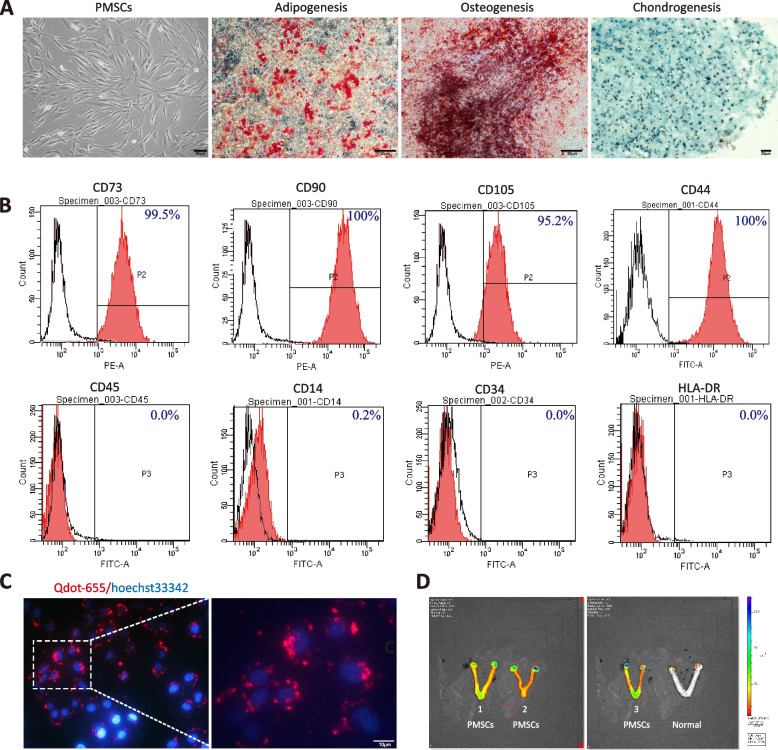
Background: Premature ovarian insufficiency (POI) is a common clinical problem, however, there are currently no effective therapies. Pyroptosis induced by the NLRP3 inflammasome is considered a possible mechanism of POI. Placental mesenchymal stem cells (PMSCs) have excellent immunomodulatory potential and offer a promising method for treating POI.

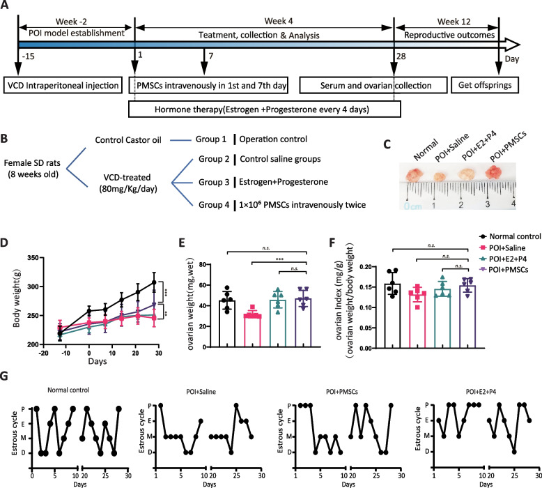
Methods: Female Sprague-Dawley rats were randomly divided into four treatment groups: control (no POI), POI with no PMSCs, POI with PMSCs transplant, and POI with hormones (estrogen + progesterone) as positive control. POI was induced by exposure to 4-vinylcyclohexene diepoxide (VCD) for 15 days. After four weeks, all animals were euthanized and examined for pathology. Hormone levels were measured and ovarian function was evaluated in relation to the estrous cycle. Levels of NLRP3 inflammasome pathway proteins were determined by immunohistochemistry and western blot.

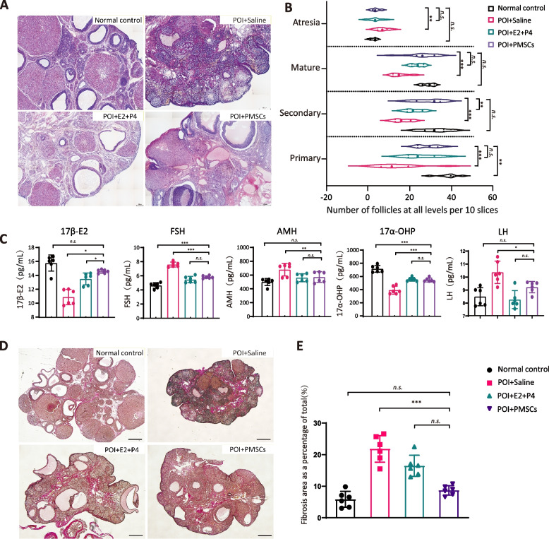
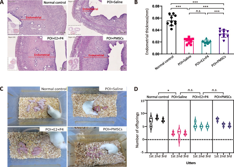
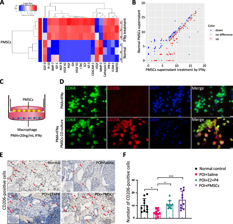
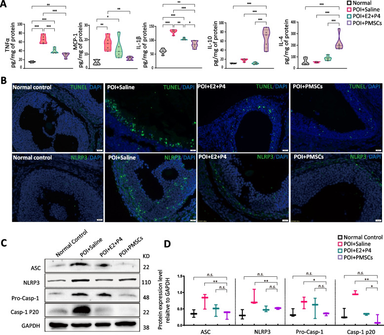
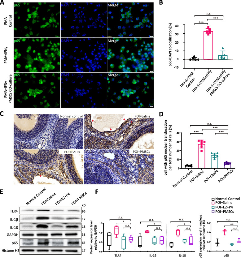
Results: VCD significantly damaged rat follicles at different estrous stages. Injection of human PMSCs improved ovarian function and reproductive ability of POI rats compared to the sham and hormone groups. Our data also showed that PMSCs markedly suppress cell pyroptosis via downregulation of the NLRP3 inflammasome, caspase-1, IL-1β and IL-18 compared to the other two groups. The human PMSCs increased the expression of IL-4 and IL-10 and decreased pro-inflammatory factors by phenotypic changes in macrophages.

Conclusions: Our findings revealed a novel mechanism of follicular dysfunction and ovarian fibrosis via activation of the NLRP3 inflammasome followed by secretion of pro-inflammatory factors. Transplantation of PMSCs into POI rats suppressed pro-inflammatory factor production, NLRP3 inflammasome formation and pyroptosis, and improved ovarian function.

Keywords: Inflammasome; Interferon-γ; Macrophage; NLRP3; Placental mesenchymal stem cells; Premature ovarian insufficiency; Pyroptosis.


Conflict of interest statement

The authors declare no competing interests.

Figures

Fig. 1
Fig. 1
Characterization and identification of human PMSCs. A Morphology of PMSCs at the fourth passage. Oil Red O staining was conducted for adipogenic differentiation, alizarin red staining was used for osteogenic identification, and Alcian blue indicated chondrogenesis. Scale bar: 50 or 200 µm. B human PMSCs were positive for CD73, CD90, CD105, CD44, and CD90, and were negative for CD34, CD45, CD14 and HLA-DR, as shown by flow cytometry analysis. C Qtracker® 655-labeled PMSCs (red) counterstained with Hoechst 33342 (blue). D Live imaging of POI model rat ovaries and uteri transplanted with Qtracker® 655-labeled PMSCs. A separate control rat, not inoculated with cells, was imaged and is shown at the far right for comparison
Fig. 2
Fig. 2
Experimental timeline and changes in ovarian morphology and function in SD rats. A Experimental timeline. After the POI model was established by VCD injection, seven days after the first treatment, MSC transplantation was performed by two injections of PMSCs. Estrogen and progesterone treatment was used as a positive control. B experimental grouping. C Ovarian size. D Body weight change. E Ovarian weight. F Ovarian index. G Estrous cycle was measured by exfoliated vaginal cell staining
Fig. 3
Fig. 3
Therapeutic effects of PMSCs in a rat model of POI. A H&E staining of ovaries in the control and POI groups. Scale bar: 50 µm. B The number of follicles at different stages in each group. C AMH, E2, LH and FSH levels were significantly elevated in four groups. D The VG staining in each group. Scale bar: 200 μm. E Fibrosis levels were significantly elevated in four groups compared. Data are presented as the mean ± SD. *p < 0.05, **p < 0.01, ***p < 0.001, ns, not significant. Data are means of three independent experiments in each group. Data are presented as the mean ± SD. *p < 0.05, **p < 0.01, ***p < 0.001, ns, not significant. Data are means of three independent experiments in each group
Fig. 4
Fig. 4
Endometrial regeneration and production of offspring by PMSC treatment. A H&E staining (scale bar, 200 μm) with B endometrial thickness of uteri. C The morphological images and D quantitative analysis of offspring. Data are presented as the mean ± SD. *p < 0.05, **p < 0.01, ***p < 0.001, ns, not significant. Data are means of three independent experiments in each group
Fig. 5
Fig. 5
The number of M2 macrophages was increased by PMSCs in vitro and in vivo. A Cluster analysis by cytokine array showing the difference in paracrine factors of PMSCs after IFNγ-stimulation. B Volcano map. C Model of co-culture of macrophages and PMSCs. D Immunofluorescence analysis of macrophage phenotype, CD206, for M2 macrophages (red) and nuclear staining (blue). E The IHC staining of CD206 in each group rat ovary. Scale bar, 200 μm. F Quantification of macrophage phenotypes. The number of M2-like macrophages increased dramatically in the PMSCs group. Data are presented as the mean ± SD. *p < 0.05, **p < 0.01, ***p < 0.001, ns, not significant. Data are means of three independent experiments in each group
Fig. 6
Fig. 6
Expression of NLRP3 inflammasome pathway and related inflammatory factors. A Inflammation-related cytokine expression detected by ELISA. B TUNEL staining for apoptosis and immunofluorescence assay for NLRP3. Scale bar, 100 µm C, D Immunoblot analysis of NLRP3 inflammasome pathway and related inflammatory factors. Data are mean ± SD. *p < 0.05, **p < 0.01, ***p < 0.001, ns, no significance. Data are means of three independent experiments in each group
Fig. 7
Fig. 7
Activation of teNFκB pathway in vitro and in vivo. A, B Immunofluorescence assay for nuclear translocation of p65 in macrophages. Scale bar: 20 µm. C, D Immunohistochemistry assay for nuclear translocation of p65 in ovarian. Scale bar: 20 µm. E, F Immunoblot analysis of NFκB pathway and related inflammatory factors. Data are mean ± SD. *p < 0.05, **p < 0.01, ***p < 0.001, ns, no significance. Data are means of three independent experiments in each group

References

    1. Chon SJ, Umair Z, Yoon MS. Premature Ovarian Insufficiency: Past, Present, and Future. Front Cell Dev Biol. 2021;9:672890. doi: 10.3389/fcell.2021.672890. - DOI - PMC - PubMed
    1. Ulin M, Cetin E, Hobeika E, Chugh RM, Park HS, Esfandyari S, Al-Hendy A. Human mesenchymal stem cell therapy and other novel treatment approaches for premature ovarian insufficiency. Reprod Sci. 2021;28(6):1688–1696. doi: 10.1007/s43032-021-00528-z. - DOI - PMC - PubMed
    1. Shin EY, Kim DS, Lee MJ, Lee AR, Shim SH, Baek SW, Han DK, Lee DR. Prevention of chemotherapy-induced premature ovarian insufficiency in mice by scaffold-based local delivery of human embryonic stem cell-derived mesenchymal progenitor cells. Stem Cell Res Ther. 2021;12(1):431. doi: 10.1186/s13287-021-02479-3. - DOI - PMC - PubMed
    1. Polonio AM, Garcia-Velasco JA, Herraiz S. Stem Cell Paracrine Signaling for Treatment of Premature Ovarian Insufficiency. Front Endocrinol (Lausanne) 2020;11:626322. doi: 10.3389/fendo.2020.626322. - DOI - PMC - PubMed
    1. Sen Halicioglu B, Saadat K, Tuglu MI. Adipose-derived mesenchymal stem cell transplantation in chemotherapy-induced premature ovarian insufficiency: the role of Connexin and Pannexin. Reprod Sci. 2022;29(4):1316–1331. doi: 10.1007/s43032-021-00718-9. - DOI - PubMed