. 2023 Jul;71(7):1683-1698.
doi: 10.1002/glia.24363. Epub 2023 Mar 21.

Low sulfated heparan sulfate mimetic differentially affects repair in immune-mediated and toxin-induced experimental models of demyelination

Affiliations

Low sulfated heparan sulfate mimetic differentially affects repair in immune-mediated and toxin-induced experimental models of demyelination

Susan L Lindsay et al. Glia. 2023 Jul.

Abstract

There is an urgent need for therapies that target the multicellular pathology of central nervous system (CNS) disease. Modified, nonanticoagulant heparins mimic the heparan sulfate glycan family and are known regulators of multiple cellular processes. In vitro studies have demonstrated that low sulfated modified heparin mimetics (LS-mHeps) drive repair after CNS demyelination. Herein, we test LS-mHep7 (an in vitro lead compound) in experimental autoimmune encephalomyelitis (EAE) and cuprizone-induced demyelination. In EAE, LS-mHep7 treatment resulted in faster recovery and rapidly reduced inflammation which was accompanied by restoration of animal weight. LS-mHep7 treatment had no effect on remyelination or on OLIG2 positive oligodendrocyte numbers within the corpus callosum in the cuprizone model. Further in vitro investigation confirmed that LS-mHep7 likely mediates its pro-repair effect in the EAE model by sequestering inflammatory cytokines, such as CCL5 which are upregulated during immune-mediated inflammatory attacks. These data support the future clinical translation of this next generation modified heparin as a treatment for CNS diseases with active immune system involvement.

Keywords: CNS repair; EAE; cuprizone; heparan sulfate; multiple sclerosis; myelination.


Figures

FIGURE 1
FIGURE 1
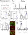
Effect of LS‐mHep7 on EAE disease course. (a) EAE was monitored over 36 days and s.c. injections of LS‐mHep7 (40 mg/kg) were administered every‐other‐day from when animals lost tail tone (score 1). LS‐mHep7 treatment caused a decrease in clinical severity compared with control animals. (b) Maximal disease scores and (c) Cumulative disease scores were both significantly reduced in LS‐mHep7 treated animals compared with PBS treated animals. (d) LS‐mHep7 treatment prevented the typical animal weight loss and promoted a faster recovery to predisease weight (red dashed line). (e) Animals required significantly less time to recover their predisease weight. (f) The percentage maximum weight loss of each animal was also significantly less in LS‐mHep7 treated animals compared with PBS injected control animals (Individual dots represent the experimental animal number, Student's unpaired two tailed t test, *p < .05, **p < .01, ***p < .001).
FIGURE 2
FIGURE 2
Effect of LS‐mHep7 on inflammatory infiltration, axonal and myelin loss, astrocyte reactivity within EAE spinal cord tissue. (a) Representative images of hematoxylin & eosin‐stained thoracolumbar spinal cord sections of animals that were injected with 40 mg/kg LS‐mHep7 every‐other‐day from loss of tail tone compared with PBS injected animals at the end of disease course. (b) Quantification of the number of nuclei present in inflammatory plaques showed that LS‐mHep7 treated animals had significantly less cellular infiltration. (c) Representative images of anti‐CD45 (shown in green), anti‐Laminin (shown in red) and DAPI (shown in blue) stained thoracolumbar spinal cord sections and (d) anti‐MBP (shown in green), anti‐SMI‐31 (shown in red) and DAPI (shown in blue). (e) Quantification of the % CD45 expression determined that LS‐mHep7 injected animals had significantly less leukocytes within the inflammatory plaques compared with PBS injected animals. (f) Quantification of the % laminin expression showed that LS‐mHep7 injected animals had significantly less expression compared with PBS injected animals. (g) Quantification of the % of abnormal axons by measuring the number of SMI‐31 lacking regions in inflammatory plaques, revealed that LS‐mHep7 injected animals had significantly less axonal loss compared with PBS injected animals. (h) Quantification of the % myelin loss by measuring the number of MBP lacking regions in inflammatory plaques, determined that LS‐mHep7 injected animals had significantly less abnormal MBP pathology compared with PBS injected animals. (i) Quantification of the % GFAP expression determined that LS‐mHep7 injected animals had similar levels of astrocyte reactivity. (j) Representative images of anti‐GFAP (shown in red) and DAPI (shown in blue) stained thoracolumbar spinal cord sections. (Individual dots represent the experimental animal number, *p < .05, **p < .01, Student's unpaired two tailed t test).
FIGURE 3
FIGURE 3
Effect of LS‐mHep7 on inflammatory infiltration, axonal, and myelin pathology 5 days posttreatment. (a) Representative images of anti‐CD45 (shown in green), anti‐Laminin (shown in red) & DAPI (shown in blue); and anti‐CD4 (shown in red) & DAPI (shown in blue), higher magnification inset boxes of area delineated by dashed white square; and anti‐IBA1 (shown in green) & DAPI (shown in blue); and anti‐MBP (shown in green), anti‐SMI‐31 (shown in red) & DAPI (shown in blue), stained thoracolumbar spinal cord sections of animals that were injected with only two doses of 40 mg/kg LS‐mHep7 every‐other‐day over 5 days from loss of tail tone compared with PBS injected animals. Scale bars represent 100 μm. (b) Disruption of the blood brain barrier was assessed by measuring blood vessel width which was significantly reduced in LS‐mHep7 treated mice compared with controls. (c) Correspondingly, there were reduced levels of laminin expression in LS‐mHep7 treated animals compared with PBS control. (d) Quantification of the % CD45 expression and (e) the % CD4 expression determined that LS‐mHep7 injected animals had significantly less leukocytes within the inflammatory plaques in white matter compared with PBS injected animals. (f) Quantification of the % of abnormal axons by measuring the number of SMI‐31 lacking/abnormal regions in inflammatory plaques, revealed no significant difference between LS‐mHep7 injected animals and PBS injected animals. (g) Quantification of the % myelin loss by measuring the number of MBP lacking regions in inflammatory plaques, revealed no significant difference between LS‐mHep7 injected animals and PBS injected animals. (h) Quantification of % IBA‐1 expression showed no difference in microglia activation in LS‐mHep7 treated animals compared with PBS controls. (Individual dots represent the experimental animal number, *p < .05, **p < .01, Student's unpaired two tailed t test).
FIGURE 4
FIGURE 4
Effect of LS‐mHep7 on PLP, MBP, OLIG2 expression in the acute cuprizone model of demyelination. (a) Representative images of anti‐PLP, anti‐MBP, and anti‐OLIG2 staining within anatomical brain region 215 of a control animal, or after being fed cuprizone diet (0.25%) for 5 weeks (Cup 5 weeks) or after treatment with PBS vehicle or LS‐mHep7 40 mg/kg via subcutaneous injections (s.c.) for 14 days post cuprizone diet removal. Dashed black line demarcates the corpus callosum (CC) region. (b) Thresholding measurements of the CC region using Image J algorithm “huang” showed no differences in the levels of PLP or (c) MBP remyelination after LS‐mHep7 treatment compared with the vehicle control group. (d) There was no difference in the OLIG2 counts after LS‐mHep7 treatment compared with control animals. (Individual dots represent the experimental animal number, *p < .05, One‐way ANOVA, Tukey's multiple post‐comparison test).
FIGURE 5
FIGURE 5
A potential role for CCL5 in LS‐mHep7 mechanism‐of‐action. (a) Control conditioned medium (Control) and demyelinated conditioned medium (DeMy CM) were collected from cultures on 25 DIV (1 day after demyelination). The conditioned media underwent LS‐mHep7 affinity chromatography, and the cytokine profile was assessed. Representative dot blots of the arrays for each conditioned media eluate are shown. (b) Quantification of dot blots based on pixel intensity. DeMy CM LS‐mHep7 eluate contained numerous factors not found in Control LS‐mHep7 eluate, including CCL5 (n = 3; technical replicates = 2). (c) Quantification of the fold change in pixel intensity showed a significant upregulation of CCL5 after demyelination (DeMy, ***p < .001, Student's t test). (d) CCL5 ELISA of conditioned media, collected from demyelinated cultures (DeMy) either immediately after demyelination (DeMy CM D0) or 5 days later (DeMy CM D5) confirmed a significant increase in CCL5 concentration compared with control cultures. (n = 3; technical replicates = 3). (e) LS‐mHep7 treatment (1 ng/mL) at 25 DIV (1 day after demyelination) promoted remyelination compared with untreated cultures. (f) CCL5 inhibits de novo myelination however, cotreatment with LS‐mHep7 (1 ng/mL) could overcome its inhibitory effect. (g) Representative images of control, CCL5 treated (100 ng/mL) or CCL5 and LS‐mHep7 treated myelinating cultures. Anti‐SMI‐31 stains axons in red, anti‐PLP stains myelin in green, Scale bar represent 100 μm. (h) Comparison of Ccl‐5 expression in spinal cords of healthy versus EAE animals and the corpus callosum of healthy vs cuprizone‐induced animals. Ccl‐5 was significantly upregulated in EAE spinal cords compared with healthy controls and was also upregulated compared with corpus callosum brain region of cuprizone fed animals. (Individual dots represent the experimental n number or individual animals in (h). One‐way ANOVA with Dunnett multiple comparison, or Two‐way ANOVA with Sikak's multiple comparison in (h) *p < .05. **p < .01, ***p < .001).

References

    1. Biancotti, J. C. , Kumar, S. , & de Vellis, J. (2008). Activation of inflammatory response by a combination of growth factors in cuprizone‐induced demyelinated brain leads to myelin repair. Neurochemical Research, 33, 2615–2628. - PubMed
    1. Billiau, A. , & Matthys, P. (2001). Modes of action of Freund's adjuvants in experimental models of autoimmune diseases. Journal of Leukocyte Biology, 70, 849–860. - PubMed
    1. Bishop, J. R. , Schuksz, M. , & Esko, J. D. (2007). Heparan sulphate proteoglycans fine‐tune mammalian physiology. Nature, 446, 1030–1037. - PubMed
    1. Bogler, O. , Wren, D. , Barnett, S. C. , Land, H. , & Noble, M. (1990). Cooperation between two growth factors promotes extended self‐renewal and inhibits differentiation of oligodendrocyte‐type‐2 astrocyte (O‐2A) progenitor cells. Proceedings of the National Academy of Sciences of the United States of America, 87, 6368–6372. - PMC - PubMed
    1. Boshans, L. L. , Sherafat, A. , & Nishiyama, A. (2020). The effects of developmental and current niches on oligodendrocyte precursor dynamics and fate. Neuroscience Letters, 715, 134593. - PMC - PubMed

Publication types

MeSH terms