[Preprint]. 2023 Mar 9:2023.03.07.23286862.
doi: 10.1101/2023.03.07.23286862.

Variants in ACTC1 underlie distal arthrogryposis accompanied by congenital heart defects

Affiliations

Variants in ACTC1 underlie distal arthrogryposis accompanied by congenital heart defects

Jessica X Chong et al. medRxiv. .

Update in

  • Variants in ACTC1 underlie distal arthrogryposis accompanied by congenital heart defects.
    Chong JX, Childers MC, Marvin CT, Marcello AJ, Gonorazky H, Hazrati LN, Dowling JJ, Al Amrani F, Alanay Y, Nieto Y, Gabriel MÁM, Aylsworth AS, Buckingham KJ, Shively KM, Sommers O, Anderson K; University of Washington Center for Mendelian Genomics; University of Washington Center for Rare Disease Research; Regnier M, Bamshad MJ. Chong JX, et al. HGG Adv. 2023 Jun 15;4(3):100213. doi: 10.1016/j.xhgg.2023.100213. eCollection 2023 Jul 13. HGG Adv. 2023. PMID: 37457373 Free PMC article.

Abstract

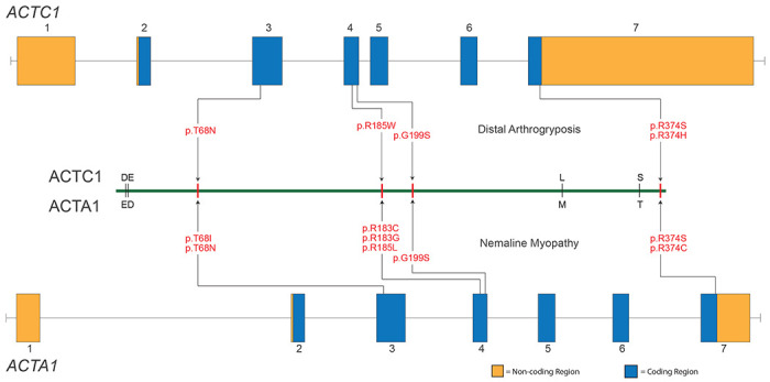
Contraction of the human sarcomere is the result of interactions between myosin cross-bridges and actin filaments. Pathogenic variants in genes such as MYH7 , TPM1 , and TNNI3 that encode parts of the cardiac sarcomere cause muscle diseases that affect the heart, such as dilated cardiomyopathy and hypertrophic cardiomyopathy. In contrast, pathogenic variants in homologous genes MYH2 , TPM2 , and TNNI2 , that encode parts of the skeletal muscle sarcomere, cause muscle diseases affecting skeletal muscle, such as the distal arthrogryposis (DA) syndromes and skeletal myopathies. To date, there have been few reports of genes (e.g., MYH7 ) encoding sarcomeric proteins in which the same pathogenic variant affects both skeletal and cardiac muscle. Moreover, none of the known genes underlying DA have been found to contain mutations that also cause cardiac abnormalities. We report five families with DA due to heterozygous missense variants in the gene actin, alpha, cardiac muscle 1 ( ACTC1 ). ACTC1 encodes a highly conserved actin that binds to myosin in both cardiac and skeletal muscle. Mutations in ACTC1 have previously been found to underlie atrial septal defect, dilated cardiomyopathy, hypertrophic cardiomyopathy, and left ventricular noncompaction. Our discovery delineates a new DA condition due to mutations in ACTC1 and suggests that some functions of actin, alpha, cardiac muscle 1 are shared in cardiac and skeletal muscle.


Conflict of interest statement

Declaration of Interests

MJB and JXC are the Editor-in-Chief and Deputy Editor of HGG Advances and were recused from the editorial handling of this manuscript. All other authors declare no competing interests.

Figures

Figure 1.
Figure 1.. Phenotypic Characteristics of Individuals with Distal Arthrogryposis due to heterozygous variants in ACTC1.
Photo is redacted for Medrxiv submission. Characteristics shown include: webbed neck, bilateral clubfoot, camptodactyly of the fingers and hypoplastic flexion creases in Family A (II-1 and III-1); camptodactyly, webbed neck, bilateral clubfoot, camptodactyly of the fingers and toes and hypoplastic flexion creases in Family B (II-2 and III-1); webbed neck, bilateral clubfoot, webbed neck, bilateral clubfoot, camptodactyly of the fingers and toes in Family C (II-2); and ptosis, webbed neck, camptodactyly of the fingers, and scoliosis in Family D (II-1). Table 1 contains a detailed description of the clinical findings of each affected individual and Figure S1 provides a pedigree for each family.
Figure 2.
Figure 2.. Genomic Model of ACTC1 and ACTA1.
Illustrated are each of the variants found in ACTC1 that underlie distal arthrogryposis and the homologous sites in ACTA1 that result in severe nemaline myopathy when mutated. ACTC1 and ACTA1 are each composed of 7 exons and consist of protein-coding (blue) and non-coding (orange) sequence. The proteins are nearly identical except for four residues (represented by single-letter amino acid codes immediately above and below the green line). The approximate location of each pathogenic variant (red text) is indicated by an arrow.
Figure 3.
Figure 3.. Molecular structures of the globular and filamentous forms of human cardiac actin.
(A) Globular (g-) actin monomers are comprised of four subdomains (subdomain 1-4, SD1-4) arranged around the nucleotide binding pocket. The g-actin monomer simulated in this study contains ATP in the binding pocket. The atoms of four residues corresponding to mutation sites examined in this study are shown as spheres: T68 (orange), R185 (magenta), G199 (purple), and R374 (blue). (B) Actin monomers polymerize into protofibrils, which then associate with one another to form filamentous (f-) actin. In this f-actin pentamer, chains A, C and E form one protofibril and chains B and D form the other. The pentamer simulated here has ADP molecules in the nucleotide binding pockets. The location of residue T68 is denoted on chain C.
Figure 4.
Figure 4.. p.Thr68Asn alters the structure and dynamics of g-actin subdomain 2.
Comparison of representative MD-derived snapshots of WT (A) and p.Thr68Asn (B) g-actin highlights the structural and dynamical changes induced by p.Thr68Asn (red ribbon). Sidechain atoms of relevant residues are shown and annotated. (C) Bar heights correspond to the fraction of time that select residue pairs spent in contact with one another, averaged over triplicate simulations (error bars correspond to st. dev.). p.Thr68Asn (orange bars) led to shifts in several amino acid interactions relative to WT (black bars). Statistically significant differences between the WT and p.Thr68Asn contact frequencies are denoted (ns: not significant, *: p ≤ 0.05, **: p ≤ 0.01) (D) p.Thr68Asn (orange) increased the α-helix secondary structure content of residues 40-50 within the D-loop relative to WT (black). (E) The p.Thr68Asn mutation (orange) led to a decrease in the solvent accessible surface area of D-loop residues (41-56) relative to WT (black). The histogram shows the SASA probability density of all replicate simulations combined.
Figure 5.
Figure 5.. DA-associated mutations lead to structural changes within subdomain 2.
The percent simulation time for which residue-residue contacts endured were compared between the four mutant simulations: p.Thr68Asn (A, orange), p.Arg185Trp (B, magenta), p.Gly199Ser (C, purple), p.Arg374Ser (D, blue) and the WT simulations (black). For each mutant-WT comparison, those residue-residue contacts that were present for statistically different percentages of the simulations have been mapped onto the reference crystal structure of g-actin. Contacts that were present more frequently in the WT simulations are denoted by black pipes and contacts present more frequently in the mutant simulations are colored orange, magenta, purple, or blue. The thickness of the pipes corresponds to the difference in percent simulation time that the contact was present between the WT and mutant simulations (larger pipes indicate a contact was observed more frequently). Although the mutations were distributed throughout the structure, they all led to statistically significant (see Table S2 for test statistics) changes in the structure of subdomain 2 (orange ribbons).

References

    1. Dowling J.J., Weihl C.C., and Spencer M.J. (2021). Molecular and cellular basis of genetically inherited skeletal muscle disorders. Nat Rev Mol Cell Bio 22, 713–732. 10.1038/s41580-021-00389-z. - DOI - PMC - PubMed
    1. Au Y. (2004). The muscle ultrastructure: a structural perspective of the sarcomere. Cell Mol Life Sci Cmls 61, 3016–3033. 10.1007/s00018-004-4282-x. - DOI - PMC - PubMed
    1. Lindskog C., Linné J., Fagerberg L., Hallström B.M., Sundberg C.J., Lindholm M., Huss M., Kampf C., Choi H., Liem D.A., et al. (2015). The human cardiac and skeletal muscle proteomes defined by transcriptomics and antibody-based profiling. Bmc Genomics 16, 475. 10.1186/s12864-015-1686-y. - DOI - PMC - PubMed
    1. Sung S.S., Brassington A.-M.E., Grannatt K., Whitby F.G., Krakowiak P.A., and Bamshad M.(2003). Mutations in Genes Encoding Fast-Twitch Contractile Proteins Cause Distal Arthrogryposis Syndromes. Am J Hum Genetics 72, 681–690. 10.1086/368294. - DOI - PMC - PubMed
    1. Sung S.S., Brassington A.-M.E., Krakowiak P.A., and Bamshad M. (2003). Mutations in TNNT3 cause multiple congenital contractures: a second locus for distal arthrogryposis type 2B. Am J Hum Genetics 73, 212–214. 10.1086/376418. - DOI - PMC - PubMed

Publication types