

This is a preprint.
Hippocampal sclerosis of aging at post-mortem is evident on MRI more than a decade prior
- PMID: 36945448
- PMCID: PMC10028863
- DOI: 10.1101/2023.03.08.531683
Hippocampal sclerosis of aging at post-mortem is evident on MRI more than a decade prior
Update in
-
Hippocampal sclerosis of aging at post-mortem is evident on MRI more than a decade prior.Alzheimers Dement. 2023 Nov;19(11):5307-5315. doi: 10.1002/alz.13352. Epub 2023 Jun 27. Alzheimers Dement. 2023. PMID: 37366342 Free PMC article.
Abstract
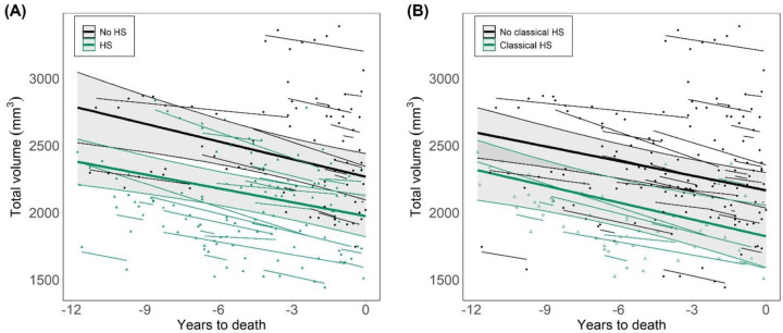
Introduction: Hippocampal sclerosis of aging (HS) is an important component of combined dementia neuropathology. However, the temporal evolution of its histologically-defined features is unknown. We investigated pre-mortem longitudinal hippocampal atrophy associated with HS, as well as with other dementia-associated pathologies.
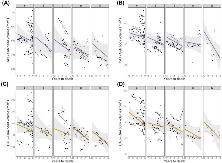
Methods: We analyzed hippocampal volumes from MRI segmentations in 64 dementia patients with longitudinal MRI follow-up and post-mortem neuropathological evaluation, including HS assessment in the hippocampal head and body.
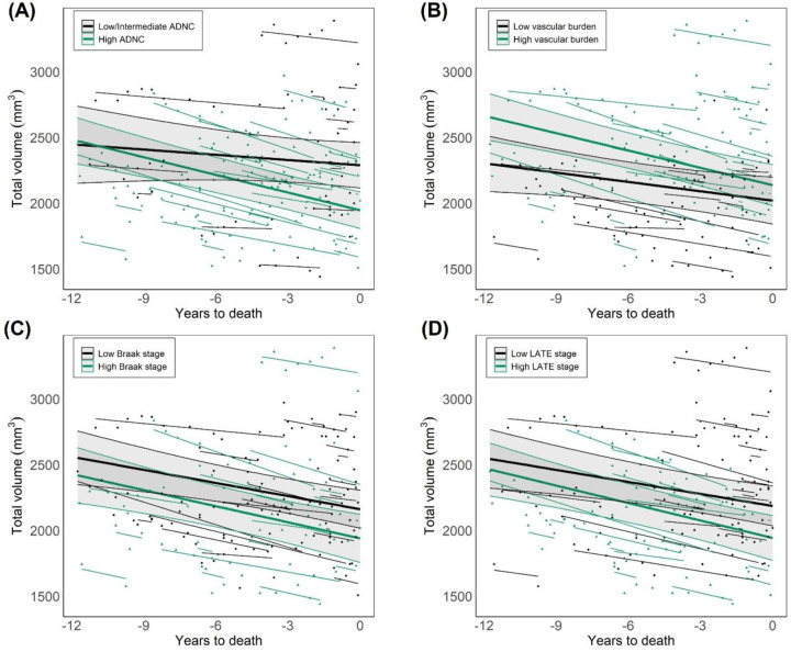
Results: Significant HS-associated hippocampal volume changes were observed thoughout the evaluated timespan, up to 11.75 years before death. These changes were independent of age and Alzheimer’s Disease (AD) burden, and specifically driven by CA1 and subiculum. AD burden, but not HS, significantly associated with the rate of hippocampal atrophy.
Discussion: HS-associated volume changes are detectable on MRI earlier than 10 years before death. These findings could contribute to the derivation of volumetric cut-offs for in vivo differentiation between HS and AD.
Conflict of interest statement
Declarations of interest: none
Figures
References
Publication types
Grants and funding
LinkOut - more resources
Full Text Sources
Miscellaneous