[Preprint]. 2023 Mar 12:2023.03.09.531970.
doi: 10.1101/2023.03.09.531970.

T-CLEARE: A Pilot Community-Driven Tissue-Clearing Protocol Repository

Affiliations

T-CLEARE: A Pilot Community-Driven Tissue-Clearing Protocol Repository

Kurt Weiss et al. bioRxiv. .

Update in

  • T-CLEARE: a pilot community-driven tissue clearing protocol repository.
    Weiss KR, Huisken J, Khanjani N, Bakalov V, Engle ML, Krzyzanowski MC, Madden T, Maiese DR, Waterfield JR, Williams DN, Wood L, Wu X, Hamilton CM, Huggins W. Weiss KR, et al. Front Bioeng Biotechnol. 2024 Sep 16;12:1304622. doi: 10.3389/fbioe.2024.1304622. eCollection 2024. Front Bioeng Biotechnol. 2024. PMID: 39351064 Free PMC article.

Abstract

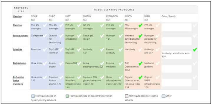
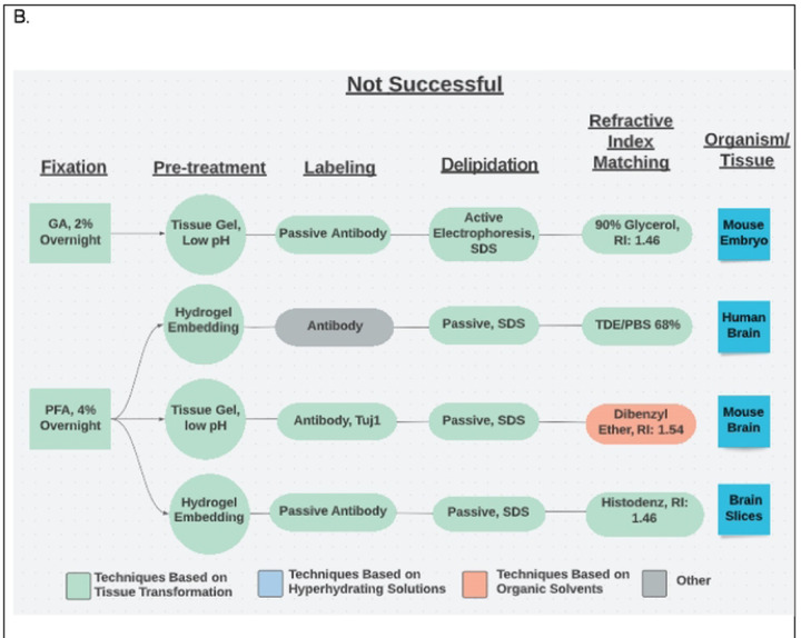
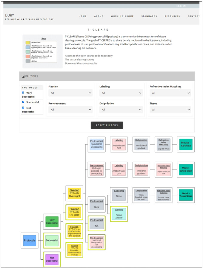
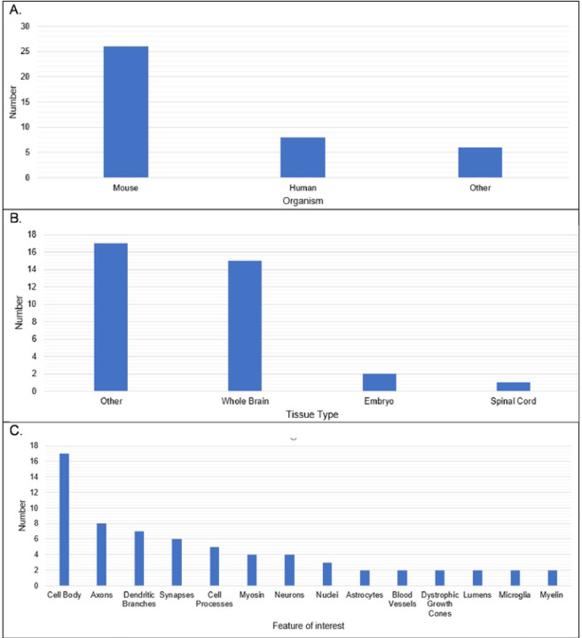
Selecting and implementing a tissue-clearing protocol is challenging. Established more than 100 years ago, tissue clearing is still a rapidly evolving field of research. There are currently many published protocols to choose from, and each performs better or worse across a range of key evaluation factors (e.g., speed, cost, tissue stability, fluorescence quenching). Additionally, tissue-clearing protocols are often optimized for specific experimental contexts, and applying an existing protocol to a new problem can require a lengthy period of adaptation by trial and error. Although the primary literature and review articles provide a useful starting point for optimization, there is growing recognition that many articles do not provide sufficient detail to replicate or reproduce experimental results. To help address this issue, we have developed a novel, freely available repository of tissue-clearing protocols named T-CLEARE (Tissue CLEAring protocol REpository; https://doryworkspace.org/doryviz). T-CLEARE incorporates community responses to an open survey designed to capture details not commonly found in the scientific literature, including modifications to published protocols required for specific use cases and instances when tissue-clearing protocols did not perform well (negative results). The goal of T-CLEARE is to provide a forum for the community to share evaluations and modifications of tissue-clearing protocols for various tissue types and potentially identify best-in-class methods for a given application.


Figures

Fig 1.
Fig 1.
Table of Commonly Used Tissue-Clearing Protocols in the Tissue-Clearing Survey The table was included in the Tissue Clearing Survey conducted by RTI in 2021–2022 (https://doryworkspace.org/tissue_clearing_feedback) Respondents identified the tissue clearing protocols used by checking boxes in the table or by entering details in the “Other, specify” column.
Fig 2.
Fig 2.
Example Flow Charts Created from Reported Tissue-Clearing Protocols A. Example flow charts of successful tissue-clearing protocols. The tissue cleared is indicated in the right box. B. Example flow charts of unsuccessful tissue-clearing protocols.
Fig 2.
Fig 2.
Example Flow Charts Created from Reported Tissue-Clearing Protocols A. Example flow charts of successful tissue-clearing protocols. The tissue cleared is indicated in the right box. B. Example flow charts of unsuccessful tissue-clearing protocols.
Fig 3.
Fig 3.
Screenshot of T-CLEARE User Interface Boxes in light blue, aqua, and light red indicate whether the mode of action for that step is based on hyperhydrating solutions, tissue transformation and organic solvents, respectively. Yellow boxes indicate a step that can be classifed according to more than one mode of action. Gray boxes indicate the respondent entered text in the “Other, specify” in the survey table.
Figure 4.
Figure 4.
Organism, Tissue Type, and Feature of Interest in Reported Tissue Clearing Protocols A. Reported organisms under study. B. Reported tissues being cleared. C. Reported feature of interest in cleared tissues (similar responses were grouped together).
Fig 5.
Fig 5.
Reported Tissue-Clearing Protocols Used Without Modification Tissue-clearing protocols that were followed without modification. iDISCO= immunolabeling-enabled three-Dimensional Imaging of Solvcnt-Cleared Organs(1) CUBIC= Clear, Unobstructed Brain Imaging Cocktails and Computational analysis(2) PACT= PAssive Clarity Technique (3) Expansion = Expansion microscopy (4) SWITCH = System-Wide control of Interaction Time and kinetics of CHemicals (3) BABB = Benzyl Alcohol/ Benzyl Benzoate(6)
Fig 6.
Fig 6.
Overall Success in Reported Tissue-Clearing Protocols Individual protocols in responses have been grouped according to mode of action (Organic Solvents, Hyperhydrating, Tissue Transformation, Other).

References

    1. Chhetri RK, Amat F, Wan Y, Hockendorf B, Lemon WC, Keller PJ. Whole-animal functional and developmental imaging with isotropic spatial resolution. Nat Methods. 2015;12(12):1171–8. - PubMed
    1. Lemon WC, Pulver SR, Hockendorf B, McDole K, Branson K, Freeman J, et al. Whole-central nervous system functional imaging in larval Drosophila. Nat Commun. 2015;6:7924. - PMC - PubMed
    1. Ahrens MB, Orger MB, Robson DN, Li JM, Keller PJ. Whole-brain functional imaging at cellular resolution using light-sheet microscopy. Nat Methods. 2013;10(5):413–20. - PubMed
    1. Vladimirov N, Mu Y, Kawashima T, Bennett DV, Yang CT, Looger LL, et al. Light-sheet functional imaging in fictively behaving zebrafish. Nat Methods. 2014;11(9):883–4. - PubMed
    1. Tomer R, Lovett-Barron M, Kauvar I, Andalman A, Burns VM, Sankaran S, et al. SPED Light Sheet Microscopy: Fast Mapping of Biological System Structure and Function. Cell. 2015;163(7):1796–806. - PMC - PubMed

Publication types