[Preprint]. 2023 Mar 7:rs.3.rs-2622363.
doi: 10.21203/rs.3.rs-2622363/v1.

Transcriptional Induction of NF-kB-Inducing Kinase by E2F4/5 Facilitates Collective Invasion of Glioma Cells

Affiliations

Transcriptional Induction of NF-kB-Inducing Kinase by E2F4/5 Facilitates Collective Invasion of Glioma Cells

Kathryn Pflug et al. Res Sq. .

Update in

Abstract

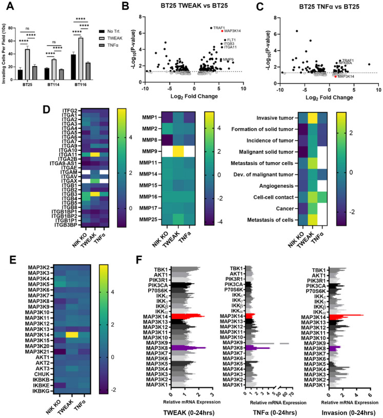
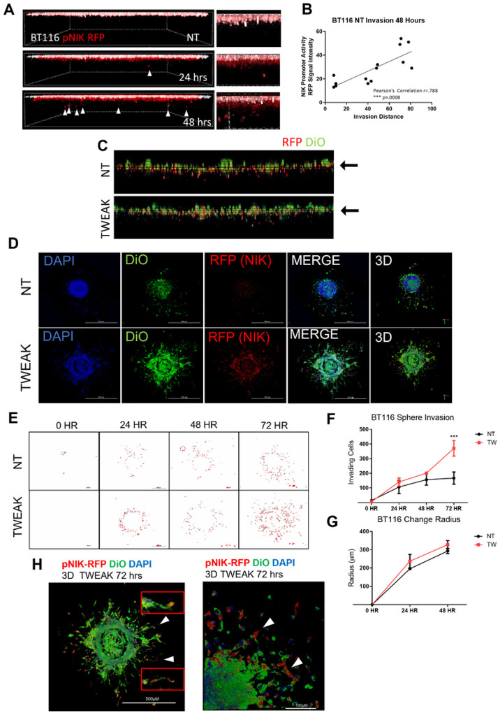
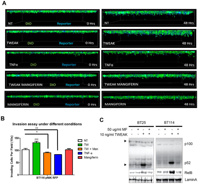
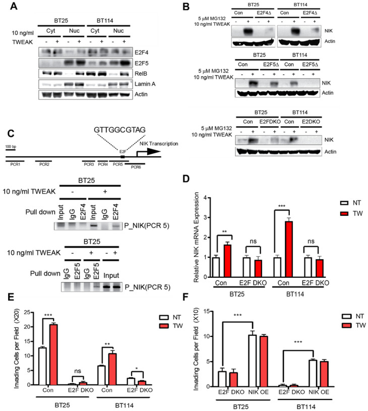
The prognosis of high-grade gliomas, such as glioblastoma multiforme (GBM), is extremely poor due to the highly invasive nature of these aggressive cancers. Previous work has demonstrated that TNF-weak like factor (TWEAK) induction of the noncanonical NF-κB pathway increases the invasiveness of glioma cells in an NF-κB-inducing kinase (NIK)-dependent manner. While NIK activity is predominantly regulated at the posttranslational level, we show here that NIK ( MAP3K14 ) is upregulated at the transcriptional level in invading cell populations, with the highest expression observed in the most invasive cells. Glioma cells with high induction of NIK gene expression demonstrate characteristics of collective invasion, facilitating invasion of neighboring cells. Furthermore, we demonstrate that the E2F transcription factors E2F4 and E2F5 directly regulate NIK transcription and are required to promote glioma cell invasion in response to TWEAK. Overall, our findings demonstrate that transcriptional induction of NIK facilitates collective cell migration and invasion, thereby promoting glioma pathogenesis.


Conflict of interest statement

Conflicts of Interest: The authors declare no conflict of interest.

Figures

Figure 1
Figure 1. Specific induction of NIK promotes invasion
A) Quantification of three-dimensional collagen invasion assay BT25, BT114, and BT116 GBM cells after 48 hours. Invasion was conducted under basal, TWEAK treatment (10 ng/mL), or TNFα (10 ng/mL). Statistical analysis was performed using two-way ANOVA p < 0.001. B-E) RNA sequencing analysis of BT25 GBM cells with either TWEAK treatment (10 ng/mL for 4 hr) or TNFα treatment (10 ng/mL for 30 minutes) on wild-type cells or NIK KO BT25 cells compared to wild-type BT25 cell gene expression. IPA software was used for pathway analyses. F) RT-qPCR analysis was utilized to further examine the expression of NF-κB proteins and MAP3Ks in BT114 GBM cells. Cells were treated with either 10 ng/mL TWEAK or 10 ng/mL TNFα at 0, 0.25, 0.5, 1, 4, 8, 16, and 24 hours. RT-qPCR analysis of cells extracted from the collagen matrix at 0, 0.5, 4, 8, and 24 hours post invasion.
Figure 2
Figure 2. NIK Expression is Upregulated in Highly Invasive Glioma Cells and Promotes Collective Invasion
A) Live-cell confocal microscopy was utilized to visualize the RFP reporter under the NIK promoter (pNIK-RFP) (red) under unstimulated conditions (NT). The monolayer (0 hrs) was pseudocolored white and overlaid to the reference invasion distance. B) Graph representation of RFP intensity after 48 hours and distance the cells invaded. Pearson’s correlation between RFP intensity and distance is r=.788 and p=.008, ***p≤.001. C) Invasion of BT116 pNIK-RFP cells labeled with DiO (green) with no treatment (NT) or treated with 10 ng/mL TWEAK. D) BT116 pNIK-RFP cells were used for a spheroid invasion assay. Spheroids were embedded in three-dimensional collagen matrix and either left untreated (NT) or treated with TWEAK (10 ng/mL). Spheres were allowed to invade for 72 hours. Confocal microscopy was used to image spheroids, labeled DiO (green), RFP reporter (red), and DAPI (blue). E) Outline of BT116 pNIK-RFP (red) spheroids embedded in three-dimensional collagen for 0–72 hr, either untreated or treated with TWEAK (TW) for 48 hours. F) The ImageJ particle analysis function was used to quantify the number of cells invading into the three-dimensional collagen matrix at various time points. G) ImageJ was used to measure the radius of the spheroids at various time points. H) Three-dimensional projection image BT116 pNIK-RFP (Red) spheroids were embedded in three-dimensional collagen and treated with TWEAK (TW) for 72 hours. Cells were labeled with DiO (green) and DAPI (blue). White arrowheads with enhanced images of cells undergoing collective invasion. Spheroid invasion assays were conducted in n>3 either untreated or treated with 10 ng/mL TWEAK.
Figure 3
Figure 3. Inhibition of NIK Reduces Glioma Invasion
A) BT116 pNIK RFP cells were labeled with DiO before being seeded on collagen matrix and allowed to invade for 48 hours. Live cell confocal microscopy was used to obtain Z-stack images over 48 hours, with the top layer depicting the monolayer at 0 hr and the bottom image depicting cell invasion at 48 hr. Cells either received no treatment (NT) or were treated with 10 ng/mL TWEAK,10 ng/mL TNF-α, 10 ng/ml TWEAK with 100 μg/mL mangiferin, or 100 μg/mL mangiferin. B) Quantification of cell invasion after 48 hours. C) Immunoblot showing decreased RelB and p52 in the nucleic fraction of glioma cells treated with mangiferin and/or TWEAK.
Figure 4
Figure 4. E2F4 and E2F5 Regulate NIK Gene Expression
A) Cytoplasmic and nuclear E2F4 and E2F5 were examined by Western blot in unstimulated vs stimulated conditions (10 ng/mL TWEAK for 4 hrs) with RelB as a positive TWEAK-inducible control and Lamin A as a nuclear marker. B) Immunoblot of NIK expression under MG132 (5 μM)- and TWEAK (10 ng/mL)-stimulated conditions in control and E2F4, E2F5, or E2F4 and E2F5 knockout cells. C) ChIP analysis performed in glioma cells treated with or without 10 ng/mL TWEAK by using E2F4 or E2F5 antibodies. D) qPCR analysis of NIK (MAP3K14) induction in double knockout E2F4 and E2F5 glioma cells (BT25 and BT114). E) Invasion assay of BT25 and BT114 double knockout E2F4/E2F5 cells. F) Invasion assay of BT25 and BT114 double knockout E2F4 and E2F5 cells rescued by NIK overexpression. D-F) Comparisons were made using glioma cells in untreated (NT) vs TWEAK-treated (10 ng/mL for 4 hrs) conditions.

References

    1. Hoelzinger D. B., Demuth T. & Berens M. E. Autocrine Factors That Sustain Glioma Invasion and Paracrine Biology in the Brain Microenvironment. JNCI: Journal of the National Cancer Institute 99, 1583–1593 (2007). - PubMed
    1. Zhang X. et al. Malignant Intracranial High Grade Glioma and Current Treatment Strategy. Curr Cancer Drug Targets 19, 101–108 (2019). - PubMed
    1. Gritsenko P. G. et al. P120-catenin dependent collective brain infiltration by glioma cell networks. Nat Cell Biol 22, 97–107 (2020). - PMC - PubMed
    1. Osswald M. et al. Brain tumour cells interconnect to a functional and resistant network. Nature 528, 93–98 (2015). - PubMed
    1. Peglion F. et al. PTEN inhibits AMPK to control collective migration. Nat Commun 13, 4528 (2022). - PMC - PubMed

Publication types

LinkOut - more resources