[Preprint]. 2023 Mar 6:rs.3.rs-2637060.
doi: 10.21203/rs.3.rs-2637060/v1.

MicroMagnify: a multiplexed expansion microscopy method for pathogens and infected tissues

Affiliations

MicroMagnify: a multiplexed expansion microscopy method for pathogens and infected tissues

Yongxin Zhao et al. Res Sq. .

Update in

Abstract

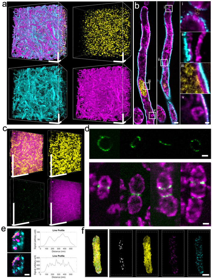
Super-resolution optical imaging tools are crucial in microbiology to understand the complex structures and behavior of microorganisms such as bacteria, fungi, and viruses. However, the capabilities of these tools, particularly when it comes to imaging pathogens and infected tissues, remain limited. We developed µMagnify, a nanoscale multiplexed imaging method for pathogens and infected tissues that are derived from an expansion microscopy technique with a universal biomolecular anchor. We formulated an enzyme cocktail specifically designed for robust cell wall digestion and expansion of microbial cells without distortion while efficiently retaining biomolecules suitable for high-plex fluorescence imaging with nanoscale precision. Additionally, we developed an associated virtual reality tool to facilitate the visualization and navigation of complex three-dimensional images generated by this method in an immersive environment allowing collaborative exploration among researchers around the world. µMagnify is a valuable imaging platform for studying how microbes interact with their host systems and enables development of new diagnosis strategies against infectious diseases.


Conflict of interest statement

CONFLICT OF INTEREST

The authors declare the following competing financial interest(s): YZ, ZC, BRG, APM and AK are inventors on several inventions related to ExM. TS is the founder of Immersive Science LLC. The remaining authors declare no competing interests.

Figures

Figure 1
Figure 1. Schematic and validation of μMagnify.
(a) Brief schematic of μMagnify chemical processing (Supplementary Figure 1). Briefly, samples were rehydrated and penetrated for format conversion. Then appropriate amount of gelling solution was added on top of the specimen for polymer synthesis. The sample-gel hybrid was mechanically homogenized in denaturing reagents followed by enzyme digestion. The homogenized gel was isotropically expanded in 1:50 diluted PBS or ddH2O for imaging. (b-g) Validations of μMagnify on representative bacteria, fungi, and bacteria infected cell culture pre- and post-expansion: (b) E. coli was stained with CellMask, (d) C. albicans with BODIPY and (f) S. aureusinfected U2OS cell culture with DAPI (red) and BODIPY (green). (b, d, f)Examples of pre-expansion images taken at 60x and processed with intensity average across 50 frames and XC2 deconvolution SOFI, compared to the post-expansion images taken at 60x for the same field of view. Post expansion images are Maximum intensity projected over 5–30 frames in z to best match the plane. Biological scales: (b) and (d), 2 μm; (f), 10 μm. Expansion factor (in 1:50 diluted PBS): (b) 4.97 ± 0.32 (n=13); (d) 6.06 ± 0.42 (n=11); (f) 5.79 ± 0.21(n=16). Right top corner images in (f) are zoomed in images of the white boxed region (length = 2.6 μm). (c, e, g) Root mean square (RMS) length measurement error as a function of measurement length for pre-expansion SOFI images versus post-expansion images for (c) E. coli (CellMask, n = 13), (e) C. albicans(BODIPY, n = 11) and (g) S. aureus infected U2OS cell (DAPI, n = 16). Solid line, mean of channel; shaded area, standard error of mean (s.e.m).
Figure 2
Figure 2. μMagnify works for a diversity of microbial cells, revealing their nanoscale structure and spatial patterns.
(a) 3D reconstruction of fully expanded Candida albicans (C. albicans) biofilm. The sample was stained with DAPI (yellow), Lycopersicon Esculentum Lectin (LEL, cyan), and DiI (magenta). Physical scale: 100 μm in x, y, z. (b) X-y section of hyphae. Zoom-in views from boxed region showing the hyphae cell junction (i), elongated mitochondria beside cell wall (ii), details of nuclear membrane, cell membrane and cell wall (iii), and lipid body (v). Physical scales: 5 μm (most left and middle columns), 1 μm (most right column). (c) 3D reconstruction of Streptococcus pneumoniae (S. pneumoniae) biofilm. Sample was stained with DAPI (yellow), LEL (green) and NHS-ester (magenta). Physical scales: 100 μm (x, y), 10 μm (z). (d) Upper part: examples of ring-like structures of peptidoglycan (PG) enrichment of dividing S. pneumonia at different orientations. PG was stained with LEL. Lower part: examples of S. pneumonia constriction by PG at different dividing stages. Cells were stained with LEL (green) and NHS-ester (magenta). Physical scale: 2 μm. (e) Viral particles imaging in E. coli. The sample was stained with Wheat Germ Agglutinin (WGA, cyan) and JCV capsid VP1 antibodies (magenta). Upper part shows spatial distribution of the virus particles in the fully expanded E. coli and its intensity profile (upper right) along the yellow dashed line. Lower part shows SRRF processed image from the same ROI and its intensity profile (lower right) along the dashed line. Physical scale: 2 μm. (f) Simultaneous protein and RNA imaging of fully expanded E. coli, revealing the spatial distribution of mNeon proteins (yellow) and its mRNAs (magenta). The bacteria are also stained with DAPI (gray) and 16s rRNA for control (cyan). Physical scale: 5 μm. All the expansion factors were characterized in Supplementary Table 1.
Figure 3
Figure 3. Expansion microscopy imaging of Candida albicans-infected FFPE mouse tongue tissue.
(a)Bright field pre-expansion image of Candida albicans (C. albicans)-infected tissue that was stained with Periodic acid-Schiff stain (PAS). The cell wall of C. albicans were shown in magenta. Nucleus of the tissue is stained in blue. Scale bar: 50 μm. (b) Confocal fluorescence image of sample (a) expanded in PBS. Sample was stained with DAPI (red), cellular sugar molecules were labeled differently by WGA (cyan), and LEL (blue). Pan-proteins were labeled with NHS-ester (yellow). Biological scale: 50 μm. (c) Zoom-in views of the boxed regions in (a) from left to the right, showing gradient density of C. albicans infection. Scale bar: 2 μm. (d) Zoom-in views of the boxed regions in (b) from left to the right. Biological scale: 2 μm. Expansion factors were characterized in Supplementary Table 1.
Figure 4
Figure 4. Nanoscale 3D characterization of various pathogen-infected cornea samples.
(a) PAS image of FFPE cornea sample of candida keratitis. Scale: 1000 μm. (b) μMagnifyimage of tissue sample cut adjacently to that from (a), taken by 4x objective. Sample were post-expansion stained with DAPI (cyan), LEL (yellow), and NHS-ester (magenta). Scale: 1000 μm. (c) μMagnify images of pointed region in (b), taken at 10x objective, showing apparent distinction of local Candida infection (yellow). Scale: 25 μm. (d) Representative images of C. albicans interactions with normal (left) and immune (right) cells residing in cornea stroma. (e-h) Single-cell level characterizations for various types of eye infections. Samples were stained with DAPI (cyan), WGA (yellow), NHS (magenta), and LEL (green, in h). Scales: 10 μm (x, y, z). (e) example image of pathogen-host interactions in Staphylococcus epidermidis(S. epidermidis, Gram-positive) keratitis eyeball sample. (f)example image of extracellular pathogen in Pseudomonas aeruginosa(P. aeruginosa, Gram-negative) keratitis cornea sample. (g)example image of pathogen-host interaction in atypical mycobacterial (neither Gram-positive nor Gram-negative) keratitis cornea sample. (h) representative images of acanthamoeba located in cornea stroma. Expansion factors were characterized in Supplementary Table 1.
Figure 5
Figure 5. Multiplexing on S. aureus-infected U2OS cell culture allows pairwise study on protein-protein interactions.
(a) Ten-color multiplexed imaging of α-toxin treated S. aureus infected wildtype U2OS cell. Sample was stained with DAPI, WGA, anti-GFP (targeting LITAF fusion with GFP), anti-CD63, anti-Vimentin, anti-α-tubulin, ConA, antiNEDD4, DiI, and NHS. Biological scale: 10 μm. (b) Single color images at different detection channels in (a). Biological scale :10 μm. (c) Ten-color multiplexed imaging of α-toxin treated S. aureus infected LITAF mutant U2OS cell. Sample was stained the same as those in (a). Biological scale: 10 μm. (d) Single color images at different detection channels in (c). Biological scale: 10 μm. (e) Colocalization matrices calculated for α-toxin treated S. aureus infected wildtype U2OS (left, n=3) and α-toxin treated S. aureus infected LITAF mutant U2OS cell samples (right, n=3) among 10 channels. For characterizing colocalization between signal1 and signal2. Colocalization coefficients are calculated as the percentage of the overlapping volume between the signal 1&2 in the volume of signal 1 or signal2. (f) Matrix of delta colocalization coefficient between Mut and Wt matrices in (e), indicating the change of pairwise signal colocalization among 10 channels. Asterisks indicated the significant levels through one-way ANOVA test, * p <0.05, **p<0.01, ***p<0.001. (g) Representative images of pair analysis for LITAF&CD63 and LITAF&NEDD4 colocalization. The first row showing images (i-ii) from boxed region in (a), the second row showing images (iii-iv) from boxed region in (c). The first column (i, iii) showing composite images of LITAF (yellow) and CD63(cyan). The second column (ii, iv) showing composite images of LITAF (yellow) and NEDD4 (magenta). Biological scales: 2 μm. Zoom-in views of arrow pointed regions (top to bottom) are listed on the right side of each image, delineating the different levels of colocalization between two signals. Biological scales: 500 nm. (h-i) Box plot of average colocalization coefficient between Wt (n=22) and Mut (n=16) for LITAF&CD63 (h) and LITAF&NEDD4 (i) in S. aureus-containing vacuoles. Middle line in the box shows the median. Bottom and top of each box show the 25th and 75th percentile of the data. Upper and bottom whiskers show the non-outlier maximum and minimum. Outliers are shown in red cross. Asterisks indicated the significant differences between Wt and Mut through one-way ANOVA test, ***P <0.001. Expansion factors were characterized in Supplementary Table 1.
Figure 6
Figure 6. Immersive visualization of multiplexing data and collaboration through ExMicroVR software.
(a) Workflow of collaboration among ExM, microbiology, and pathology research groups. Microbiologist and pathologist provide samples of infections along with list of potential biomarkers. ExM lab uses μMagnify to expand the samples and acquire multiplexed images. Images are converted to ExMicroVR-compatible format followed by data examination and interpretation through immersive visualization and real-time discussion in ExMicroVR space. (b) A representative user view of collaborative data examination through ExMicroVR. 3D multi-color Image data are presented and adjusted real-time among joined users. (c) Fast multi-channel data adjustments through ExMicroVR. Example images from mutant cell (figure 5c) that was stained with DAPI, WGA, antiGFP (targeting LITAF fusion with GFP), antiCD63, antiVimentin, antiAtubulin, ConA, antiNEDD4, DiI, and NHS. Biological scales 1 μm in x, y, z. Each channel is easily adjusted and color coded. Composite images can be made to study the interactions between different channels. (d) Size-adjustable excluder applies to inspection of thick biofilm data (figure 2a), Physical scale: 100 μm.

References

    1. Weiser J. N. The battle with the host over microbial size. Current Opinion in Microbiology (2013). doi: 10.1016/j.mib.2013.01.001 - DOI - PMC - PubMed
    1. Wegel E. et al. Imaging cellular structures in super-resolution with SIM, STED and Localisation Microscopy: A practical comparison. Sci. Rep. 6, 1–13 (2016). - PMC - PubMed
    1. Ku T. et al. Multiplexed and scalable super-resolution imaging of three-dimensional protein localization in size-adjustable tissues. Nat. Biotechnol. (2016). doi: 10.1038/nbt.3641 - DOI - PMC - PubMed
    1. Wang X. et al. Three-dimensional intact-tissue sequencing of single-cell transcriptional states. Science (80-.). (2018). doi: 10.1126/science.aat5691 - DOI - PMC - PubMed
    1. Truckenbrodt S. et al. X10 expansion microscopy enables 25-nm resolution on conventional microscopes. EMBO Rep. (2018). doi: 10.15252/embr.201845836 - DOI - PMC - PubMed

Publication types

LinkOut - more resources