

[Intermittent heat exposure induces thoracic aorta injury in spontaneously hypertensive rats by activating the AMPK/mTOR/ULK1 pathway]
- PMID: 36946037
- PMCID: PMC10034555
- DOI: 10.12122/j.issn.1673-4254.2023.02.05
[Intermittent heat exposure induces thoracic aorta injury in spontaneously hypertensive rats by activating the AMPK/mTOR/ULK1 pathway]
Abstract
Objective: To investigate the effects of different manners of heat exposure on thoracic aorta injury in spontaneously hypertensive rats (SHRs) and explore the underlying mechanism.
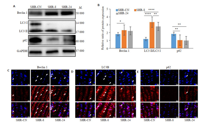
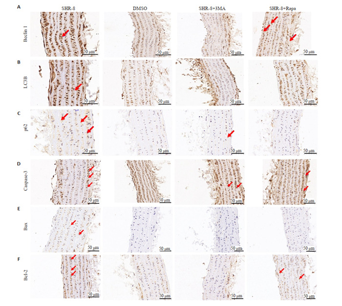
Methods: Normal 6 to 7-week-old male SHRs were randomized into control group (cage at room temperature), intermittent heat exposure group (SHR-8 group, exposed to 32 ℃ for 8 h daily for 7 days) and SHR-24 group (with continuous exposure to 32 ℃ for 7 days). After the treatments, the pathologies of the thoracic aorta of the rats were observed with HE staining, and the expressions of Beclin1, LC3B and p62 were detected with Western blotting and immunofluorescence assay; TUNEL staining was used to observe cell apoptosis in the thoracic aorta, and the expressions of caspase-3, Bax, and Bcl-2 were detected using Western blotting. The effects of intraperitoneal injections of 3-MA (an autophagy agonist), rapamycin (an autophagy inhibitor) or compound C 30 min before intermittent heat exposure on the expressions of proteins associated with autophagy, apoptosis and the AMPK/mTOR/ULK1 pathway in the aorta were examined with immunohistochemistry.
Results: In SHR-8 group, the rats showed incomplete aortic intima with disordered cell distribution and significantly increased expressions of Beclin1, LC3II/LC3I and Bax, lowered expressions of p62 and Bcl-2, and increased apoptotic cells in the thoracic aorta (P < 0.05). Pretreatment with 3-MA obviously inhibited the expressions of autophagy- and apoptosis-related proteins, whereas rapamycin promoted their expressions. Compared with the control group, the rats in SHR-8 group had significantly down-regulated p-mTOR and up-regulated p-AMPK and p-ULK1 expression of in the aorta; Treatment with compound C obviously lowered the expressions of p-AMPK and p-ULK1 and those of LC3B and Beclin1 as well.
Conclusion: In SHRs, intermittent heat exposure causes significant pathologies and promotes autophagy and apoptosis in the thoracic aorta possibly by activating the AMPK/mTOR/ULK1 pathway.
目的: 探讨不同热暴露方式对自发性高血压大鼠胸主动脉血管损伤的影响及其机制。
方法: 选用自发性高血压大鼠(SHR),构建热暴露大鼠模型,分为对照组SHR-CN(n=9,饲养在24 ℃室温下),间断性热暴露组SHR-8(n=9,每天接受任意连续8 h热暴露,其余时间饲养在与对照组相同的条件下)和持续性热暴露组SHR-24(n=9,持续暴露在32 ℃高温下)。进行自噬和通路干预的SHRs,分别于热暴露前30 min腹腔注射自噬激活剂Rapa,抑制剂3-MA和AMPK抑制剂Compound C(CC)。。通过苏木素-伊红染色(HE)观察大鼠胸主动脉形态的变化;利用Western blot和免疫荧光(IF)分别检测高血压大鼠胸主动脉自噬相关因子Beclin1、LC3B和p62的表达情况;利用TUNEL检测高血压大鼠胸主动脉凋亡的情况,Western blot检测Caspase-3,Bax和Bcl-2的表达情况;用自噬的激活剂和抑制剂干预高血压大鼠后,检测自噬和凋亡相关因子的表达情况;免疫组化(IHC)检测通路相关因子的表达情况,大鼠腹腔注射AMPK抑制剂Compound C(CC)后,进一步观察通路及自噬的表达情况。
结果: HE染色显示,SHR-8组胸主动脉内膜排列分布不完整,细胞分布紊乱。Western blot显示Beclin1、LC3II/LC3I表达增高,p62降低,差异均具有统计学意义(P < 0.05)。TUNEL结果表明SHR-8组中凋亡阳性细胞数明显高于对照组。Western blot表明热暴露后Bax表达升高,Bcl-2表达降低,差异具有统计学意义(P < 0.05)。3-MA抑制自噬的表达,同时凋亡表达降低,Rapamycin促进自噬和凋亡的表达。IHC显示,与SHR-CN相比,SHR-8组p-mTOR明显降低,p-AMPK和p-ULK1表达升高。腹腔注射CC降低了p-AMPK和p-ULK1的表达,同时自噬相关因子LC3B和Beclin1表达降低。
结论: 不同热暴露下,间断性热暴露导致自发性高血压大鼠胸主动脉形态、自噬和凋亡相关因子表达变化显著,其作用机制可能是通过激活AMPK/mTOR/ULK1信号通路来实现的。
Keywords: apoptosis; autophagy; heat exposure; spontaneously hypertensive rat.
Figures


References
-
- Zhou B, Carrillo-Larco RM, Danaei G, et al. Worldwide trends in hypertension prevalence and progress in treatment and control from 1990 to 2019: a pooled analysis of 1201 population-representative studies with 104 million participants. Lancet. 2021;398(10304):957–80. doi: 10.1016/S0140-6736(21)01330-1. [Zhou B, Carrillo-Larco RM, Danaei G, et al. Worldwide trends in hypertension prevalence and progress in treatment and control from 1990 to 2019: a pooled analysis of 1201 population-representative studies with 104 million participants[J]. Lancet, 2021, 398(10304): 957-80.] - DOI - PMC - PubMed
-
- Spronck B, Ferruzzi J, Bellini C, et al. Aortic remodeling is modest and sex-independent in mice when hypertension is superimposed on aging. J Hypertens. 2020;38(7):1312–21. doi: 10.1097/HJH.0000000000002400. [Spronck B, Ferruzzi J, Bellini C, et al. Aortic remodeling is modest and sex-independent in mice when hypertension is superimposed on aging[J]. J Hypertens, 2020, 38(7): 1312-21.] - DOI - PMC - PubMed
-
- Pearson AC, Guo R, Orsinelli DA, et al. Transesophageal echocardiographic assessment of the effects of age, gender, and hypertension on thoracic aortic wall size, thickness, and stiffness. Am Heart J. 1994;128(2):344–51. doi: 10.1016/0002-8703(94)90488-X. [Pearson AC, Guo R, Orsinelli DA, et al. Transesophageal echocardiographic assessment of the effects of age, gender, and hypertension on thoracic aortic wall size, thickness, and stiffness[J]. Am Heart J, 1994, 128(2): 344-51.] - DOI - PubMed
-
- de Rezende LMT, Brito LC, Moura AG, et al. Core temperature circadian rhythm across aging in Spontaneously Hypertensive Rats. J Therm Biol. 2021;97:102807. doi: 10.1016/j.jtherbio.2020.102807. [de Rezende LMT, Brito LC, Moura AG, et al. Core temperature circadian rhythm across aging in Spontaneously Hypertensive Rats [J]. J Therm Biol, 2021, 97: 102807.] - DOI - PubMed
Publication types
MeSH terms
Substances
LinkOut - more resources
Full Text Sources
Research Materials
Miscellaneous