

MRI-based morphometric analysis of corpus callosum dimensions of adults in Southeast Nigeria
- PMID: 36946121
- PMCID: PMC10035938
- DOI: 10.1080/19932820.2023.2188649
MRI-based morphometric analysis of corpus callosum dimensions of adults in Southeast Nigeria
Abstract
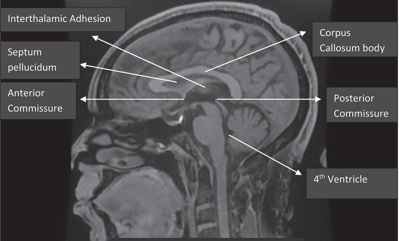
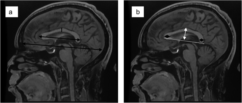
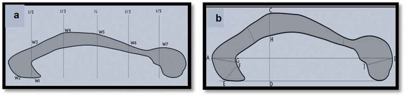
The Corpus callosum (CC) is the largest commissural fibre tract, ensuring swift information transfer and integration in both cerebral hemispheres. Variations in morphometry exist. There is a paucity of data on CC dimensions in our subregion, and no standardized reference is available. The study aims to determine the CC dimensions among the adult population in southeast Nigeria. The result will provide reference ranges and form a benchmark for comparisons of CC-related pathologies. A retrospective study of CC morphometric dimensions in normal subjects who had cranial MRI over two years in Memfys Hospital, Enugu, Southeast Nigeria, using a 1.5T GE© 16 channel machine. The CC was segmentalized into seven subregions using the modified Witelson method with special computer software. All measurements were taken twice from the T1 mid-sagittal image, and the mean was used for computation. The results were analyzed using descriptive and inferential statistics. A total of 200 subjects were recruited for the study. The mean length and height of the CC were 75.58 ± 4.52 mm and 24.64 ± 3.40 mm, respectively. The width dimensions of the genu, body, rostrum and splenium were 10.88 ± 1.81 mm, 5.66 ± 1.32 mm, 3.65 ± 1.25 mm, and 10.02 ± 1.70 mm, respectively. No gender variations were noted among the different dimensions of CC (P = 0.90). The length and height of CC increase gradually with age and show a positive correlation. The width dimensions of the genu and splenium increase till middle age and subsequently decreases in line with brain atrophy (p = 0.0000& p = 0.004). Using Pearson's correlation test, no correlation was noted in the dimensions of the body and rostrum of the corpus callosum when related to age and sex. (P = 0.92 & p = 0.66). Reference ranges of CC dimensions in our subregion were presented, and variations exist in its different morphometric dimensions which are affected by brain atrophy. Gender does not influence the dimensions in our subpopulations.
Keywords: Corpus callosum; Southeast Nigeria; morphometric dimensions; reference ranges; subregion.
Conflict of interest statement
No potential conflict of interest was reported by the authors.
Figures
References
-
- Mourgela S, Anagnostopoulou S, Sakellaropoulos A, et al. An MRI study of sex- and age-related differences in the dimensions of the corpus callosum and brain. Neuroanatomy. 2007;6:63–65. www.neuroanatomy.org
-
- Junle Y, Youmin G, Yanjun G, et al. A MRI quantitative study of corpus callosum in normal adults. J Med Coll PLA. 2008;23(6):346–351.
-
- Driesen NR, Raz A. The influence of sex, age, and handedness on corpus callosum morphology: a meta-analysis. Psychobiology. 1995;23(3):240–247. DOI:10.3758/BF03332028 - DOI
MeSH terms
LinkOut - more resources
Full Text Sources
Medical