. 2023 Mar 28;120(13):e2221874120.
doi: 10.1073/pnas.2221874120. Epub 2023 Mar 22.

A c-di-GMP binding effector controls cell size in a cyanobacterium

Affiliations

A c-di-GMP binding effector controls cell size in a cyanobacterium

Xiaoli Zeng et al. Proc Natl Acad Sci U S A. .

Abstract

Cyclic-di-GMP (c-di-GMP) is a ubiquitous bacterial signaling molecule. It is also a critical player in the regulation of cell size and cell behaviors such as cell aggregation and phototaxis in cyanobacteria, which constitute an important group of prokaryotes for their roles in the ecology and evolution of the Earth. However, c-di-GMP receptors have never been revealed in cyanobacteria. Here, we report the identification of a c-di-GMP receptor, CdgR, from the filamentous cyanobacterium Anabaena PCC 7120. Crystal structural analysis and genetic studies demonstrate that CdgR binds c-di-GMP at the dimer interface and this binding is required for the control of cell size in a c-di-GMP-dependent manner. Different functions of CdgR, in ligand binding and signal transmission, could be separated genetically, allowing us to dissect its molecular signaling functions. The presence of the apo-form of CdgR triggers cell size reduction, consistent with the similar effects observed with a decrease of c-di-GMP levels in cells. Furthermore, we found that CdgR exerts its function by interacting with a global transcription factor DevH, and this interaction was inhibited by c-di-GMP. The lethal effect triggered by conditional depletion of DevH or by the production of several point-mutant proteins of CdgR in cells indicates that this signaling pathway plays critical functions in Anabaena. Our studies revealed a mechanism of c-di-GMP signaling in the control of cell size, an important and complex trait for bacteria. CdgR is highly conserved in cyanobacteria, which will greatly expand our understanding of the roles of c-di-GMP signaling in these organisms.

Keywords: c-di-GMP; c-di-GMP effector; cell size; cyanobacteria; signal transduction.


Conflict of interest statement

The authors declare no competing interest.

Figures
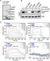
Fig. 1.
Fig. 1.
CdgR is a c-di-GMP effector in Anabaena. (A) SDS-PAGE analysis of proteins enriched by pull-down assay from Anabaena lysates with c-di-GMP conjugated (+) or control (-) sepharose beads. The indicated protein band (in square) was identified as CdgR by MALDI-TOF/TOF-MS (SI Appendix, Table S1). (B) Streptavidin blotting assay. Purified CdgR protein was incubated with biotinylated c-di-GMP (Bio-c-di-GMP) in the absence or presence of different nucleotides as competitors (c-di-GMP, cGMP, c-di-AMP, and cAMP). The binding of Bio-c-di-GMP was visualized by streptavidin-HRP. Coomassie brilliant blue (CBB) staining showed equal amounts of CdgR were included for each reaction. (C) LSPR analysis of the binding between CdgR and different nucleotides. His-CdgR was covalently immobilized to an NTA sensor. A series of indicated concentrations of nucleotides were applied as analytes. N.D., no detected.
Fig. 2.
Fig. 2.
Crystal structure of Syn_CdgR in complex with c-di-GMP. (A) Overall architecture of Syn_CdgR-(c-di-GMP) complex with two c-di-GMP located at each end of the dimer interface. The two CdgR subunits are shown in cartoon representation with different colors (magenta and yellow). (B) Zoom-in view of the dimer interface reveals the polar interactions between Y106, N64, and E67 from both subunits. (C) A Fo-Fc electron density map of c-di-GMP contoured at 2.0 σ. (D) Details of the c-di-GMP binding site with relevant residues indicated. c-di-GMP is shown in cyan/orange-colored sticks with key interactions to the residues indicated by dashed lines. (E) Details of the interactions between Syn_CdgR and c-di-GMP. The figure was produced using Ligplot (34). Hydrogen bonds are indicated by dashed lines between the atoms involved, while nonligand residues involved in direct hydrophobic contacts with c-di-GMP are shown as partial spoked circles in red.
Fig. 3.
Fig. 3.
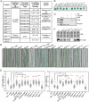
CdgR is the c-di-GMP receptor involved in the c-di-GMP signaling pathway for cell size control. (A) A table listing the Kd values of CdgR variants for c-di-GMP binding determined by LSPR. For detailed sensorgrams, see SI AppendixFig. S4. (B) Bacterial two-hybrid (BACTH) assay to test the self-interaction of CdgR and CdgR variants. The gene encoding the leucine zipper region of the GCN4 protein (ZIP) and empty vectors (EV) were used as positive and negative controls, respectively. (C) In vitro cross-linking of CdgR in the absence or the presence of c-di-GMP. Protein bands corresponding to monomeric and dimeric CdgR are indicated. EGS: succinimidyl succinate. (D) Micrographs of Anabaena filaments of different strains: WT (wild type), ΔcdgR (cdgR markless deletion mutant), OECT-cdgR (cdgR overexpression strain, under the control of the CT promoter), ΔcdgS (cdgS markless deletion mutant), ΔcdgSΔcdgR (cdgS and cdgR double mutant), OECT-yhjH (overexpression yhjH in WT), ΔcdgR::OECT-yhjH (overexpression of yhjH in ΔcdgR), and different strains with point mutation in cdgR (cdgRY97A, cdgRR101A, cdgRY106A, cdgRT109A, and cdgRY115A). cdgRWT is the WT cdgR used as a positive control. All overexpression strains were cultured in BG11 for 48 h after induction with 2 mM TP and 0.5 μM Cu2+. (Scale bars: 15 µm.) (E) Cell length and cell width distribution of indicated strains as in panel D. 500 cells of each strain from three independent experiments were measured and quantified. The boxplots enclose the 25th and 75th percentile with the black line representing the median value, red dot the mean value, and whiskers the SD. ***: P value <0.001 in two-sided Student’s t test, in comparison to WT or between the two strains connected by bracket at the top. n.s.: not significant (P > 0.05). (F) Western blotting analysis of protein levels in the indicated strains. Similar amounts of total proteins extracted from different strains were loaded on the gel, stained with coomassie brilliant blue (Top, CBB), or probed with polyclonal antibody against CdgR (Bottom).
Fig. 4.
Fig. 4.
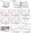
Apo-CdgR interacts with the essential transcription factor DevH. (A) SDS-PAGE analysis of proteins obtained by pull-down assays from Anabaena lysates with NTA columns saturated with either CdgR or CdgR-(c-di-GMP) complex. The two protein bands (indicated by arrows) were identified by MALDI-TOF/TOF-MS (SI Appendix, Table S2). (B) Copurification assays of DevH and CdgR in the presence (+) or absence (−) of c-di-GMP. Purified Strep-CdgR and His-Pros2-DevH were used, and the proteins were captured by Strep-Tactin column (IBA) after incubation. The eluted proteins (Top panel) and the input proteins (Bottom) were subjected to SDS-PAGE analysis and stained with coomassie brilliant blue (Left). The ratios between His-Pros2-DevH and Strep-CdgR under different conditions were quantified by densitometry and shown as histograms at the right panels. Control: relative intensity of His-Pros-DevH in lane 1 and Strep-DevH in lane 2. c-di-GMP (−): relative intensity of the two protein bands in lane 3. c-di-GMP (+): relative intensity of the two protein bands in lane 4. M: maker. (CG) BLI analysis of binding properties of DevH and CdgR at different states. Biotinylated-CdgR or CdgR-(c-di-GMP) complex or CdgR variants were immobilized on streptavidin-coated biosensors. A series of indicated concentrations of Pros2-DevH were applied as analytes. Experimental data are shown in lines with different color. The data were fitted into a simple 1:1 binding model, and the fitted lines are shown in red. (H) Immunoblot analysis of the DevH level in the PcoaT-devH strain during the time course of Co2+ (inducer for the expression of devH) depletion. Similar amounts of total proteins extracted from different strains were loaded on the gel and stained with coomassie brilliant blue (Top, CBB) and probed with polyclonal antibody against DevH (Bottom). (I) The growth curves of PcoaT-devH and WT strain in BG11 medium with or without Co2+. Absorbance at 750 nm was measured at the indicated times. To better remove traces of Co2+, the cultures were reinoculated once in medium free of Co2+ after 7 d of incubation. All values are showed as mean ± SD, calculated from triplicate data. (J) Cell length and cell width distribution of the PcoaT-devH strain during the time course of Co2+ depletion. The cell length and cell width were measured based on images as shown in SI Appendix, Fig. S7B. Cells of each strain from three independent experiments were measured. Number above each box indicates the number of cells used for quantification. The boxplots enclose the 25th and 75th percentile with the black line representing the median value, red dot the mean value, and whiskers the SD. ***: P value<0.001 in two-sided Student’s t test, in comparison to WT. n.s.: not significant (P > 0.05).
Fig. 5.
Fig. 5.
A model for c-di-GMP mediated cell size control in Anabaena. (A) After sensing unknown signals, the histidine kinase CdgK becomes autophosphorylated (CdgK~P) and then phosphorylates CdgS. The phosphorylated form of CdgS (CdgS~P) promotes its DGC activity and maintains the c-di-GMP pool in cells at a basal or a high level. Under such conditions, c-di-GMP binds to CdgR. The CdgR-(c-di-GMP) complex shows a poor affinity with the transcription factor DevH, allowing DevH to control the expression of genes required for maintenance of standard cell size. (B) When CdgK is in its unphosphorylated state, CdgS exists in an unphosphorylated form, leading to less c-di-GMP synthesis. At low c-di-GMP level, the apo form of CdgR predominates, which favors interaction with DevH. The CdgR-DevH complex would alter the gene expression pattern of the devH regulon, leading to changes of cell size.

Similar articles

Cited by

References

    1. Young K. D., The selective value of bacterial shape. Microbiol. Mol. Biol. Rev. 70, 660–703 (2006). - PMC - PubMed
    1. Martins B. M. C., Tooke A. K., Thomas P., Locke J. C. W., Cell size control driven by the circadian clock and environment in cyanobacteria. Proc. Natl. Acad. Sci. U.S.A. 115, E11415–E11424 (2018). - PMC - PubMed
    1. Campos M., et al. , A constant size extension drives bacterial cell size homeostasis. Cell 159, 1433–1446 (2014). - PMC - PubMed
    1. Weart R. B., et al. , A metabolic sensor governing cell size in bacteria. Cell 130, 335–347 (2007). - PMC - PubMed
    1. Velazquez-Suarez C., Luque I., Herrero A., The inorganic nutrient regime and the mre genes regulate cell and filament size and morphology in the phototrophic multicellular bacterium anabaena. mSphere 5 (2020). - PMC - PubMed

Publication types

MeSH terms

LinkOut - more resources