. 2023 Apr 13;66(7):5041-5060.
doi: 10.1021/acs.jmedchem.2c02132. Epub 2023 Mar 22.

Discovery of Nanomolar DCAF1 Small Molecule Ligands

Affiliations

Discovery of Nanomolar DCAF1 Small Molecule Ligands

Alice Shi Ming Li et al. J Med Chem. .

Abstract

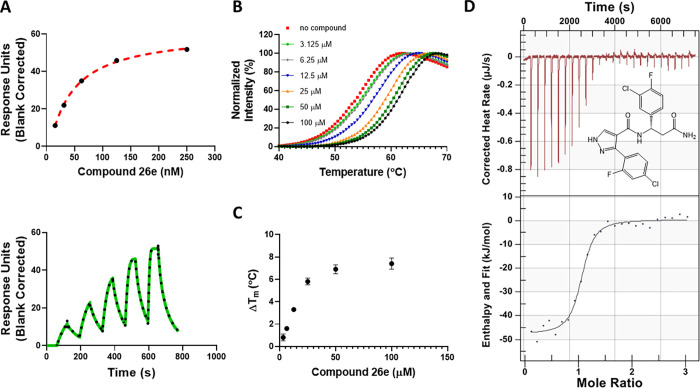
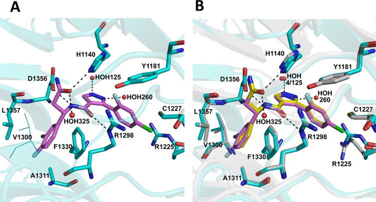
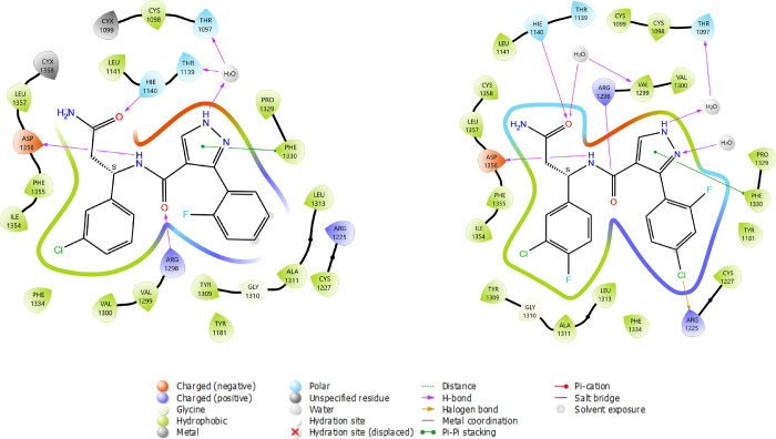
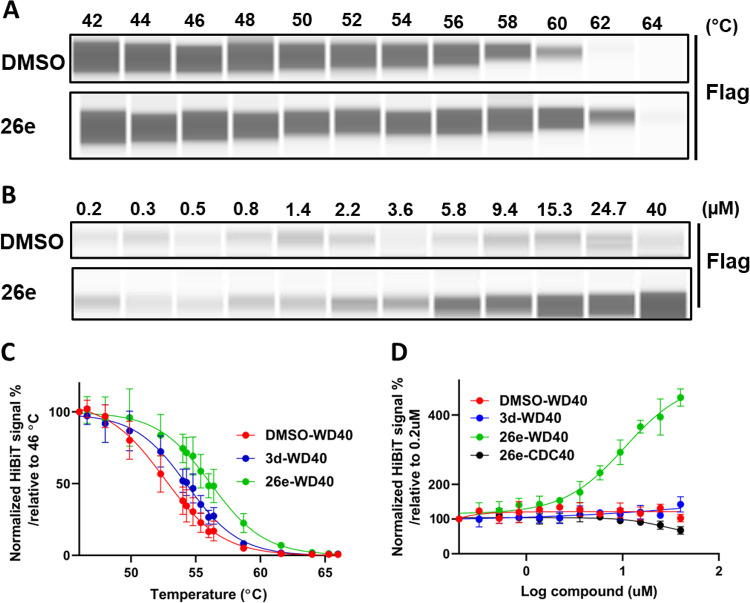
DCAF1 is a substrate receptor of two distinct E3 ligases (CRL4DCAF1 and EDVP), plays a critical physiological role in protein degradation, and is considered a drug target for various cancers. Antagonists of DCAF1 could be used toward the development of therapeutics for cancers and viral treatments. We used the WDR domain of DCAF1 to screen a 114-billion-compound DNA encoded library (DEL) and identified candidate compounds using similarity search and machine learning. This led to the discovery of a compound (Z1391232269) with an SPR KD of 11 μM. Structure-guided hit optimization led to the discovery of OICR-8268 (26e) with an SPR KD of 38 nM and cellular target engagement with EC50 of 10 μM as measured by cellular thermal shift assay (CETSA). OICR-8268 is an excellent tool compound to enable the development of next-generation DCAF1 ligands toward cancer therapeutics, further investigation of DCAF1 functions in cells, and the development of DCAF1-based PROTACs.


Conflict of interest statement

The authors declare the following competing financial interest(s): J.P.G, Y.Z., M.A.G., A.D.K. are employees of X-Chem. M.v.R., J.S.D, B.L.S. and J.W.C. are or were (C.J.M.) employees of Relay Therapeutics and were employees of X-Chem at the time of writing. Employees and past employees may hold stocks and shares. All other authors declare no conflict of interest.

Figures

Figure 1
Figure 1
Confirmation of Z1391232269 binding to the DCAF1 WDR domain by biophysical methods. (A) Z1391232269 stabilized the WDR domain of DCAF1 at 400 μM (green trace) compared with the WDR domain with no compound (black trace), and (B) the effect was concentration dependent. (C) SPR confirmed its binding to DCAF1 with a KD of 11.5 ± 4.2 μM. Experiments were performed in triplicate.
Scheme 1
Scheme 1. Synthesis of Compounds 3a and 3cd
Reagents and conditions: (a) EDCI, DIEA, 0 °C (5 min), rt (18 h).
Figure 2
Figure 2
Biophysical characterization of compounds 3c and 3d. Binding of compound 3c and 3d to DCAF1 WD40 domain was assessed using (A) SPR (KD values of 490 ± 90 nM for 3d and 13.5 ± 0.2 μM for 3c) and (B) DSF (ΔTm of 2.8 ± 0.05 and 3.8 ± 0.08 °C at 25 and 50 μM for 3d, respectively, and no stabilization effect for 3c even at 50 μM). Compound 3d was orthogonally confirmed by (C) ITC with a KD value of 932 ± 150 nM. The chemical structure of 3d is shown. Experiments were performed in triplicate.
Figure 3
Figure 3
Co-crystal structure of DCAF1 WDR domain in complex with compound 3d. (A) The top and side views of the DCAF1 WDR domain shown as cartoon representation in gray bound to 3d shown as yellow sticks. (B) Close-up view of the 3d binding site in chain A of DCAF1-3d. 3d forms a network of interactions with the protein and a water molecule within the central channel of DCAF1 WDR domain ring. The compound is shown as yellow sticks, and the protein residues involved in the interactions are rendered as gray sticks. The hydrogen bonds are shown as black dashes, and an interacting water molecule is shown as a red sphere. (C) An overlay of the DCAF1-3d structure (shown in surface representation in light gray and 3d in yellow sticks indicated by black arrow) onto the DCAF1 WDR domain in complex with lentiviral Vpx (shown in cartoon representation in green) and SAMHD1 (not shown) (PDB ID: 5AJA), showing the compound binding site relative to that of Vpx.
Scheme 2
Scheme 2. Synthesis of Compounds 5ab
Reagents and conditions: (a) MsCl, DIEA, DCM, 0 °C (10 min), then 1d, 0 °C (30 min).
Scheme 3
Scheme 3. Synthesis of Compound 5c
Reagents and conditions: (a) HATU, DIEA, DMF, MeNH2; (b) HCl in 1,4-dioxane; (c) TsCl, Py; (d) HATU, DIEA, DMF, 8; (e) KOH (aq.), THF.
Scheme 4
Scheme 4. Synthesis of Compounds 14aj
Reagents and conditions: (a) BOC2O, DIEA, DCM; (b) EDCI, NH4Cl, DIEA, DCM; (c) HCl in 1,4-dioxane; (d) EDCI, DIEA, DCM 0 °C (5 min), rt (18 h).
Scheme 5
Scheme 5. Synthesis of Compounds 15ab, 15f, 15j
Reagents and conditions: (a) EDCI, DIEA, 0 °C (5 min), rt (18 h).
Scheme 6
Scheme 6. Synthesis of Intermediates 1no
Reagents and conditions: (a) PPTS, MgSO4, DCM; (b) Zn, CuCl, THF; (c) 7 N NH3 in MeOH; (d) HCl in 1,4-dioxane.
Scheme 7
Scheme 7. Synthesis of Compounds 26ae
Reagents and conditions: (a) NaH, SEMCl, THF; (b) PdCl2(PPh3)2, Cs2CO3, 1,4-dioxane, H2O, μw, 110 °C; (c) KOH, THF/MeOH/H2O, 70 °C; (d) HATU, DIEA, DMF; (e) TFA, DCM.
Figure 4
Figure 4
Biophysical characterization of compound 26e (OICR-8268) with the DCAF1 WDR domain. Binding of compound 26e to the DCAF1 WDR domain was tested by SPR and DSF assays. (A) SPR showed 26e binding to DCAF1 potently with a KD of 38 ± 1.5 nM, and (B) DSF showed that the compound stabilizes DCAF1 in a concentration-dependent manner (C) with increases in the protein’s melting temperature, indicating binding. (D) Orthogonal binding confirmation of 26e by ITC with a KD of 216 ± 76 nM. All experiments were performed in triplicate.
Figure 5
Figure 5
Co-crystal structure of DCAF1 WDR domain in complex with 26e. (A) A close-up view of 26e binding site in the central tunnel of the DCAF1 WDR domain. Compound 26e retains the network of interactions observed in DCAF1-3d (Figure 3B) but also makes additional interactions with two extra water molecules, as well as additional hydrophobic interactions with the protein residues. Compound 26e is shown as magenta sticks, and the protein residues involved in the interactions are rendered as cyan sticks. The hydrogen bonds are shown as black dashes, and interacting water molecules are shown as red spheres. (B) An overlay of DCAF1-3d and DCAF1-26e structures showing the location of the additional atoms in the 26e compound and the binding site differences in the two proteins. Compounds and active site residues are rendered as described in Figure 3B and in panel A here.
Figure 6
Figure 6
2D interaction diagrams of 3d and 26e ligands in complex with DCAF1 protein. The most important interaction classes, including water-mediated hydrogen bonds, are represented. All protein residues within 5.0 Å from ligand are displayed. This picture has been generated using the LID tool from Maestro software (Schrodinger) from the prepared respective X-ray structures (7UFV and 8F8E).
Figure 7
Figure 7
26e engages the WDR domain of DCAF1 in cells. In a cellular thermal shift assay (CETSA) as described in the materials and methods, the amount of unaggregated flag tagged WDR domain of DCAF1 was quantified. (A) 26e significantly stabilized the Flag-Hb-DCAF1-WD at 40 μM. (B) Titration of 26e showed a dose-dependent pattern of DCAF1-WD stability. Different compound concentrations (40 to 0.2 μM) of 26e were heated at 61 °C for 3.5 min. (C) When compared to the DMSO treatment, 26e thermally stabilized the WDR domain of DCAF1 to a greater extent than 3d, causing a 3.6 and 1.7 °C shift in the aggregation temperature of Flag-Hb-DCAF1_WD at 40 μM. Results shown as average ± SD (n = 3) (D) 26e thermally stabilized Flag-Hb-DCAF1_WD in a dose-dependent manner with an EC50 value of 10.5 μM. Results shown as average ± SD (n = 3). CDC40 was used as a negative control. Note that in C, cells are subjected to a gradient of temperatures (46 to 66 °C). Across this range of temperatures, 3d thermally stabilizes the WDR domain of DCAF1, but the stabilization does not extend beyond 61 °C. In D, the protein is kept at 61 °C, and consistent with our observation in C, 3d was not expected to stabilize the WDR domain at any concentration.

References

    1. Khan S.; He Y.; Zhang X.; Yuan Y.; Pu S.; Kong Q.; Zheng G.; Zhou D. PROteolysis TArgeting Chimeras (PROTACs) as emerging anticancer therapeutics. Oncogene 2020, 39, 4909–4924. 10.1038/s41388-020-1336-y. - DOI - PMC - PubMed
    1. Nalawansha D. A.; Crews C. M. PROTACs: An Emerging Therapeutic Modality in Precision Medicine. Cell Chem. Biol. 2020, 27, 998–1014. 10.1016/j.chembiol.2020.07.020. - DOI - PMC - PubMed
    1. Zheng N.; Shabek N. Ubiquitin Ligases: Structure, Function, and Regulation. Annu. Rev. Biochem. 2017, 86, 129–157. 10.1146/annurev-biochem-060815-014922. - DOI - PubMed
    1. Zou Y.; Ma D.; Wang Y. The PROTAC technology in drug development. Cell Biochem. Funct. 2019, 37, 21–30. 10.1002/cbf.3369. - DOI - PMC - PubMed
    1. Ishida T.; Ciulli A. E3 Ligase Ligands for PROTACs: How They Were Found and How to Discover New Ones. SLAS Discovery 2021, 26, 484–502. 10.1177/2472555220965528. - DOI - PMC - PubMed

Publication types

LinkOut - more resources