Review
. 2023 Apr 12;123(7):3493-3542.
doi: 10.1021/acs.chemrev.3c00033. Epub 2023 Mar 22.

Plate-Like Colloidal Metal Nanoparticles

Affiliations
Review

Plate-Like Colloidal Metal Nanoparticles

Leonardo Scarabelli et al. Chem Rev. .

Abstract

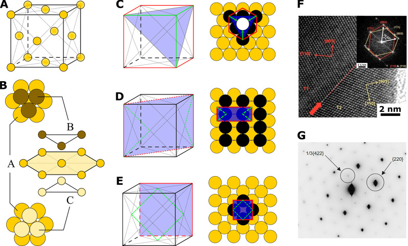
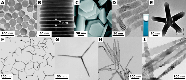
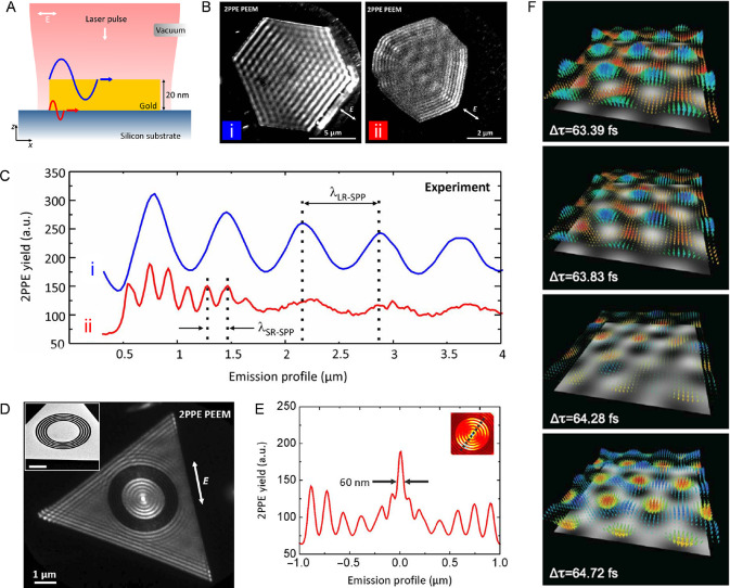
The pseudo-two-dimensional (2D) morphology of plate-like metal nanoparticles makes them one of the most anisotropic, mechanistically understood, and tunable structures available. Although well-known for their superior plasmonic properties, recent progress in the 2D growth of various other materials has led to an increasingly diverse family of plate-like metal nanoparticles, giving rise to numerous appealing properties and applications. In this review, we summarize recent progress on the solution-phase growth of colloidal plate-like metal nanoparticles, including plasmonic and other metals, with an emphasis on mechanistic insights for different synthetic strategies, the crystallographic habits of different metals, and the use of nanoplates as scaffolds for the synthesis of other derivative structures. We additionally highlight representative self-assembly techniques and provide a brief overview on the attractive properties and unique versatility benefiting from the 2D morphology. Finally, we share our opinions on the existing challenges and future perspectives for plate-like metal nanomaterials.


Conflict of interest statement

The authors declare no competing financial interest.

Figures

Figure 1
Figure 1
(a) Image from original 1951 Turkevich paper on gold nanoparticle synthesis showing numerous anisotropic nanoplate impurities. Magnification is indicated in the original manuscript as 50 000 diameters. Reproduced with permission from ref (1). Copyright 1951 Royal Society of Chemistry. (b) Calculation of anisotropicity value (α, defined in text) for a plate (gold solid line) and a rod (blue solid line) both with circular cross-section. Aspect ratio is defined as length/width.
Figure 2
Figure 2
fcc crystallographic habit. (A,B) Three-dimensional spatial arrangement of the unit cell in an fcc crystal. (C–E) Projection perspective (left) an top view (right) representations of the three main crystallographic directions in order of stability (from top to bottom) from the most stable: {111} (C), {100} (D), and {110} (E). (F) High resolution transmission electron microscopy (HRTEM) analysis of a nanocrystal presenting a single twin- defect. Adapted with permission from ref (37). Copyright 2016 Wiley-VCH. (G) Diffraction pattern of an fcc nanoplate, showing the forbidden that in1/3{422} reflections, indicating the formation of stacking faults. Adapted with permission from ref (39). Copyright 2005 Wiley-VCH.
Figure 3
Figure 3
(A) Plot showing the relative distribution of the twin structure of Pd nanocrystals as a function of the initial reaction rate (r0) in a synthesis. Reproduced with permission from ref (52). Copyright 2015 American Chemical Society. (B) Proposed growth pathway of Au nanoplates through oxidative etching. Reproduced with permission from ref (65). Copyright 2014 American Chemical Society. (C) Schematic representation of the experimental procedures for oxidative etching of Au nanoplates and Au nanorods to obtain seeds with selected size (12 and 20 nm) and crystallinity (monotwinned and single-crystal), and subsequent seeded growth under specific conditions (different iodide concentrations) to investigate the effect of size and twinning on the shape evolution of Au nanoparticles. Reproduced with permission from ref (46). Copyright 2017 Royal Society of Chemistry under CC BY-NC 3.0.
Figure 4
Figure 4
(A) Scheme showing the single-twin growth mechanism responsible for the formation of triangular platelets; concave and convex edges of the platelet are marked with “A” and “B,” respectively. Reproduced with permission from ref (74) Copyright 2016 American Chemical Society. (B–D) Schematic illustrations of the (B) double-twinned growth mechanism of hexagons, (C) triply twinned primary growth mechanism of nonequilateral hexagons, and (D) secondary growth on triply twinned particles that yields nonagons or nonequilateral hexagons. Reproduced with permission from ref (75). Copyright 2011 American Institute of Physics. The red circles indicate the positions of re-entrant grooves.
Figure 5
Figure 5
(A) Citrate ions selectively protect the {111} basal facets of Ag nanoplates and only allow lateral overgrowth. Reproduced with permission from ref (28). Copyright 2010 American Chemical Society. (B) Role of iodide ions in the growth of Au nanoplates enclosed by {111} facets: Presence of iodide ions leads to the formation of Au nanoplates through seeded-growth, while its absence yields nanorods when iodide-free CTAB is used as a surfactant. Reproduced with permission from ref (90). Copyright 2018 American Chemical Society. (C) Schematic illustration showing the synthesis of Al nanosheets, where selective binding of O2 on {111} basal planes promotes the formation of ultrathin Al nanosheets. Reproduced with permission from ref (89). Copyright 2020 Cell Press.
Figure 6
Figure 6
(A) Proposed photomediated growth pathways of Ag nanoplates from spherical nanoparticles. Reproduced with permission from ref (6). Copyright 2001 AAAS. (B) Proposed photovoltage mechanism for light conversion of citrate-stabilized Ag nanocrystal seeds into large nanoplates. Reproduced with permission from ref (91). Copyright 2008 American Chemical Society. (C) Liquid cell TEM observation of 2D nanoplate growth, mediated by plasmon-enhanced nanoparticle coalescence. Reproduced with permission from ref (93). Copyright 2020 American Chemical Society.
Figure 7
Figure 7
Tomographic sequential orthoslices of Pt islands in epitaxial contact with the Au nanoplate surface indicating the root, trunk, and cap island morphologies. Reproduced from ref (111). Copyright 2016 American Chemical Society.
Figure 8
Figure 8
(A) Synthetic scheme for Au@Pt nanodisks. (B–E) SEM images of Au nanodisks (B), rim-preferentially Pt-coated Au nanodisks (Au@Pt(rim) nanodisks) (C–E) with increasing the concentration of Pt precursors. Atomic percent of Au and Pt is indicated at the upper right side of each image, which was obtained from EDS analysis. The insets in B and E show side views. Adapted with permission from ref (117). Copyright 2014 The Royal Society of Chemistry.
Figure 9
Figure 9
Scheme of Au–Ag alloy triangular nanoframe synthesis. Reproduced with permission from ref (118). Copyright 2003 American Chemical Society.
Figure 10
Figure 10
(a) SEM image of particles-in-a-frame nanostructures (PIAFs). Au PIAFs containing one inner nanoparticle are marked by orange circles. (b) TEM image of Au PIAFs containing one inner nanoparticle. (c) HAADF-STEM image and corresponding EDX elemental mapping image of an Au PIAF (scale bar: 10 nm). (d) Representative TEM images of nanostructures in the samples collected at different reaction times during the formation of Au PIAFs: (d1) 0, (d2) 5, (d3) 60, (d4) 300 s. Scale bars: 20 nm. (e) Illustration of hypothesized growth pathway of Au PIAFs. Adapted with permission from ref (120). Copyright 2019 Wiley-VCH.
Figure 11
Figure 11
(A) Schematic illustration of the experimental procedures for synthesizing Pt@Au nanorings. (B) SEM images of Pt@Au circular nanorings (a1–a3), Pt@Au triangular nanorings (b1–b3), and Pt@Au hexagonal nanorings (c1–c3), having different thickness. Adapted with permission from ref (123). Copyright 2014 American Chemical Society.
Figure 12
Figure 12
(A) eqs 1 and 2 represent the competing reactions during the particle growth process. When given amounts of Au ions and reducing agent are added stepwise, k2 < k1. However, when the same amount is added “all-in-one”, k2 > k1. Adapted from ref (125). Copyright 2006 Wiley-VCH. (B) Schematic Illustration of the anisotropic seeded growth of Ag nanoplates based on selective ligand adhesion. (C) Schematic Illustration of the consequences of seeded growth at different reaction rates. Adapted from ref (28). Copyright 2010 American Chemical Society.
Figure 13
Figure 13
SEM images of Ag nanoplates synthesized in the presence of (A) trisodium citrate and (B) poly(vinylpyrrolidone) (PVP) as capping agents, respectively. (C,D) Plots of both edge length and thickness of the Ag nanoplates as a function of the number of growth rounds for (C) trisodium citrate and (D) PVP capping ligands. Adapted from ref (129). Copyright 2011 Wiley-VCH. (E) Scheme of core@shell Au@Ag nanostructure formation with and without added iodide. Adapted from ref (130). Copyright 2011 American Chemical Society.
Figure 14
Figure 14
(A) Reduction potential diagram of the shape transformation reaction. Adapted from ref (131). Copyright 2021 American Chemical Society. (B) The Δμ for NC growth can be tuned to favor corner- and edge-selective growth. Δμ is defined as the difference between chemical potential of the solute in solution (e.g., Au0 atoms) and that of the solid crystal (e.g., Au seed). (C) Pictures depicting different regions [corners (c), edges (e), and faces (f)] of a Au nanoplate with varying curvatures and, therefore, different ligand distributions. The higher the curvature (K), the larger the average distance between the ligands (δ) (here, Kc > Ke > Kf such that δc > δe > δf). (D) Schematic showing the general energy profile for nanocrystal growth, which is dictated by the degree of particle curvature Ec < Ee < Ef. (E) SEM images showing corner- and edge-selective nucleation on Au nanoplates. Scale bars: 100 nm. Adapted from ref (132). Copyright 2021 AAAS under CC BY 4.0 http://creativecommons.org/licenses/by/4.0/.
Figure 15
Figure 15
Periodic table indicating the most stable crystal structure at standard temperature and pressure for each element; nonmetal and synthetic/radioactive elements are not included in this review.
Figure 16
Figure 16
fcc nanoplates, part 1: (A,B) Cu nanodisks prepared in ultrapure water balancing oxidative O2 gas and reductive glucose. Adapted with permission from ref (162). Copyright 2022 American Chemical Society. (C) Pd nanoplates synthesized via a solvothermal/PVP approach. Adapted with permission from ref (173). Copyright 2015 Royal Society of Chemistry. (D) Pd nanodisks prepared in aqueous environment controlling oxidative etching from I/O2 pairs. Adapted with permission from ref (184). Copyright 2017 Royal Society of Chemistry). (E) Pt pentatwinned planar nanostars obtained combining oleylamine and hydrogen gas as solvent/stabilizer and reducing agent, respectively. Adapted with permission from ref (212). Copyright 2012 Wiley-VCH). (F,G) Pt planar tripod synthesized using adamantanecarboxylic acid and hexadecylamine as a double capping agent system. Adapted with permission from ref (209). Copyright 2006 Royal Society of Chemistry). (H,I) Pt tribranched nanoribbons produced by the addition of ammonium fluoride to the reaction mixture. Adapted with permission from ref (213). Copyright 2009 American Chemical Society.
Figure 17
Figure 17
fcc nanoplates, part 2. (A) Ni nanotriangles synthesized in a mixture of oleic acid and oleylamine as capping agents and iron pentacarbonyl as shape-directing agent. Adapted with permission from ref (239). Copyright 2007 ACS. (B) Rh platelets prepared in N,N-dymethylformamide using PVP as capping agent and carbon monoxide as shape inducing agent. Adapted with permission from ref (259). Copyright 2015 Wiley-VCH under CC BY 4.0 http://creativecommons.org/licenses/by/4.0/). (C) Monoatomically thin Rh sheets obtained using formaldehyde and PVP as capping agent. Adapted with permission from ref (262). Copyright 2014 Springer. (D) Al platelets prepared exploiting a mixture of oxygen and nitrogen gas in the growth solution to regulate the concentration of oxygen during growth. Adapted with permission from ref (89). Copyright 2020 Cell Press. (E) Ir nanoflakes synthesized under UV light irradiation of a basic 1,2-dihydroxynaphthalene solution, in the presence of CTAB. Adapted with permission from ref (289). Copyright 2011 Elsevier. (F) Pb platelets prepared combining PVP and CTAB. Adapted with permission from ref (298). Copyright 2007 Wiley-VCH).
Figure 18
Figure 18
hcp crystallographic habit. (A,B) 3-Dimensional spatial arrangement of the unit cell of an hcp crystal (A) and its primitive cell (B), in orthogonal view (left) and top view (right). (C) Representation of the unit cell crystallographic facets and multiple low-energy twin planes. Adapted with permission from ref (307). Copyright 2020 American Chemical Society under CC BY 4.0 http://creativecommons.org/licenses/by/4.0/). (D) Predicted Wulff shapes for single-crystalline and monotwinned structures with predominant (0001) facets. Adapted with permission from ref (307). Copyright 2020 American Chemical Society under CC BY 4.0 http://creativecommons.org/licenses/by/4.0/.
Figure 19
Figure 19
hcp nanoplates. (A,B) HAADF-STEM images and corresponding Mg and O STEM-EDS maps of a Mg nanoplate synthesized using Li naphthalide as reducing agent. The platelet is oriented (A) perpendicular and (B) parallel to the electron beam. Adapted with permission from ref (331). Copyright 2018 American Chemical Society. (C) Co nanodisks prepared by thermal decomposition of Co2(CO)8 in a mixture of oleic acid and trioctylphosphine oxide in dichlorobenzene. Adapted with permission from ref (339). Copyright 2002 American Chemical Society. (D) Co nanoplates synthesized through the reduction of CoCl2 by NaH2PO2 in a basic aqueous solution. Inset: SAED pattern of the flat face of a single nanoplate. Adapted with permission from ref (341). Copyright 2009 Elsevier. (E,F) Zn hexagonal nanoplates synthesized via LiN(SiMe3)2 reduction, in the presence of a noncoordinating solvent such as octadecene. Adapted with permission from ref (348). Copyright 2015 American Chemical Society. (G) Ru capped nanocolumns prepared by hydrothermal decomposition of RuCl3 in the presence of formaldehyde and sodium oxalate. Adapted with permission from ref (354). Copyright 2012 American Chemical Society. (H) Ultrathin Ru nanoplates obtained as the main product of the hydrogenation of [Ru(COD)(COT)] in the presence of both hexadecylamine, and lauric acid. Adapted with permission from ref (359). Copyright 2022 American Chemical Society. (I) High-resolution HAADF-STEM image of a Ru 3-fold star. Inset: image at lower magnification showing two 3-fold stars with overlapping branches, Adapted with permission from ref (359). Copyright 2022 American Chemical Society.
Figure 20
Figure 20
bcc crystallographic habit. (A) Three-dimensional spatial arrangement of the unit cell in a bcc crystal. (B) Hard-sphere model of a bcc unit cell; the atoms are in contact along the body diagonal (bottom). (C) Representation of the distribution of nearest neighbors (giving a CN of 8) and next-nearest neighbors (Nnn) in a bcc crystal. The high number of Nnn and their close distance (only 15% higher compared to the nearest neighbors) contributes significantly to the overall stability. (D–F) Top view representation of the three main crystallographic directions in the order of stability, from the most to the less stable: {110} (D), {100} (E), and {111} (F); in F, the atom at the volumetric center of the cubic unit cell is not visible.,
Figure 21
Figure 21
bcc nanoplates. (A) Electrochemical growth in a solid-state open-cell configuration of a hexagonal ultrathin Li nanoplate imaged by TEM at two different times. Adapted with permission from ref (369). Copyright 2021 Wiley. (B) SAED pattern of the extended face of the growing nanoplate, confirming the bcc crystal structure. Adapted with permission from ref (369). Copyright 2021 Wiley. (C,D) SEM (C) and TEM (D) imaging of amorphous Fe nanoplates obtained by chemical reduction of FeSO4 by NBH4 assisted by an external magnetic field. Inset: SAED pattern showing no diffraction points, thus confirming the amorphous nature of the Fe nanoparticles. Adapted with permission from ref (371). Copyright 2012 Royal Society of Chemistry.
Figure 22
Figure 22
Nanoplates with noncubic crystallography. (A–C) In nanotriangles synthesized via InCl3 reduction by NaBH4 in the presence of PVP; SAED analysis (C) confirmed the expected tetragonal crystal structure. Adapted with permission from ref (385). Copyright 2017, Royal Society of Chemistry under CC BY-NC 3.0. (D,E) Bi hexagonal nanoplates obtained by solvent-free thermolysis of Bi thiolate precursors in the presence of excess 1-dodecanoethiol. SAED analysis (E) confirmed the expected rhombohedral crystal structure. Adapted with permission from ref (399). Copyright 2010 American Chemical Society). (F) Single-crystal Bi nanotriangles resulting from the hydrothermal reduction of Bi(NO3)3 in alkaline conditions by ascorbic acid, in the presence of Na2EDTA, and carboxylic acid-terminated PVP. Adapted with permission from ref (402). Copyright 2022 Elsevier.
Figure 23
Figure 23
Self-assembly of nanoplates. (A) Depletion attraction-driven self-assembly of Au nanotriangles under different initial CTAC concentrations ranging from 0.1 to 10 mM. Schematics below show different configurations of self-assemblies. Scale bars: 100 nm. Reproduced with permission from ref (406). Copyright 2017 American Chemical Society. (B) TEM images of binary superlattices self-assembled from nanotriangles and nanospheres. The schematics at the corner illustrate the proposed unit cell. Reproduced with permission from ref (414). Copyright 2006 Nature Publishing Group. (C) Schematic process and resulting product of large-area template-assisted self-assembly of Au nanotriangles. Scale bars: 500 nm. Reproduced with permission from ref (411). Copyright 2014 American Chemical Society. (D) Schematic self-assembly procedure of Au bowties directed by DNA origami (left), and the TEM and AFM images of the resulting Au bowties (right). Reproduced with permission from ref (409). Copyright 2018 Wiley-VCH.
Figure 24
Figure 24
Factors that influence the LSPR mode of 2D plasmonic nanoparticles. (A) Dielectric functions of bulk Au (i), Pt (ii), and Al (iii), and experimentally measured extinction spectra of random arrays of Au (iv), Pt (v), and Al (vi) nanodisks. ε1 and ε2 are the real and imaginary parts of the dielectric function. Reproduced with permission from ref (418). Copyright 2011 American Chemical Society. (B) Schematic diagrams of the charge oscillation corresponding to in-plane dipolar mode (i), out-of-plane dipolar mode (ii), in-plane quadrupolar mode (iii), and out-of-plane quadrupolar mode (iv). Reproduced with permission from ref (436). Copyright 2005 American Chemical Society. (C) Simulated extinction spectra of Ag nanotriangles with different edge lengths (left) and snips (right). Reproduced with permission from ref (437). Copyright 2003 American Chemical Society. (D) Schematic diagram, digital photographs, and extinction spectra of Ag nanoplates reshaping from triangular to circular shape, while the thickness of the nanoplates increases. Reproduced with permission from ref (440). Copyright 2009 Wiley-VCH.
Figure 25
Figure 25
Characterization of characteristic LSPR modes. (A) Single-particle dark-field scattering measurement of vertically and horizontally oriented hexagonal Au nanoplates. (top) Comparison of the SEM and dark-field images of individual Au nanoplates deposited on an ITO substrate. (bottom) Dark-field scattering spectra and schematic diagrams of vertically and horizontally oriented hexagonal Au nanoplates. Reproduced with permission from ref (450). Copyright 2018 Royal Society of Chemistry. (B) Experimental EELS amplitude distributions of a triangular Ag nanoplate. The three maps centered at different energies were resolved from EEL spectra excited at the corner, the center, and the edge of the Ag nanoplate. Reproduced with permission from ref (454). Copyright 2007 Nature Publishing Group. (C) Schematic diagram of SNOM (left top), near-field transmission images measured from a hexagonal Au nanoplate at different near-field probe tip–sample distances (left bottom), and spatial distributions of out-of-plane and in-plane LSPR modes (right). Reproduced with permission from ref (451). Copyright 2019, American Chemical Society. (D) Comparison of EELS and CL spectra taken at the low left tip (T) and left side (S) of the exact same individual Au nanoplate (left), and the EELS and CL maps of the dipolar and higher-order LSPR modes (right). Reproduced with permission from ref (457). Copyright 2013 American Institute of Physics.
Figure 26
Figure 26
Surface plasmon polaritons supported by Au platelets. (A) Schematic diagram showing Au platelets deposited on silicon substrates. Long-range (LR) SPPs are indicated at the Au–vacuum interface (blue), and short-range (SR) SPPs are indicated at the Au–Si interface (red). (B) 2PPE PEEM images measured from a 120 nm thick Au platelet (i) and a 37 nm thick Au platelet (ii) at normal incidence. (C) PEEM emission profiles from the two platelets. Profile (i) shows LR- SPP pattern, whereas profile (ii) illustrates the superposition of LR-and SR-SPP. (D) 2PPE PEEM image of an Au platelet patterned with a circular grating of 150 nm period and a central disk with a diameter of 2 μm, under the excitation of 800 nm laser. Inset shows the SEM image. (E) Emission profile perpendicular to occurring wavefronts. (A–E) Reproduced with permission from ref (458). Copyright 2017 AAAS under CC BY 4.0 http://creativecommons.org/licenses/by/4.0/. (F) Time dependence of the SPP skyrmion lattice on an Au platelet with grooves for the excitation of SPPs. Reproduced with permission from ref (460). Copyright 2020 AAAS.
Figure 27
Figure 27
Light–matter interaction based on 2D plasmonic nanoparticles. (A) Strong coupling between single Ag nanoplate and molecular J aggregates at ambient conditions. (i) Extinction spectrum (in water), schematics, and cryo-TEM image of TDBC J aggregates. Inset shows the chemical structure of TDBC monomer. (ii) Scattering spectrum, schematics, and SEM image of the Ag nanoplate. (iii) Scattering spectrum, schematics, and SEM image of a single Ag nanoplate strongly coupled to J aggregates. Reproduced with permission from ref (462). Copyright 2015 American Institute of Physics. (B) Plasmonic nanolasers based on 3D-bowtie nanoarray. (i) Schematic diagram of the 3D-bowtie array with IR-140 dye as gain materials. (ii,iii) SEM images of 3D-bowtie array with period of 1200 nm (ii) and 600 nm (iii), respectively. Reproduced with permission from ref (423). Copyright 2012 American Chemical Society. (C) Comparison between chemically synthesized Au flakes and vapor-deposited Au film. (i–iii) SEM images of the chemically synthesized Au flakes (i), smooth surface of a crystalline Au flake with the rectangular area milled by FIB (ii), rough surface of a vapor-deposited Au film (iii). (iv) SEM image of bowtie antennas fabricated by FIB on a crystalline Au flake (left) and vapor deposition (right). (v) TPPL map of the same area shown in (iv). Reproduced with permission from ref (466). Copyright 2010 Springer Nature. (D) Tilted-nanocavity-coupled system comprising an upconverting nanoparticle embedded in the nanogap between an Ag nanocube (AgNC) and an Ag microplate (AgMP). Reproduced with permission from ref (483). Copyright 2022 Springer Nature.
Figure 28
Figure 28
Sensing based on 2D plasmonic nanoparticles. (A) Dependence of the dipolar LSPR wavelengths on the refractive index for Au nanoplates, nanorods, and bipyramids. Reproduced with permission from ref (128). Copyright 2016 Wiley-VCH. (B) Schematic diagram, simulated electric field profile, and fluorescence enhancement factor of Au bowtie nanoantenna. The red and blue dots denote excitation light polarized parallel/perpendicular to the long axis of the bowtie. The black dashed lines connect results measured from the same molecule. Reproduced with permission from ref (420). Copyright 2009 Springer Nature. (C) Schematic diagram of a TERS setup. Light is focused at the apex of an Ag-coated TERS tip approaching the Au nanoplate surface, functionalized by a self-assembled molecular monolayer. Reproduced with permission from ref (489). Copyright 2020 Springer Nature under CC BY 4.0 http://creativecommons.org/licenses/by/4.0/. (D) Schematic illustration of light-directed reversible assembly of Au nanoplates, and their release or redispersion due to plasmon-enhanced thermophoresis and electrostatic repulsive interaction. Reproduced with permission from ref (490). Copyright 2016 American Chemical Society. (E) Schematic diagram of a plasmonic biosensor based on enzymatic etching on triangular Au nanoplates in the presence of H2O2. Reproduced with permission from ref (491). Copyright 2020 Springer.
Figure 29
Figure 29
Catalysis. (A) Mapping of specific catalytic activity correlated with the SEM image of a single Au@SiO2 nanoplate. Reproduced with permission from ref (503). Copyright 2013 American Chemical Society. (B) The schematic atom models of the PtPb@Pt core–shell nanoplate viewed from the top and the side interface. (C) High-resolution HAADF image at the edge of the PtPb@Pt core–shell nanoplates, together with the overlapped schematic atom model. (D) Mass and specific activities of PtPb nanoplates/C, PtPb nanoparticles/C, and commercial Pt/C catalysts. Reproduced with permission from ref (430). Copyright 2016 AAAS. (E) Catalytic FAOR activities of the Ag@Au@Pt nanoplates measured by cyclic voltammetry in 0.5 m H2SO4 + 0.25 m HCOOH. (F) Mass activities of the catalysts in the FAOR (left axis) and degree of CO poisoning in the FAOR (right axis). (G) Mass activities of Pt ensembles with 3–4 contiguous Pt atoms in the methanol oxidation reaction (MOR). Reproduced with permission from ref (432). Copyright 2022 Wiley-VCH
Figure 30
Figure 30
Mechanical properties of 2D plasmonic nanoparticles. (A) Single-particle pump–probe spectroscopy for the measurement of acoustic vibrations. Reproduced with permission from ref (426). Copyright 2017 American Chemical Society. (B) Illustration, optical image, and SEM image of the self-assembled optical cavity consisting of two Au nanoplates. (C) Schematic of the experimental setup (left) for active tuning of the empty cavity and plasmon–exciton coupling system, and the resulting time-resolved reflection spectra (right). Reproduced with permission from ref (428). Copyright 2021 Springer Nature. (D) Local deformation of nanoplates via van der Waals interactions. (i) Illustration showing the local deformation of Ag nanoplates over a spherical template nanoparticle. (ii,iii) Representative TEM images showing the bend contour patterns of deformed Ag nanoplates. (iv) AFM topographical map of a deformed Ag nanoplate. Reproduced with permission from ref (18). Copyright 2021 American Chemical Society.
Figure 31
Figure 31
Optical trapping based on 2D plasmonic nanoparticles. (A) Schematic and experimental results of optical trapping of a 30 nm Ag nanoparticle with the interferometric optical tweezers. Reproduced with permission from ref (525). Copyright 2014 American Chemical Society. (B) Schematic of pulling and pushing a hexagonal Au plate on a tapered fiber. (C) Experimental optical images showing a back-and-forth movement of a single Au nanoplates on a tapered fiber. Reproduced with permission from ref (526). Copyright 2017 American Institute of Physics.

References

    1. Turkevich J.; Stevenson P. C.; Hillier J. A Study of the Nucleation and Growth Processes in the Synthesis of Colloidal Gold. Discuss. Faraday Soc. 1951, 11, 55–75. 10.1039/df9511100055. - DOI
    1. Frens G. Controlled Nucleation for the Regulation of the Particle Size in Monodisperse Gold Suspensions. Nat. Phys. Sci. 1973, 241, 20–22. 10.1038/physci241020a0. - DOI
    1. Sun Y.; Xia Y. Shape-Controlled Synthesis of Gold and Silver Nanoparticles. Science 2002, 298, 2176–2179. 10.1126/science.1077229. - DOI - PubMed
    1. Jana N. R.; Gearheart L.; Murphy C. J. Wet Chemical Synthesis of High Aspect Ratio Cylindrical Gold Nanorods. J. Phys. Chem. B 2001, 105, 4065–4067. 10.1021/jp0107964. - DOI
    1. Nikoobakht B.; El-Sayed M. A. Preparation and Growth Mechanism of Gold Nanorods (NRs) Using Seed-Mediated Growth Method. Chem. Mater. 2003, 15, 1957–1962. 10.1021/cm020732l. - DOI